978-1-4244-1764-3/08/$25.00 ©2008 IEEE ... the pixels {g| g â G} located inside a ring with radius r is Ï ... Thus for G located in the uniform background we find.
BACKGROUND ESTIMATION FOR MICROSCOPIC CELLULAR IMAGES Nezamoddin N. Kachouie and Paul Fieguth Department of Systems Design Engineering University of Waterloo, 200 University Ave. West, Waterloo, Canada ABSTRACT Background modelling is a key task in tracking applications. Our interest in this paper is the accurate estimation of static backgrounds in scientific imaging, such as those in automated stem cell tracking. In this paper, an effective background estimation method is proposed. First, the segmentation results are used to remove the foreground objects, then the background is robustly estimated over the resultant 3-Dimensional residual image sequence. We do spatio-temporal background estimation over a local neighbourhood with a robust trimmed mean. The experimental results generated by the proposed method are quite promising. Index Terms— Background Estimation, Spatio-Temporal Analysis, Statistical Detection, Tracking, Biomedical Imaging. 1. INTRODUCTION In most video analysis applications, the goal is tracking one or multiple moving objects over the data stream such as human tracking, traffic control, and medical imaging. Although most of the televised videos involve frequent scene cuts and camera motion, a great deal of imaging such as medical and biological imaging are based on a fixed camera which yields a static background and a dynamic foreground. Moreover, in most tracking problems the dynamic foreground is of interest, hence an accurate removal of the background is desired. Removing the estimated background leaves us with foreground on a plain background. The estimated background might be composed of random temporal noise, temporal illumination variations, spatial distortions caused by CCD camera pixel non-uniformities, and stationary or quasi-stationary clutter and background structures. There are different methods for background estimation using different image features at each pixel location. In most of them spectral features of each pixel representing gray level intensity or colour information of the pixel have been used to model the background [1, 2]. Some of them have used spatial features to model the local structures of the background image [3, 4]. Methods which employ spatial and spectral This research has been funded by the Natural Science and Engineering Research Council of Canada (NSERC).
978-1-4244-1764-3/08/$25.00 ©2008 IEEE
3040
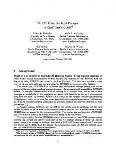
Phenotype 1
Phenotype 2
Phenotype 3
Fig. 1. Different stem cell phenotypes: (Phenotype 1) Bright boundary and dark interior. (Phenotype 2) Uniformly bright. (Phenotype 3) Poor contrast. features have a good performance when the background image consists of stationary objects with static pixels but they demonstrate a poor performance when the background image consists of non-stationary objects with dynamic pixels. A robust vision system can accurately model the non-stationary elements of the background if it could effectively use the temporal features [1, 5, 6]. Among the methods which use temporal features, Gaussian mixture model has been widely used and performed well to estimate non-stationary temporal background pixel distributions [5]. Different extensions of Gaussian mixture models have been introduced to improve its performance and reduce the running time [7, 8]. 2. PROPOSED METHOD Most tracking problems have an implicit, nonparametric model of the background; by developing a model for the background it is possible to find a classifier that labels each image pixel as background/not background. That is, the foreground is identified as that which is not background. In contrast, our cell tracking problem admits an explicit model of the foreground. We have developed a cell localizing model [9], however because of the low SNR of our problem, it is desired to remove all deterministic non-cell variations in the image (i.e. the background) before localizing the cells. Although cell localization appears to be a foreground/background classifier, there is a difference; we do not need to actually segment the image, only to identify the cell locations. Therefore we do not need to reliably classify each pixel definitively as fore-
ICIP 2008
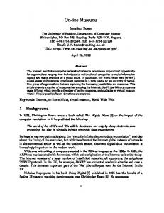
HSC Phenotype 1 - Dividing cells
(a)
(b)
(c)
(d)
(e)
(f)
Fig. 2. (a) Original HSC image. (b) Application of a circular mean square template. (c) Classification of circular mean square to cell and background classes by minimizing the inter-class variance. (d) Euclidean distance of cell pixels from the background. (e) Product of Circular mean square and Euclidean distance. (f) Cell center locations after thresholding the maxima of (e). ground or background, with unavoidable error around the cell margins. Rather to accurately estimate the background we only need to identify most background pixels, most of the time. 3. CELL LOCALIZATION Let I = (I1 , I2 , · · · , IK ) be a set of K images, each frame having N × L pixels. As the foreground cells are essentially outliers relative to the background statistics, to estimate the background we need to identify and remove the foreground, thus we remove the pixels which are associated to the located cells in each frame, specifically all pixels inside a rectangular m box with side length 2rkm , centred at (xm k , yk ). The image after removing the objects for a typical frame is depicted in Figs. 3(c). Except for Phenotype 1, the other HSC phenotypes cannot be modelled as an object with dark interior and bright boundary, therefore the proposed method in [9] performs poorly to detect HSCs of Phenotype 2 and Phenotype 3. To design a general method that could be applicable for detecting different HSC phenotypes investigated in this research, some common features among different HSC phenotypes must be extracted. All HSC phenotypes in this work can be characterized as an approximately circular object. Cell pixels have also high intensity variations against a uniform background. HSCs are modelled as a circular anomaly which is represented by a set of pixels with significant intensity variations against the uniform background. Assuming (x, y) and r as center coordinates and radius of a cell respectively, we construct the set G(zkm , Ik ), which returns the inside cell pixels G(z, I) = {Iij | (x − i)2 + (y − j)2 ≤ (r)2 }
(1)
from which we extract the sample mean of square intensities � ¯= G
g∈G
|G|
g2
To recognize cells from the uniform background, first (2) is ¯ is computed. The variance of applied to the cell image and G the pixels {g| g ∈ G} located inside a ring with radius r is � � � 2 2 2 g∈G g − [1,|G|] μ g∈G (g − μ) 2 = (3) σ = |G| |G| after simplification we have � 2 g∈G g ¯ G= = σ 2 + μ2 |G|
Thus for G located in the uniform background we find ¯ bkg = σ 2 whereas for G located inside a cell we have G bkg ¯ cell = σ 2 + μ2 For all of the different cell phenotypes G cell cell 2 , μ2cell are significantly higher than one or both of the σcell ¯ cell and as ¯ bkg T }
(19)
where � · � counts the number of elements in the set, and the threshold T is selected based on background statistics as T ≤ D < [size of clique = n2 ∗ K]
(20)
where a precise estimation of background would be possible. Eventually the estimated spatial illumination variations B is subtracted out from the temporal corrected sequence g as Fˆ = ˆ g − B.
(B1)
(B2)
(Proposed)
Fig. 4. The estimeted background by applying: B1, B2 (The method proposed in [6]), and Our proposed method. The proposed method estimated a uniform background in which cell boundary pixels are not presented, hence restoring the dynamic foreground robustly. 5. RESULTS We have applied the proposed cell detection and background estimation method to different sequences and different phenotypes of phase contrast HSC images. The results obtained by the proposed method is compared with: i) Spatio-temporal pixel-wise version of [1] that we call it B1. ii) The proposed method by Heikkila and Pietikainenin [6] as the most recent background modelling method with very promising results that we call it B2. iii) Frame-difference segmentation method. iv) Morphological averaging background estimation method in [10]. The proposed method outperforms the present background estimation methods including [1, 10], and the most recent background modelling approach based on texture information [6]. The original frames and located cells are depicted in Figs. 2 and 3 for dividing and non-dividing stem cells respectively. As we can observe nondividing and more challenging dividing cells are localized perfectly applying the proposed method. The estimated background images applying the proposed method, B1, and B2 are depicted in Figs. 4(a), (b) and (c). As it can be observed in Figs. 4(a) and (b), B1 and B2 methods fail to precisely estimate the background in the spatial locations where cells have slow motion dynamics. As a result cell boundary pixels are visible in the estimated background by these methods. In contrast, as we can observe in the estimated background by the proposed method, not only the well boundaries are precisely estimated, but there are very smooth variations over the background image. The proposed method also estimates background in locations where cells have slow motion dynamics precisely as we can observe in 4(c). 6. CONCLUSIONS A novel algorithm for cell detection/background estimation is proposed. The proposed method employs the detection re-
3043
sults to remove the foreground objects and estimates the background over 3-D residual sequence. The proposed method is applied to different HSC image sequences and generated promising results. Using temporal detection information, the proposed method outperforms the present background estimation methods including the most recent background modelling based on texture information [6]. The future work focused to design a recursive foreground segmentation-background estimation version of the proposed method in which the segmentation and estimation results will recursively be used to improve the performance of each other. 7. REFERENCES [1] C.R. Wren, A. Azarbayejani, T. Darrell, and A.P. Pentland, “Pfinder: Real-time tracking of the human body,” IEEE Tran. on PAMI, vol. 19, no. 7, pp. 780–785, 1997. [2] A. Elgammal, D. Harwood, and L. Davis, “Nonparametric model for background subtraction in proc. eur. conf. , 2000.,” in ECCV, 2000. [3] J. Kato, T. Watanabe, S. Joga, and A. Blake, “An hmm-based segmentation method for traffic monitoring movies,” IEEE Tran. on PAMI, vol. 24, no. 9, pp. 1291– 1296, 2002. [4] L. Li and M. Leung, “Integrating intensity and texture differences for robust change detection,” IEEE Transactions on Image Processing, vol. 11, pp. 105–112, 2002. [5] C. Stauffer and W.E.L. Grimson, “Adaptive background mixture models for real-time tracking,” in IEEE CVPR, 1999, pp. 246–252. [6] M. Heikkila and M. Pietikainen, “A texture-based method for modeling the background and detecting moving objects,” IEEE Tran. on PAMI, vol. 28, no. 4, pp. 657–662, 2006. [7] Z. Zivkovic, “Improved adaptive gaussian mixture model for background subtraction,” in IEEE ICPR, 2004, pp. 28–31. [8] D. S. Lee, “Effective gaussian mixture learning for video background subtraction,” IEEE Tran. on PAMI, vol. 27, no. 5, pp. 809830, 2005. [9] N. N. Kachouie, P. Fieguth, J. Ramunas, and E. Jervis, “Probabilistic model-based cell tracking,” International Journal of BioImaging, vol. 2006, pp. 1–10, 2006. [10] D. Koller, J. Weber, T. Huang, J. Malik, G. Ogasawara, B. Rao, and S. Russel, “Toward robust automatic traffic scene analysis in real-time,” in International Conference Pattern Recognition (ICPR), 1994, pp. 126–131.