Benchmarks Cloning: A Laboratory Manual. CSH Laboratory Press, Cold Spring Harbor, New York. 3.Treco, D. and V. Lundblad. 1997. Basic techniques of yeast genetics, p. 13.1.1-13.1.7. In F. Ausubel, R. Brent, R. Kingston, D. Moore, J. Seidman, J. Smith, and K. Struhl (Eds.), Current Protocols in Molecular Biology. John Wiley and Sons, New York.
This work was supported in part by National Institutes of Health grant no. DK02501 and by the American Digestive Health Foundation. This work is dedicated to the memory of Daniel Nathans and his trainees. Special thanks to Tim Schaefer for helpful discussions and to Phil Hieter for his gift of yeast strain YPH499. Address correspondence to Dr. Mark T. Worthington, MR-4 Building, Room 1036, 1 Lane Road, UVAHSC, Charlottesville, VA 22908, USA. e-mail:
[email protected] Received 2 August 2000; accepted December 2000.
Mark T. Worthington, Roger Qi Luo, and Jared Pelo University of Virginia Health Sciences Center Charlottesville, VA, USA
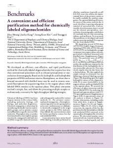
Incorporation of Sodium Sulfite into Extraction Protocol Minimizes Degradation of Acacia DNA BioTechniques 30:742-748 (April 2001)
The isolation of high-quality DNA from plants, especially woody plants, is often difficult. Although there are many different methods for DNA extraction in use, it is not unusual to find plant species for which commonly used extraction methods do not produce goodquality DNA. The main reason for the inability to obtain high-quality genomic DNA from Australian native plants is the high levels of cytosolic compounds 742 BioTechniques
such as polysaccharides, polyphenolics, and tannins. A reliable DNA extraction procedure for eucalypts (5) was developed by modification of a method of Wagner et al. (9) and has been successfully used to extract high-quality DNA from other Australian native species such as Lambertia orbifolia (4) and Macrozamia riedlei (6). The procedure is based on a crude organelle isolation from which the DNA is extracted using a cetyltrimethylammonium bromide (CTAB) method. Modifications to this protocol have been used to obtain high-quality DNA from other species. The addition of high levels of salt to the extraction protocol, followed by a caylase treatment, resulted in the removal of polysaccharide contamination from Acacia mangium DNA (2). In Melaleuca alternifolia, degradation of DNA during restriction enzyme digestion was prevented by the incorporation of a differential solvent precipitation into the extraction procedure (3). The use of the modified Wagner method (5) to isolate DNA from a number of Acacia species (A. acuminata, A. anfractuosa, A. lobulata, A. sciophanes, and A. verricula) resulted in degradation of DNA immediately after extraction, as determined by agarose gel electrophoresis (Figure 1). Several modifications to the protocol were tested, including the ones noted above, but the best results were obtained by the addition of 0.1 M sodium sulfite to the two buffers used in the extraction procedure (the initial extraction buffer and the buffer used to resuspend the organelle pellet). No other modifications to the modified Wagner extraction procedure were necessary when sodium sulfite was used, and no other modifications were successful when sodium sulfite was not used. The addition of sodium sulfite resulted in extraction of highquality DNA that was not degraded, while the standard protocol produced DNA that was degraded (Figure 1). The DNA extracted with sodium sulfite was suitable for use in restriction fragment length polymorphism (RFLP) analyses. Figure 2 shows the effect on RFLP analysis of the DNA extracted with and without the addition of sodium sulfite to the buffers. The high molecular weight DNA extracted with the addition of
sodium sulfite to the extraction buffers gives a clean hybridization profile, while the degraded DNA obtained from the standard method gives a poor hybridization profile with smeared lanes and a weak hybridization signal. The direct action of sodium sulfite in the extraction procedure is unclear. Sodium sulfite is a reducing agent for polyphenol oxidase, and its presence will prevent the production of polyphenolic compounds. It seems likely that there is a nuclease present in the extraction solution that becomes bound with a phenolic compound and is not removed by the organic extraction. Prevention of phenolic formation by the addition of sodium sulfite would leave the nuclease free to be removed during the organic extraction. Alternatively, there may be a compound in the phenolic pathway that has nuclease properties. Other studies of Acacia species using RFLP techniques have also encountered problems with the quality of DNA. Polysaccharide contamination was problematic in A. mangium(2) and a number of other Acacia species (1), and another study required purification of DNA through cesium chloride gradi-
Figure 1. A. verricula DNA extracted with and without addition of sodium sulfite to the buffers in the modified Wagner extraction method. Lanes 1–5, DNA extracted from the Merredin population without using sodium sulfite in the extraction buffers. Lanes 6–10, DNA extracted from the Merredin population with the addition of sodium sulfite to the extraction buffers. Vol. 30, No. 4 (2001)
Benchmarks ents, although the authors did not indicate why such purification was necessary (7). Degradation of DNA was not a problem in DNA extractions from A. mangium (2), and the problem of polysaccharide contamination that was encountered in A. mangiumdid not occur in the Acacia species discussed here. These species are from the southwest of Western Australia and are taxonomically quite distinct from the tropical A. mangium(Bruce Maslin, personal communication), and it is not surprising that they would contain different cytosolic compounds. These results highlight that it is not valid to make assumptions
about DNA isolation for previously untested plant species, even when they are members of the same genus. The addition of sodium sulfite in buffers during DNA extraction is a small modification to the method, yet it had a significant effect on the quality of the DNA extracted, such that it was suitable for RFLP analysis and could be stored without degradation. The use of sodium sulfite during DNA extraction is recommended in plant species that contain high levels of polyphenolics and tannins and exhibit isolation of poor-quality DNA. REFERENCES
Figure 2. Hybridization profiles of A. verricula DNA extracted with and without addition of sodium sulfite to the buffers in the modified Wagner extraction method. DNA was digested with EcoRV, electrophoresed for 16 h at 30 V, transferred to nylon membrane, and hybridized with a petunia chloroplast probe P6 (8). Lanes 1–5, DNA extracted with sodium sulfite added to the extraction buffers. Lanes 6–10, DNA extracted without adding sodium sulfite to the buffers.
1.Bukhari, Y.M., K. Koivu, and P.M.A. Tigerstedt. 1999. Phylogenetic analysis of Acacia (Mimosaceae) as revealed from chloroplast RFLP data. Theor. Appl. Genet. 98:291-298. 2.Butcher, P.A., G.F. Moran, and H.D. Perkins. 1998. RFLP diversity in the nuclear genome of Acacia mangium. Heredity 81:205-213. 3.Butcher, P.A., M. Byrne, and G.F. Moran. 1995. Variation within and among the chloroplast genomes of Melaleuca alternifolia and M. linariifolia (Myrtaceae). Plant Syst. Evol. 194:69-81. 4.Byrne, M., B. Macdonald, and D. Coates. 1999. Divergence in the chloroplast genome and nuclear rDNA of the rare Western Australian plant Lambertia orbifolia Gardner (Proteaceae). Mol. Ecol. 8:1789-1796. 5.Byrne, M., G.F. Moran, and W.N. Tibbits. 1993. Restriction map and maternal inheritance of chloroplast DNA in Eucalyptus nitens. J. Heredity 84:218-220. 6.Byrne, M., M. Waycott, A.A. Hobbs, and S.H. James. 1997. Variation in ribosomal DNA within and between populations of Isotoma petraea and Macrozamia riedlei. Heredity 79:578-583. 7.Robinson, J. and S.A. Harris. 2000. A plastid DNA phylogeny of the genus Acacia Miller (Acacieae, Leguminoseae). Bot. J. Linnean Soc. 132:195-222. 8.Sytsma, K.J. and L.D. Gottlieb. 1986. Chloroplast DNA evolution and phylogenetic relationships in Clarkia sect. Peripetasma (Onagraceae). Evolution 40:1248-1261. 9.Wagner, D.B., G.R. Furnier, M.A. SaghaiMaroof, S.M. Williams, B.P. Dancik, and R.W. Allard. 1987. Chloroplast DNA polymorphisms in lodgepole and jack pines and their hybrids. Proc. Natl. Acad. Sci. USA 84:2097-2100.
Address correspondence to Dr. Margaret Byrne, CALMScience, W.A. Herbarium, Department of Conservation and Land Management, Locked Bag 104, Bentley Delivery Centre, WA 6983 Australia. e-mail:
[email protected] Vol. 30, No. 4 (2001)
Benchmarks Received 27 July 2000; accepted 27 November 2000.
Margaret Byrne, Bronwyn Macdonald, and Michael Francki 1 Department of Conservation and Land Management Bentley Delivery Centre 1The University of Western Australia Crawley, WA, Australia
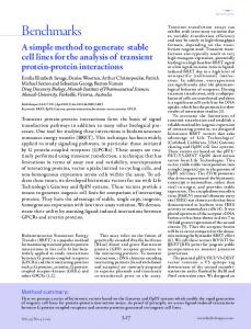
Use of Inexpensive Dyes to Calibrate and Adjust Your Microarray Printer BioTechniques 30:748 (April 2001)
A critical factor in the preparation of cDNA microarrays is the calibration and adjustment of the array printer to optimize spotting. Here are a couple techniques to help you detect the quality of your print, the proper calibration, and the condition of your printing pins. A simple way to determine the quality of the array spots is to examine the salt deposits remaining after the droplet of printer material has dried. However, this method does not necessarily reflect the morphology of the nucleic acid on the array because of the drying effects. Another common method includes the use of free Cy 3-dCTP/dUTP or Cy 5dCTP/dUTP dyes (Amersham Pharmacia Biotech, Piscataway, NJ, USA) or dyes containing oligonucleotides, followed by fluorescence scanning (1). Here, we describe an inexpensive alternative to using expensive fluorescent probes for the calibration and adjustment of pin-type array printers. Using a Stanford cDNA microarray printer (http://cmgm.stanford.edu/pbrown/ mguide/index.html) with ChipMaker 3 spotting pins (TeleChem International, Sunnyvale, CA, USA), we compared the results of printing under two conditions, with and without DNA. The goal of the calibration and adjustment steps is to ob748 BioTechniques
Figure 1. Salt deposition ± DNA (at 0.25 mg/mL). (A) Zoomed area printed without DNA. (B) Zoomed area printed with 0.25 mg/mL of sheared Salmon sperm DNA. Spots in panel B seem to be more uniform due to additional viscosity from DNA.
tain uniform and consistent spots. We used plain red and blue food coloring dye in 1:1000 dilution of 3× standard saline citrate (SSC) with sheared salmon sperm DNA at 0.25 mg/mL to mimic the actual printing conditions. All fluorescent scans were performed on a Packard/GSI Lumonics 3000 scanner (GSI Lumonics, Northville, MI, USA), with 85% laser and 90% PMT settings (Figure 1). The quality of the DNA spots was further examined with DNA binding dyes (data not shown). In the future, with the help of food coloring dye, we are going to compare different types of printing buffers such as 50% dimethyl sulfoxide (DMSO) with and without DNA, formamide, and a new printing buffer that recently became available from Mosaic Technologies (Waltham, MA, USA) and Clontech Laboratories (Palo Alto, CA, USA).
REFERENCES 1.Hegde, P., R. Qi, K. Abernathy, C. Gay, S. Dharap, R. Gaspard, J.E. Hughes, E. Snesrud et al. 2000. A concise guide to cDNA microarray analysis. BioTechniques 29:548-562.
Address correspondence to Gregory Khitrov, Director of Gene Array Facility, The Rockefeller University Gene Array Facility, 1230 York Ave., Box 203, New York, NY 10021, USA. e-mail: khitrog@ rockefeller.edu Received 17 October 2000; accepted 27 November 2000.
Gregory Khitrov Rockefeller University New York, NY, USA
Vol. 30, No. 4 (2001)