Benchmarks ments of T7 DNA and determination of molecular weights by electrophoresis in neutral and alkaline gels. J. Mol. Biol. 110:119-146. 18.Myers, T.W. and D.H. Gelfand. 1991. Reverse transcription and DNA amplification by Thermus thermophilus DNA polymerase. Biochem. 30:7661-7666. 19.Myers, T.W. and C.L. Sigua. 1995. Amplification of RNA: high-temperature reverse transcription and DNA amplification with Thermus thermophilus DNA polymerase, p. 58-68. In M.A. Innis, D.H. Gelfand, and J.J. Sninsky (Eds.), PCR Strategies. Academic Press, New York. 20.Wu, W., L.E. Henderson, T.D. Copeland, R.J. Gorelick, W.J. Bosche, A. Rein, and J.G. Levin. 1996. Human immunodeficiency virus type 1 nucleocapsid protein reduces reverse transcriptase pausing at a secondary structure near the murine leukemia virus polypurine tract. J. Virol. 70:7132-7142.
Address correspondence to Dr. Gary F. Gerard, Transgenomic, Inc., 11 Firstfield Road, Suite E, Gaithersburg, MD 20878, USA. e-mail:
[email protected] Received 31 May 2002; accepted 9 July 2002.
Gary F. Gerard, Sarah Collins, and Michael D. Smith Invitrogen Frederick, MD, USA For reprints of this or any other article, contact
[email protected]
Cloning Method for Taxonomic Interpretation of T-RFLP Patterns BioTechniques 33:990-992 (November 2002)
Terminal RFLP (T-RFLP) is a recently introduced PCR-based tool for studying diversity among amplified DNA molecules with the same size (4). Terminal restriction fragment (TRF) patterns obtained using the T-RFLP technique are generated and analyzed in a procedure that combines PCR with a labeled primer, restriction digestion of the amplified DNA, and separation of fragments in an automated se990 BioTechniques
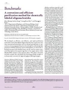
quencer. One of the most interesting for Soil (BIO101, Qbiogene, Carlsapplications of T-RFLP is its use for the bad, CA, USA) from 0.5 g fresh soil description of genetic diversity within a collected in a wood of cypress near Flobacterial community (2,3,7). In this rence (Italy). The 16S rDNA was amcase, the PCR is performed using plified in a 50-µL volume with 2 U Taq primers annealing to conserved seDNA polymerase (Dynazyme II; quences of 16S rRNA gene, and the Finnzyme, Espoo, Finland) using 27f TRF pattern obtained represents a “picprimer labeled with TET (4,7,2′,7′ture” of the number of different 16S tetrachloro-6-carboxyfluorescein) and rRNA genes present in the community 1495r primer (6). The amplified prod(i.e., different operating taxonomic ucts were purified with a QIAquick units). One of the main limits of this PCR purification kit (Qiagen, Valencia, technique is the difficulty of obtaining CA, USA), and 600 ng were digested taxonomic information of the organwith 20 U MspI or HhaI (New England isms responsible for a particular TRF. Biolabs, Beverly, MA, USA) for 3 h at To allow the interpretation of TRFs, an 37°C. The digested products (200 ng) online tool has been developed called were resolved by capillary electroTAP (T-RFLP Analysis Program; Refphoresis on an ABI 310 Genetic Anaerence 5) that performs in silico PCR lyzer using TAMRA 500 as size stanamplification and restriction of the 16S dard for GenScan (all from Applied rDNA sequences present in the RibosoBiosystems, Foster City, CA, USA) mal Database (RDPII, Center for analysis. The restricted products (200 Microbial Ecology at Michigan State ng) were ligated with 10 pmol compleUniversity, http://rdp.cme.msu.edu/ mentary double-stranded adapter with html/TAP-trflp.html#program). How10 U T4 DNA ligase (New England Biever, database matching of TRF sizes is olabs) for 10 h at 14°C in a 10-µL volimprecise and may not produce speume. One microliter of the ligation recies- or even genus-specific assignment action was PCR-amplified using 27f (2), and results can be experimentally verified indirectly only after a long screening of a 16S rDNA library. Here we present a simple and fast method that allows for an empirical taxonomic interpretation of TRFs. This method would be particularly useful when a detailed taxonomic description of a bacterial community is needed. Figure 1 depicts the flow chart of the methodology. The method is based on the ligation of the TRFs with oligonucleotide adapters, subsequent PCR-amplification, cloning, and final sequencing of the single TRFs. Figure 1. Schematic representation of the cloning system. The We used this tech- nucleotide sequences of the double-stranded adapters used are as folnique to identify the bac- lows. For MspI, 5′-CGCTCGTAGACTGCGTACC-3′ and 3′-GAGterial groups represented CATCTGACGCATGGTTAA-5′. For HhaI, 5′-GAGCATCTGACGin a TRF pattern ob- CATGGTTAA-3′ and 3′-GCCTCGTAGACTGCGTACC-5′. The amplification of the ligated products was performed using the tained from soil DNA. PCR following temperature profile in a Primus 96 Thermal Cycler DNA was extracted with (MWG-AG-Biotech, Ebersberg, Germany): 2 min at 94°C, followed the Fast Prep DNA Kit by 35 cycles of 30 s at 94°C, 30 s at 55°C, and 30 s at 72°C. Vol. 33, No. 5 (2002)
Benchmarks and pMspI (5′-GGTACGCAGTCTACGAGCGG-3′) or pHhaI (5′-CCATGCGTCAGATGCTCCGC-3′) reverse primers, which were designed on the sequence of the double-stranded adapter and on the half restriction site of MspI and HhaI, respectively. The amplified fragments were resolved on a 2% agarose gel, and the bands at molecular weights from 30 to 300 bp were eluted from the gel (MinElute Gel Extraction Kit; Qiagen) and cloned in the pDrive vector (Qiagen PCR Cloning Kit; Qiagen). This procedure allows for great reduction and faster screening of the clone library needed, when compared with cloning the original 16S rDNA products before digestion. A rapid screening for clones of appropriate size corresponding to specific TRF peaks was performed by PCR, using the standard M13 primer pair (M13forward primer labeled with TET), and the size of the cloned TRFs was verified by capillary electrophoresis of the amplified products in the ABI 310 Genetic Analyzer. All cloned and sequenced
fragments were represented in the TRFLP profile. The most abundant fragments in the original T-RFLP were found to be predominant also in the library. However, a bias toward a higher content of fragments around 100 bp was found, probably due to PCR amplification. Twelve selected clones (six for each of the two enzymes) were then sequenced using the BigDye Terminator (v.2) chemistry (Applied Biosystems). Sequences were used to perform a BLASTn search (1) on the GenBankEMBL-DDBJ database. All the obtained fragments were found to be significantly similar to sequences present in GenBank, with score and E-value varying from 44 and 1e-3 for the smallest fragment (33 bp) to 321 and 1e-85 for larger fragments (202 bp). In general, the longer the fragment, the higher the score is. The taxonomic interpretation derived from BLAST matching was then compared with that obtained by using the TAP tool; in four cases out of the 12 analyzed (TRFs produced with MspI at 64, 202, 212, and 33 bp), TAP failed to show any matching, while the BLAST matching of the sequences found that those fragment were similar to 16S rDNA bacterial sequences present in GenBank (with score and E-values varying from 46 and 1e-3 to 321 and 1e-85). In two cases, the BLAST matching of the sequenced TRF was different from the matching produced by TAP (TRFs produced with MspI at 174 bp and with HhaI at 38 bp). In the remaining cases, the two procedures were well in agreement (TRFs produced with MspI at 68 and 145 bp and with HhaI at 82, 86, 89, and 95 bp). The reported results demonstrate that the direct cloning of TRFs could be a useful tool, complementary to TAP matching, for the taxonomic interpretation of TRFLP community profiles. REFERENCES 1.Altschul, S.F., W. Gish, W. Miller, E.W. Myers, and D.J. Lipman. 1990. Basic local alignment search tool. J. Mol. Biol. 215:403410. 2.Kitts, C.L. 2001. Terminal restriction fragment patterns: a tool for comparing microbial communities and assessing community dynamics. Curr. Issues Intes. Microbiol. 2:17-25. 3.Liu, W.-T., T.L. Marsh, H. Cheng, and L.J., Forney. 1997. Characterization of microbial
diversity by determining terminal restriction fragment length polymorphism of genes encoding 16 rRNA. Appl. Env. Microbiol. 63:4516-4522. 4.Marsh, T.L. 1999. Terminal restriction fragment length polymorphism (T-RFLP): an emerging method for characterizing diversity among homologous populations of amplification products. Curr. Op. Microbiol. 2:323-327. 5.Marsh, T.L., P. Saxman, J. Cole, and J. Tiedje. 2000. Terminal restriction fragment length polymorphism analysis program, a Web-based research tool for microbial community analysis. Appl. Env. Microbiol. 66:3616-3620. 6.Mengoni, A., R. Barzanti, C. Gonnelli, R. Gabbrielli, and M. Bazzicalupo. 2001. Characterization of nickel-resistant bacterial communities isolated from serpentine soil. Environ. Microbiol. 3:691-698. 7.Osborn, A.M., E.R.B. Moore, and K.N. Timmis. 2000. An evaluation of terminal-restriction fragment length polymorphism (TRFLP) analysis for the study of microbial community structure and dynamics. Environ. Microbiol. 2:39-50.
Address correspondence to Dr. Alessio Mengoni, Dipartimento di Biologia Animale e Genetica, Università di Firenze, via Romana 17, I-50125 Firenze, Italy. e-mail:
[email protected] Received 10 June 2002; accepted 5 August 2002.
Alessio Mengoni, Eva Grassi, and Marco Bazzicalupo Università di Firenze Firenze, Italy
Molecular Cloning Using the Interrupted Recognition Sequence of BplI to Select for Recombinant Molecules BioTechniques 33:992-996 (November 2002)
The cloning of DNA fragments into a uniquely restricted target vector is a common procedure for constructing genomic DNA or cDNA libraries. The recovery of recombinant molecules is Vol. 33, No. 5 (2002)