Changes in surface roughness and work function of indium-tin-oxide due to KrF excimer laser irradiation Yow-Jon Lina兲 Institute of Photonics, National Changhua University of Education, Changhua 500, Taiwan, Republic of China
Iain D. Baikie KP Technology, Ltd., Wick, Caithness, KW1 5EH, United Kingdom
Wei-Yang Chou and Shih-Ting Lin Institute of Electro-Optical Science and Engineering, National Cheng Kung University, Tainan 701, Taiwan, Republic of China
Hsing-Cheng Chang Department of Automatic Control Engineering, Feng Chia University, Taichung 407, Taiwan, Republic of China
Yao-Ming Chen and Wen-Fung Liu Department of Electrical Engineering, Feng Chia University Taichung 407, Taiwan, Republic of China
共Received 21 January 2005; accepted 17 May 2005; published 22 July 2005兲 In this study, from the observed x-ray photoelectron spectroscopy and atomic force microscopy results, and Kelvin probe measurements, it is suggested that the induced indium-tin-oxide 共ITO兲 surface chemical changes by KrF excimer laser irradiation had strong effects on the surface work function 共SWF兲 and surface roughness of ITO. During the laser irradiation, the incorporation of O22− peroxo species or the dissolution of oxygen species near the ITO surface leads to the reduction of the surface roughness and an increase in the SWF. In addition, it is worth noting that the laser-irradiated ITO sample has an excellent stability in the SWF. © 2005 American Vacuum Society. 关DOI: 10.1116/1.1953670兴
I. INTRODUCTION Indium-tin oxide 共ITO兲 with high work function 共⬃5.1– 5.2 eV兲 共Refs. 1–3兲 is a transparent degenerate semiconductor which is often used as an anode material for optoelectronics such as organic light emitting diodes 共OLEDs兲4 and GaN-related light emitting diodes.5,6 The surface work function 共SWF兲 of ITO is very critical to the performance of OLED because it affects the energy barrier height at the interface of ITO with the organic semiconductors, playing a role in reducing the operating voltage of devices.7–9 Consequently, studies of ITO surface modification for the purpose of enhancing hole injection are plentiful, with a focus on understanding how the SWF is altered by passivation with surface-active species or by acids, bases, or plasma cleaning procedures.9–13 The possibility to tailor the ITO SWF by various methods is of great interest in the fabrication of optoelectronic devices where ohmic charge injection is desired. Simple ways of tuning the SWF to match the organic energy levels is of technological importance. On the other hand, the control of surface morphology of ITO is one of the most serious problems to be resolved for the fabrication of OLEDs with a long lifetime.14,15 Because all the functional organic thin films are deposited on ITO, the surface morphology of ITO is directly transferred to the morphology of the organic a兲
Author to whom all correspondence should be addressed; electronic mail:
[email protected]
1305
J. Vac. Sci. Technol. A 23„5…, Sep/Oct 2005
layers. Therefore, the uneven surface of the hole injecting electrode can have negative effects on device performance by affecting the morphology of the organic thin film.14 In this study, we report the variation of the ITO SWF and the change in the surface roughness of ITO, due to KrF excimer laser irradiation.
II. EXPERIMENTAL PROCEDURES ITO samples, which were purchased from Wintek Corporation, with the film thickness of 26 nm and sheet resistance of 71.38 ⍀ / 䊐, were used in this work. All samples were first cleaned by ultrasonic agitation in ethanol and subsequently in acetone followed by mechanical scrubbing in deionized water using a detergent. The samples were then cleaned in an ultrasonic bath using a water solution of the same detergent and rinsed several times by ultrasonic agitation in water. After drying under a dry nitrogen flow 共referred to as ascleaned ITO兲, some of the as-cleaned ITO samples were irradiated in air for 25 min, by multiple pulses from a KrF excimer laser 共referred to as 25-min-irradiated ITO兲. A KrF 共 = 248 nm兲 excimer laser was used as the source of irradiation. Its energy density is 26 mJ/ cm2 and pulse duration is 50 ns at repetition rate of 50 Hz. The morphology of the irradiated and nonirradiated ITO samples was analyzed using an atomic force microscopy 共AFM兲. A Kelvin probe 共KP兲
0734-2101/2005/23„5…/1305/4/$22.00
©2005 American Vacuum Society
1305
1306
Lin et al.: Changes in surface roughness and work function
1306
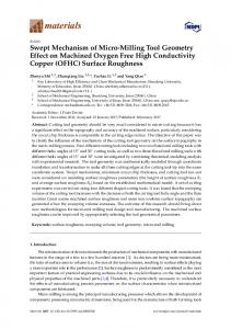
FIG. 1. AFM images of 共a兲 as-cleaned ITO surface and 共b兲 25-min-irradiated ITO surface.
and x-ray photoelectron spectroscopy 共XPS兲 were employed to examine the changes in the SWF and surface chemical bonding states of ITO. III. EXPERIMENTAL RESULTS AND DISCUSSION Figure 1 shows two AFM images 共1 m ⫻ 1 m兲 of ITO films. The root-mean-square surface roughness 共Rrms兲 for 25min-irradiated ITO films is 0.51 nm compared to a value of 1.04 nm for as-cleaned ITO films. This indicates that the 25-min-irradiated ITO shows smaller Rrms than that of the as-cleaned ITO. Park et al. have suggested that the reduction of surface roughness could lead to the enhancement of the performance of OLEDs.16 Figure 2 shows the observed SWF image of ITO films by KP. The model number of the equipment is KP Technology, Scanning Kelvin probe, SKP0401. Prior to scan for ITO samples, the gold 共Au兲 surface was scanned via the tip. We find that the Au work function is higher than that of the tip 共262.0± 5.0 meV兲. Next, the as-cleaned and 25-minirradiated ITO samples were placed together. The as-cleaned ITO is on the right-hand side and the 25-min-irradiated ITO is on the left-hand side. In Fig. 2, we find that the SWF of the as-cleaned ITO is higher than that of the tip 共185.0± 40.1 meV兲 and the SWF of the 25-min-irradiated ITO is higher than that of the tip 共311.6± 8.5 meV兲. If the work function of Au is assumed to be equal to 5.1 eV, the SWF of the as-cleaned and 25-min-irradiated ITO samples is calculated to be 5.02 and 5.15 eV, respectively. It is noteworthy that the 25-min-irradiated ITO sample has an excellent SWF stability. SWF data are homogeneous coverage, almost metal-like Fermi level, as shown in Fig. 2共b兲. On the contrary, the as-cleaned ITO sample does not have a good SWF stability. SWF data are due to inhomogeneous coverage, as shown in Fig. 2共a兲. J. Vac. Sci. Technol. A, Vol. 23, No. 5, Sep/Oct 2005
In order to clarify the surface chemical changes of ITO after laser irradiation, the XPS spectra of In 3d5/2, Sn 3d5/2, O 1s, and C 1s were measured via XPS. Charging effects were corrected by referencing the binding energies to that of the adventitious C 1s line at 285 eV.17 Figures 3共a兲 and 3共b兲 show normalized XPS spectra of O 1s core level for ITO samples with and without laser irradiation, respectively. Figure 3共c兲 shows the difference between the O 1s spectrum of the as-cleaned ITO and the O 1s spectrum of the 25-minirradiated ITO. It can be seen that a few O-In bonds 共530.6
FIG. 2. 共Color online兲. Observed SWF image of ITO films by KP. 关共a兲 as-cleaned ITO; 共b兲 25-min-irradiated ITO; ⌬WF: the difference in work function between the tip and the observed object兴.
1307
Lin et al.: Changes in surface roughness and work function
1307
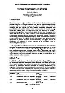
radiated samples was plotted versus the detection angle 共兲. In this work, the exact sensitivity factor for ITO was not known, so the observed O / 共In+ Sn兲 ratio of the as-cleaned sample at the angle of ⬃0° was taken as 1. The O / 共In + Sn兲 ratio increased after laser irradiation, and the increase was more pronounced. According to the experimental results shown in Figs. 1–4, we deduce that the increase of the SWF and the reduction of Rrms are deeply related to the oxygen enrichment near the ITO surface. For 25-min-irradiated samples, the O / 共In+ Sn兲 ratio near the surface region increases, due to the incorporation of O22− peroxo species or the dissolution of oxygen species near the ITO surface, as shown in Fig. 4. We further deduce that induced oxidation of ITO by laser irradiation might be adequate to beneficially effect its electrode properties. These changes provide a rational guideline for development of new processing methodologies to enhance ITO-based optoelectronic devices. FIG. 3. XPS spectra of O 1s core level for 共a兲 as-cleaned and 共b兲 25-minirradiated ITO samples. 共c兲 The difference energy spectrum 共25-minirradiated minus as cleaned兲.
eV兲 共Refs. 9 and 18兲 existed on the as-cleaned and 25-minirradiated ITO surfaces. XPS studies of the 25-min-irradiated sample showed different features compared to those observed in the sample without laser irradiation. In Fig. 3共c兲, we can see that the difference energy spectrum is a peak posited at ⬃532 eV, which is attributed to the adsorption and/or incorporation of O22− peroxo species near the ITO surface.9,19 Choi and Thompson have suggested that the peak at ⬃532 eV may be attributed to oxygen species dissolved in the material or to adsorbed oxygen.20 However, Ranke and Kuhr indicated that O22− may desorb at 300 K.19 As a result, we deduced that oxygen species may be dissolved in the ITO or O22− peroxo species incorporated into the ITO during the laser irradiation, which might lead to an increase in the intensity of the peak at ⬃532 eV and the O-rich formation near the ITO surface region. In Figure 4, the O / 共In+ Sn兲 ratio for irradiated and nonir-
FIG. 4. Change of relative atomic ratio 关O / 共In+ Sn兲兴 on the ITO surfaces as a function of detection angle. JVST A - Vacuum, Surfaces, and Films
IV. SUMMARY In conclusion, XPS, KP, and AFM measurements have been carried out to investigate changes in the surface atomic composition, SWF and Rrms of ITO by the surface treatment using laser irradiation. The laser irradiation not only led to an increase in the SWF, but resulted in the reduction of Rrms, which could be useful for enhancing the performance of OLEDs. In addition, it is worth noting that the 25-minirradiated ITO sample has an excellent stability in the SWF. ACKNOWLEDGMENTS This project is supported by National Science Council of Taiwan, Republic of China. The KP measurements were kindly provided by KP Technology, Ltd. 1
L. Zuppiroli, L. Si-Ahmed, K. Kamaras, F. Nűesch, M. N. Bussac, D. Ades, A. Siove, E. Moons, and M. Grätzel, Eur. Phys. J. B 11, 505 共1999兲. 2 J. S. Kim, B. Lägel, E. Moons, N. Johansson, I. D. Baikie, W. R. Salaneck, R. H. Friend, and F. Cacialli, Synth. Met. 111/112, 311 共2000兲. 3 E. Moons, A. Goossens, and T. Savenije, J. Phys. Chem. B 101, 8492 共1997兲. 4 J. Shinar, Organic Light-Emitting Devices 共Springer, Berlin, 2003兲. 5 S. Y. Kim, H. W. Jang, and J. L. Lee, Appl. Phys. Lett. 82, 61 共2003兲. 6 Y. C. Lin, S. J. Chang, Y. K. Su, C. S. Chang, S. C. Shei, J. C. Ke, H. M. Lo, S. C. Chen, and C. W. Kuo, Solid-State Electron. 47, 1565 共2003兲. 7 C. C. Wu, C. I. Wu, J. C. Strum, and A. Kahn, Appl. Phys. Lett. 70, 1348 共1997兲. 8 M. G. Mason, L. S. Hung, C. W. Tang, S. T. Lee, K. W. Wong, and M. Wang, J. Appl. Phys. 86, 1688 共1999兲. 9 K. H. Lee, H. W. Jang, K. B. Kim, Y. H. Tak, and J. L. Lee, J. Appl. Phys. 95, 586 共2004兲. 10 A. L. Swint and P. W. Bohn, Appl. Phys. Lett. 84, 61 共2004兲. 11 D. J. Milliron, I. G. Hill, C. Shen, A. Kahn, and J. Schwartz, J. Appl. Phys. 87, 572 共2000兲. 12 F. Nűesch, L. J. Rothberg, E. W. Forsythe, Q. T. Le, and Y. Gao, Appl. Phys. Lett. 74, 880 共1999兲. 13 I. M. Chan, W. C. Cheng, and F. C. Hong, Appl. Phys. Lett. 80, 13 共2002兲. 14 K. B. Kim, Y. H. Tak, Y. S. Han, K. H. Baik, M. H. Yoon, and M. H. Lee, Jpn. J. Appl. Phys., Part 2 42, L438 共2002兲. 15 H. Kim, A. Piqué, J. S. Horwitz, H. Mattoussi, H. Murata, Z. H. Kafafi, and D. B. Chrisey, Appl. Phys. Lett. 74, 3444 共1999兲. 16 N. G. Park, M. Y. Kwak, B. O. Kim, O. K. Kwon, Y. K. Kim, B. You, T.
1308
Lin et al.: Changes in surface roughness and work function
W. Kim, and Y. S. Kim, Jpn. J. Appl. Phys., Part 1 3, 1523 共2002兲. P. J. Hartlieb, A. Roskowski, R. F. Davis, and R. J. Nemanich, J. Appl. Phys. 91, 9151 共2002兲. 18 J. F. Moulder, W. F. Strickle, P. E. Sobol, and K. D. Bomben, Handbook 17
J. Vac. Sci. Technol. A, Vol. 23, No. 5, Sep/Oct 2005
1308
of X-Ray Photoelectron Spectroscopy 共Perkin-Elmer, Eden Prairie, MN, 1992兲. 19 W. Ranke and H. J. Kuhr, Phys. Rev. B 39, 1595 共1989兲. 20 J. G. Choi and L. T. Thompson, Appl. Surf. Sci. 93, 143 共1996兲.