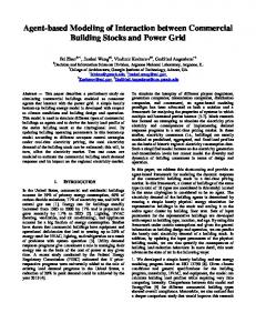
superfamily enzymes which play crucial roles in aldehyde metabolism from ..... Structure of the aldehyde dehydrogenase and docking validation. (A). Surface ...
International Journal of Computer Applications (0975 – 8887) Volume 179 – No.35, April 2018
Computer-Aided Modeling of Interaction between Aldehyde Dehydrogenase and Garcinia Biflavonoids Tomisin Happy Ogunwa Centre for Bio-computing and Drug Development, Adekunle Ajasin University, Akungba-Akoko, Ondo State, Nigeria Department of Biochemistry, Adekunle Ajasin University, Akungba-Akoko, Ondo State, Nigeria
ABSTRACT The Garcinia-derived biflavonoids GB1, GB2, kolaflavanone (together known as kolaviron) and morelloflavone have been reported for various bioactivities including protection of cellular tissues against damages from toxic compounds. In this study, computer-aided procedures were used to model the interaction of these naturally-occurring biflavonoids with aldehyde dehydrogenase (ALDH). This study sought to validate these compounds as potential inhibitors of the protein towards treatment/prevention of ALDH related pathophysiologically-associated diseases. Detailed observation of the results obtained divulged that the Garcinia biflavonoids actually inserted only one of their monoflavonoid subunits into the putative substrate-binding pocket while the other subunit occupied the hydrophobic binding region in a manner that can prevent substrate access. Some amino acid residues found in the protein loop flanking the ligands within the putative binding pocket established interactions with the biflavonoids via hydrophilic bonds. Several hydrophobic interactions between the aromatic rings of the dimeric form of flavonoids and non-polar residues of the protein were observed to play crucial role in stabilizing the biflavonoids within the active site. Phe314 might further participate in π-π stacking with the biflavonoids aromatic rings. The relatively large size of the biflavonoids enhances their occupation of the binding pocket, however having less interference with the solvent-exposed region. The compounds are therefore predicted as unique competitive inhibitors of ALDH.
Keywords Biflavonoids, Garcinia species, molecular docking, Aldose dehydrogenase, interaction
1. INTRODUCTION Aldehyde dehydrogenases (ALDHs) are members of superfamily enzymes which play crucial roles in aldehyde metabolism from exogenous and endogenous sources. Human ALDH participates in important physiological and toxicological reactions [1], [2]. The Aldehyde dehydrogenases 2 (ALDH2) is a significant enzyme in the oxidation of acetaldehyde, an essential step in alcohol metabolism. Accumulation of acetaldehyde, which can occur after ethanol consumption, may result into development of unpleasant physiological effects comprising nausea, facial flushing, and tachycardia [1], [3]. This well-established function of ALDH in alcohol metabolism became a bedrock that drove research efforts towards identification of ALDH inhibitors. Moreover, ALDH isozymes have been reported for their importance in oxidizing lipid peroxidation-derive aldehydes which are reactive towards cellular components [4]. Hence, the enzymes contribute to maintenance of cellular homeostasis [5]. In addition, the proteins have the ability to interfere with neural function, most importantly in dopaminergic nerves [6], [7]. Both ALDH1A1 and ALDH2 have been implicated in this
regard. Over the years, there have been observed correlation between elevated expression and activity of ALDH isozymes in various human cancers [1]. The effects have also been associated with cancer relapse [8]. Hence, the search for inhibitors of these enzyme to treat human diseases is a direct consequence of their significant physiological and toxicological roles. The activity of ALDH is constitutively expressed in various mammalian organs [9]. To date, the highest reported level is in the liver which is followed by the kidney, uterus and the brain [9]. Recent reports linked ALDH to a multidrug resistant efflux transporter (MDR) to drugs which remains a major challenge in the chemotherapeutic treatment [10]. The protein was associated with drug resistance [11]. Increased ALDH activity during cancer treatment has been demonstrated to be a mechanism of cytostatic drug resistance as well as an indicator of patient poor survival [1], [7] [12]. Hence, ALDH specific, competitive inhibitor that may not require enzymatic activation is believed to be suitable for in vivo inhibition of this protein in pathophysiological response that underlie some human diseases [12]. Members of the Garcinia genus are important plants with numerous reports on their medicinal application in the literature [13], [14]. Garcinia kola, an African indigenous plant, has been used for folkloric medicine over many decades. The major flavonoid extract of this plant is known as kolaviron, containing mainly biflavonoids such as Garcinia biflavonoid 1 (GB1), Garcinia biflavonoid 2 (GB2) and kolaflavanone [15], [16], [17]. The various beneficial health effects and bioactivities of kolaviron have been described in recent time. These include anticancer, anti-inflammatory, antidiabetic, hepatoprotective, anti-toxic, anti-oxidative, antiviral, antimicrobial, anti-aging and anti-neurodegenerative disease effects [18], [19], [20], [21], [22], [23]. Morelloflavone, another biflavonoid identified in Garcinia species like Garcinia morella, Garcinia dulcis, Garcinia kola, Garcinia subelliptica, Garcinia livingstonei, Garcinia multiflora and Garcinia brasiliensis, is a potent biologically active chemical compound for which diverse pharmacological properties have been ascribed. Morelloflavone was reported to inhibit HMG-COA reductase enzyme as anti-hyperlipidemic protein [24], [25]. The biflavonoid is also said to block the enzymatic functions of phospholipase A2, tyrosinase, trypsin, cruzain, fatty acid synthase and papain [26], [27], [28], [29]. Moreover, the anti-fungal, anti-plasmodia, antitumor, antioxidant, antiviral and anti-inflammatory activities of morelloflavone have been documented [26], [30], [31], [32], [33], [34]. Since efforts in the search for natural inhibitors of ALDH is still encouraged due to their cheap cost and possible reduced adverse effects, the current study is aimed at elucidating the molecular mechanism of interaction between Garcinia biflavonoids (GB1, GB2, kolaflavanone and morelloflavone) and human aldehyde dehydrogenase.
18
International Journal of Computer Applications (0975 – 8887) Volume 179 – No.35, April 2018
2. MATERIALS AND METHOD 2.1 Preparation of target protein The crystal structure of human ALDH (PDB ID: 6B5I) used in this research was retrieved from the Brookhaven protein data bank (http://www.rcsb.org/pdb). The was visualized using the molecular graphics program PyMol intended for the structural visualization of proteins and was found in complex with inhibitor CM121, a potent competitive inhibitor of the enzyme. Crystallographic water molecules which were found with structure were then deleted prior molecular docking procedures. The active site of the protein was identified with reference to the co-crystallized ligand.
2.2 Ligands selection, preparation and optimization The five (5) ligands used in this docking study were identified and selected from the literature [18], [25]. Out of these compounds, four (4) were biflavonoids obtained from Garcinia species while the other ligand was the co-crystallized ligand which was adopted as a reference ligand. The chemical structures of these compounds: Garcinia GB1 (CID: 161087), Garcinia GB2 (CID: 161259), kolaflavanone (CID: 155169) and morelloflavone (CID: 5464454) were retrieved from NCBI PubChem database (http://www.ncbi.nlm.nih.gov/pccompound) and prepared using ChemAxon software (https://www.chemaxon.com). Marvin-Sketch v15.11.30 was used to sketch the 2Dcoordinates of the ligands. The structures were then cleaned up in 2D and converted to 3D geometry using the Conformers suit of the software based on the Merck molecular force field (MMFF94). The MDL SDfile (.sdf) format of the ligands were finally docked into the active site of the targets using the AutoDock 4.2.
2.4 Validation of docking protocol Molecular docking validation was carried out as previously described [24], [35]. Briefly, the ligand found at the binding site of the experimentally determined protein crystal was deleted. Then, the ligand (.sdf format) was separately prepared using Marvin sketch as described above and redocked into the active site. The binding pose and molecular interaction pattern was compared to that of the x-ray diffraction crystal structure.
2.5 Molecular docking and scoring For ligand docking and target-ligand complex analysis, Autodock Vina suite on PYMOL was used [36], [37]. First, based on the already present co-crystallized ligand in the pdb file, the inhibitor binding site was defined with grid parameters and coordinate of origin (x, y and z) set as described previously [38] to include all the amino acid residues at the active site. This gives enough space to enhance adequate ligand rotation and translation. The spacing between grid points was maintained at 0.375 angstroms. All optimized ligands were docked to the active site of the protein. Throughout this experiment, the rotatable bonds of the ligands were set to be free, however the protein molecule was treated as rigid structure. A total of ten (10) docking runs were performed for each ligand with the number of modes set to 10 so as to achieve more accurate and reliable results. The best results obtained based on the binding configuration and binding affinity were chosen for further analysis.
2.6 Data analysis Protein-ligand complex visualization and snapshots were achieved using PYMOL while Ligplot was used to depict details of protein-ligand interactions [39].
3. RESULTS AND DISCUSSION Both synthetic and natural ALDH inhibitors have gained significant attention from researchers over the years with the intention of optimally utilizing such inhibitors for the treatment of disease states in which ALDH activity has been implicated in their pathophysiology [40], [41], [42], [43]. ALDHs remain as one of the most characterized proteins till date, and their structure and substrate profiles have been ascertained with exquisite precision [44], [45]. The structure of the protein is shown in Figure 1a with its co-crystallized ligand binding to the active site. This ligand (CM121) was used to validate the accuracy and reliability of the docking results by comparing the docked binding poses to the crystallographically-determined conformation (Figure 1b). The comparable binding configuration obtained indicates that the docking procedures adopted in this study could predict the binding mode of the ligands investigated [46]. Recent trend of evidence in literature has indicted a possible link between ALDH and Garcinia constituents which is becoming increasingly imaginable. The medicinal plant Garcinia and its bioactive components have been observed for their hepatoprotective effects in alcohol, paracetamol and other pernicious compounds-induced toxicity [47], [48], [49], [50]. However, the precise interaction mechanism and affinity of such bioactive ingredients with the enzyme still remain unexplored. Previously, our research team has explored the molecular interaction of biflavonoids as inhibitors of important protein targets in various human diseases/disorders including tyrosinase, α-amylase, 3-hydroxy-3-methyl-glutaryl coenzyme A (HMG-COA) reductase [24], [38], [51]. As a continuation of these research activities, the current study features computational experimental approach to depict the interaction of biflavonoids obtained from Garcinia plants as potential competitive antagonists against ALDH protein. Herein, Table 1 summarizes the binding energy values estimated for each of the Garcinia-derived biflavonoids against ALDH and their molecular interaction revealing the essential amino acid residues that contributed to the ALDHbiflavonoids complex formation and stability. The binding energy ranges from -8.7 kcal/mol to -9.0 kcal/mol compared to the reference ligand (-11.6 kcal/mol) which correlates with its IC50 value (0.54 µM) [4], thus indicating the relatively moderate potential of the biflavonoids in inhibiting the functions of ALDH. The binding energy can provide insights into the affinity of the biflavonoids to the active site of ALDH. It can also be useful in determining the stability of the complex [49]. The reduced potential of the natural compounds may not be unconnected to their bulky structure and the type of linkage between the two flavonoid monomers, preventing the biflavonoids from gaining a deeper penetration into the binding pocket within the active site. As flavone-flavone dimers with 3',8" linkage, they could suffer steric hindrance at the binding site [52]. The chemical structure of these compounds are presented in Figure 2. They possess numerous hydroxyl groups which participate in their hydrophilic interactions with protein targets while the aromatic rings are often seen enjoying hydrophobic interactions and possible π-π stacking with the aromatic amino acids at the active site of their targets. The chemical scaffolds can also enjoy van der Waal interactions with residues at a protein targets’ binding site. Their physicochemical parameters have been robustly discussed previously [24]. As shown in Figure 3, the
19
International Journal of Computer Applications (0975 – 8887) Volume 179 – No.35, April 2018 flavonoid dimers exhibit varying affinity to the target. It has been confirmed that substrate access channel size is a crucial determinant of ALDH function and inhibitor binding [53]. Accordingly, size selection is a fundamental property of ligand or substrate binding by the ALDH channel leading directly to the active-site binding pocket. In addition, while the large ALDH1 channels has been associated with a capability to accommodate bulky aldehydes such as retinaldehyde, the intramolecular cavities that direct aldehydes to the catalytic sites of ALDH2 enzyme is constricted to support abilities to detoxify small aldehydes [53]. Based on the ALDH-biflavonoids generated through in silico analysis and molecular docking in the current study, it was found that all the flavone-flavone structures showed occupancy of the active site, inserting a monoflavonoid subunit into the binding pocket and the other subunit barricades the hydrophobic region as well as the solventexposed region. The bulky structure of the dimeric flavonoids could be an influencing factor in the binding pattern seen with them on the active site of ALDH [52]. Out of all the compounds selected for this experiment, GB1 showed the highest affinity (least binding energy) as illustrated in Figure 3. To elucidate the binding cavity in details and the critical residues participating in ALDH-biflavonoids binding, the best binding pose obtained after molecular docking was further analyzed using Ligplot tool and the results are given in Figure 4. GB1 interacted at the active site of ALDH establishing hydrogen bonding interactions with Lys145 having bond length of 3.06 Å and hydrophobic bonds with residues Gln141, Gln310, Asn475, Ala476, Asn478 and Val138. Morelloflavone and kolavananone both occupied the active site with similar binding orientation and energy value (-8.7 kcal/mol) where they formed two hydrogen bonds each; one common bond with residue Lys145 as well as Glu141 and Asn475 respectively. The nitrogen and hydroxyl moieties of these residues associated with O11 atom of C6 in morelloflavone while O11 of C8 and O7 of C18 connected with nitrogen atoms of Lys145 and Asn475 respectively via hydrophilic interactions. Hence, they displayed competitive type of inhibition relative to the substrate. This observation is compatible with earlier reports claiming competitive inhibition pattern as the predominant type among the ALDH proteins studied so far [54]. The ALDH-GB2 complex on the
A
other hand is stabilized by the hydrogen with residue Lys145 and, hydrophobic interactions as well as Van der Waals bonding with amino acid residues Tyr474, Phe314, Gly310, Asn475 and Ala476 within the active site. A π-π stacking with residue Phe314 by the biflavonoids aromatic rings is also not impossible. As estimated by Auto Dock Vina, GB2 displayed good binding affinity with a minimum binding energy of -8.8 kcal/mol. As shown in Figure 4, these compounds are fixed to both the active pocket and the hydrophobic region of the active site with less perturbation of the solvent exposed region. The binding region of the natural biflavonoids on the enzyme is located in the interior hydrophobic cavity of the protein. This revealed that Garcinia biflavonoids (GB1, GB2, kolaflavanone and morelloflavone) are good compounds which dock well with ALDH. Usually, the human ALDHs are known to display different (narrow or large) substrate entry channels with a constricted entrance [53], [54], [55]. This observation, coupled with their relatively reduced number of interaction and higher energy values suggest that their interaction with the protein may be transient or reversible, a desirable type of inhibition in ALDH related pathophysiology. These results are in agreement with the recent report of Kolawole et al. [12] revealing the capacity of kolaflavanone to interact with ALDH reversibly in experimental and computational modeling experiments. GB2 also displayed a favourable and comparatively moderate binding to ALDH having a single hydrophilic bond and binding poses similar to GB1. The increase in its energy value on the protein binding pocket may be due to the extra OH group in GB2 which influences its binding orientation and made it become slightly different from GB1 (Figure 4). The unique interaction pattern of the biflavonoids in the current study could give information on their potency and selectivity. Since ALDHs participate in the metabolism of a variety of carbonyl compounds, the effects of naturally-occurring biflavonoids on such metabolism should be considered. As an example, previous reports have indicated that aspirin and its major metabolite salicylate can block the activities of human ADH family to widely varying levels [54]. Hence, precautions might be required in the combined use of Garcinia products (kolaviron) and this drug. However, the required clarification of this proclivity should be an essential part of further studies.
B
Fig. 1. Structure of the aldehyde dehydrogenase and docking validation. (A). Surface representation of the enzyme showing the binding pocket. (B). The co-crystallized ligand in stick representation (magenta) bound to the ALDH active site. The enzyme is shown as cartoon (grey). (C). Validation of the docking protocol. The binding pose regenerated for the co-crystallized ligand (red) by docking procedure is comparable to the experimentally-determine structure (yellow).
20
International Journal of Computer Applications (0975 – 8887) Volume 179 – No.35, April 2018 Table 1. Binding energy and molecular interactions of Garcinia biflavonoids against Aldehyde dehydrogenase Biflavonoids Binding energy No of hydrogen Hydrogen bond Bond length Residues value bonds interacting involved in (Å) residues Hydrophobic (kcal/mol) interactions
GB1
1
Lys145
3.06
Asn475, Val138, Gln310, Gln141, Ala476, Asn478,
1
Lys145
3.22
Tyr474, Phe314, Gly310, Asn475, Ala476
2
Asn475, Lys145
3.30
Ala476, Leu477, Phe314, Phe188
-9.0
GB2
-8.8
Kolaflavanone
-8.7
2.87 Morelloflavone
-8.7
2
Gln141, Lys145
2.90 3.31
CM121 (Reference ligand)
-11.6
3
Kolaflavanone
GB2
Thr312,
3.16
Lys320
3.29
Thr312
3.06
Ala476, Gln310, Leu191, Phe188 Gly142, Leu477, Asn478, Phe314
GB1
Morelloflavone
Fig. 2. 2D structure of Garcinia biflavonoids used as ligands in the this study.
21
International Journal of Computer Applications (0975 – 8887) Volume 179 – No.35, April 2018
Fig. 3. Estimated binding-affinity ranking of Garcinia biflavonoids to ALDH.
A
The hydrophobic binding region
B
C
D
Fig. 4. Molecular interaction of selected Garcinia biflavonoids with ALDH at the active site. In Ligplot, only important residues for binding within the active site that are shown; carbons are in black, oxygens in red and nitrogens in blue.
22
International Journal of Computer Applications (0975 – 8887) Volume 179 – No.35, April 2018
Ligand bond Non-ligand bond
Non-ligand residues involved in hydrophobic contact(s) Corresponding atoms involved in hydrophobic contact(s)
Hydrogen bond and its length Equivalent Residues
4. CONCLUSION In the current study, the interaction between Garcinia biflavonoids and ALDH at the molecular level was studied using in silico approach. Results obtained from molecular docking experiments revealed the capacity of the compounds to conveniently occupy the hydrophobic portion as well as the active-site pocket with moderate affinity compared to the cocrystallized ligand. While one of the monoflavonoid subunits was directly inserted to block the putative binding pocket, the other subunit interacted with essential amino acid residues within the hydrophobic pocket. GB2, a component of Garcinia kolaviron, displayed the highest binding affinity to the enzyme at the active site while morelloflavone and kolaflavanone were the least potent compounds against ALDH according to this study. Insights on the contributions of hydrophilic, hydrophobic and van der Waal interaction in ALDH inhibition by the Garcinia biflavonoids were provided. This work identified these biflavonoids as potential competitive inhibitors of ALDH which might be useful in the treatment of pathophysiological diseases associated with the enzyme functions while the mode of interaction unraveled here may provide insights into the biflavonoids potency (IC50) and effectiveness. Therefore, future in vitro, in vivo and X-ray crystallographic determination of the ALDH-Garcinia biflavonoids complexes are suggested to elucidate more on their exact molecular structures after interaction.
5. ACKNOWLEDGMENTS The technical support received from all researchers at the Centre for Bio-computing and Drug Development (CBDD), Adekunle Ajasin University, Akungba-Akoko, Ondo state, Nigeria is hereby acknowledged. The financial support from Akintade Omotayo (USA) is also deeply appreciated.
6. REFERENCES [1] Koppaka, V., Thompson, D.C., Chen, Y., Ellermann, M., Nicolaou, K.C., Juvonen R.O. 2012. Aldehyde dehydrogenase inhibitors: a comprehensive review of the pharmacology, mechanism of action, substrate specificity, and clinical application. Pharmacol. Rev. 64:520-539 [2] Kikonyogo, A., Pietruszko, R. 1996. Aldehyde dehydrogenase from adult human brain that dehydrogenates gammaaminobutyraldehyde: Purification, characterization, cloning and distribution. Biochem. J. 316:317-324. [3] Chen, Y.C., Peng, G.S., Tsao, T.P., Wang, M.F., Lu, R.B., Yin, S.J. 2009. Pharmacokinetic and pharmacodynamic basis for overcoming acetaldehydeinduced adverse reaction in Asian alcoholics, heterozygous for the variant ALDH2*2 gene allele. Pharmacogenet Genomics. 19(8):588-99. [4] Chen, Y., Zhu, J.Y., Hong, K.H., Mikles, D.C., Georg, G.I., Goldstein, A.S., Amory, J.K., Schönbrunn, E. 2018. Structural basis of ALDH1A2 inhibition by irreversible and reversible small molecule inhibitors. ACS Chem Biol. doi: 10.1021/acschembio.7b00685.
[5] Nene, A., Chen, C., Disatnik, M., Cruz, L., MochlyRosen, D. 2017. Aldehyde dehydrogenase 2 activation and coevolution of its εPKC-mediated phosphorylation sites. J. Biomed. Sci. 24: 3. [6] Yao, L., Fan, P., Arolfo, M., Jiang, Z., Olive, M.F., Zablocki, J., Sun, H.L., Chu, N., Lee, J.J., Kim, H.Y., Leung, K., Shryock, J., Blackburn, B., Diamond, I. 2010. Inhibition of aldehyde dehydrogenase-2 suppresses cocaine seeking by generating THP, a cocaine use dependent inhibitor of dopamine synthesis. Nat. Med. 16: 1024–1028. [7] Grunblatt, E., Riederer, P. 2016. Aldehyde dehydrogenase (ALDH) in Alzheimer's and Parkinson's disease. J. Neural Transm. 123(2): 83-90. [8] Jiang, F., Qiu, Q., Khanna, A., Todd, N.W., Deepak, J., Xing, L., Wang, H., Liu, Z., Su, Y., Stass, S.A., Katz, R.L. 2009. Aldehyde dehydrogenase 1 is a tumor stem cell-associated marker in lung cancer, Mol. Cancer Res. 7:330-338. [9] Alnouti, Y., Klaassen, C.D. 2008. Tissue distribution, ontogeny, and regulation of aldehyde dehydrogenase (Aldh) enzymes mRNA by prototypical microsomal enzyme inducers in mice. Toxicol. Sci. 101:51-64. [10] Ota, N., Ohno, J., Seno, K., Taniguchi, K., Ozeki, S. 2014. In vitro and in vivo expression of aldehyde dehydrogenase 1inoral squamous cell carcinoma. International J. Oncol. 44:435-442. [11] Januchowski, R., Wojtowicz, K.A., Zabel, M. 2013. The role of aldehyde dehydrogenase (ALDH) in cancer drug resistance. Biomed. Pharmacother. 67: 669-68. [12] Kolawole, A.N., Akinladejo, V., Elekofehinti, O.O., Akinmoladun, A.C., Kolawole, A.O. 2018. Experimental and Computational Modeling of Interaction of kolavironkolaflavanone with Aldehyde dehydrogenase. Bioorg. Chem. doi: https://doi.org/10.1016/j.bioorg.2018.02.012 [13] Farombi, E.O., Owoeye, O. 2011. Antioxidative and chemopreventive properties of Vernonia amygdalina and Garcinia biflavonoid. Int. J. Environ. Res. Public Health. 8, 2533-2555. [14] Esiegwu, A.C., Okoli, I.C., Emenalom, O.O., Esonu, B.O., Udedibie, A.B.I. 2014. The emerging nutriceutical benefits of the African wonder nut (Garcinia Kola Heckel): A review, Global J. Animal. Sci. Res. 2(2):173180. [15] Olivier, T.T., Martin, S., Armel, J.S., Francis, N.T. 2013. Review on traditional uses, phytochemical and pharmacological profiles of Garcinia kola Heckel, Merit Res. J. Med. Med. Sci. 4(11):480-489. [16] Ijomone, O.M., Obi, A.U. 2013. Kolaviron, isolated from Garcinia kola, inhibits acetylcholinesterase activities in the hippocampus and striatum of wistar rats, Ann. Neurosci. 20(2):42-46.
23
International Journal of Computer Applications (0975 – 8887) Volume 179 – No.35, April 2018 [17] Okunji, C., Komarnytsky, S., Fear, G., Poulev, A., Ribnicky, D.M., Awachie, P.I., Ito, Y., Raskin, I. 2007. Preparative isolation and identification of tyrosinase inhibitors from the seeds of Garcinia kola by high-speed counter-current chromatography, J. Chromatography A 1151:45-50. [18] Iwu, M.M., Igboko, O.A., Okunji, C.O., Tempest, M.S. 1990. Antidiabetic and aldose reductase activities of biflavanones of Garcinia kola. J. Pharm. Pharmacol. 42:290-292. [19] Olaleye, S.B., Farombi, E.O., Adewoye, E.A., Owoyele, B.V., Onasanwo, S.A., Elegbe, R.A. 2000. Analgesic and anti-inflammatory effects of kolaviron (a Garcinia kola seed extract). Afr. J. Biomed. Res. 3: 171-174 [20] Odukanmi, O.A., Oluwole, F.S., Olaleye, S.B. 2014. Effects of kolaviron, a Garcinia Kola biflavonoid, on rat intestinal glucose absorption and alpha amylase inhibitory activities, Arch. Basic App. Med. 2:161-167. [21] Onasanwo, S.A., Ilenre, K.O., Faborode, S.O. 2015. The impact of kolaviron (a bioflavonoid of Garcinia Kola seed) on depression status in laboratory rodents: Roles of monoaminergic systems, Ann. Depress. Anxiety 2(1):1042. [22] Farombi, E.O., Tahnteng, J.G., Agboola, A.O., Nwankwo, J.O., Emerole, G.O. 2000. Chemoprevention of 2 acetylaminofluorene-induced hepatotoxicity and lipid peroxidation in rats by kolaviron-a Garcinia kola seed extract. Food Chem. Toxicol. 38:535-541. [23] Adaramoye, O.A., Nwaneri, V.O., Anyanwu, K.C., Farombi, E.O., Emerole, G.O. 2005. Possible antiatherogenic effect of kolaviron (A Garcinia kola seed extract) in hypercholesterolemic rats. Clin. Exp. Pharmacol. Physiol. 32(1-2):40-46. [24] Ogunwa, T.H., Ayenitaju, F.C. 2017. Molecular Binding Signatures of Morelloflavone and Its Naturally Occurring Derivatives on HMG-COA Reductase. International J. Biol. Sci. Appl. 4(5): 74-81 [25] Tuansulong, K., Hutadilok-Towatana, N., Mahabusarakam, W., Pinkaew, D., Fujise, K. 2011. Morelloflavone from Garcinia dulcis as a novel biflavonoid inhibitor of HMG-CoA reductase. Phytother. Res. 25: 424-428. [26] Gil, B., Sanz, M.J., Terencio, M.C., Gunasegaran, R., Paya, M., Alcaraz, M.J. 1997. Morelloflavone, a novel biflavonoid inhibitor of human secretory phospholipase A2 with anti-inflammatory activity. Biochem. Pharmacol. 53:733-740. [27] Pereañez, J.A., Patiño, A.C., Núñez, V., Osorio, E. 2014. The biflavonoid morelloflavone inhibits the enzymatic and biological activities of a snake venom phospholipase A2. Chem. Biol. Interact. 220:94-101. [28] Vanessa, S.G., Jaqueline, P.J., Wagner, A.J., Alyne, A.A., Ingridy, R.C., Diego, M.A., Maria, A.J., Ihosvany, C., Marcos, J.M., Claudio, V.J., Marcelo, H.S. 2015. Morelloflavone and its semisynthetic derivatives as potential novel inhibitors of cysteine and serine proteases. J. Med. Plant Res. 9(13):426-434. [29] Li, X.C., Joshi, A.S., ElSohly, H.N., Khan, S.I., Jacob, M.R., Zhang, Z., Khan, I.A., Ferreira, D., Walker, L.A., Broedel, S.E. (Jr), Raulli, R.E., Cihlar, R.L. 2002. Fatty
acid synthase inhibitors from plants: isolation, structure elucidation and SAR studies. J. Nat. Prod. 65:1909-1914. [30] Verdi, L.G., Pizzolatti, M.G., Montanher, A.B., Brighente, I.M., Smânia, J.A., Smânia Ed E.,F., Simionatto, E.L., Monache, F.D. 2004. Antibacterial and brine shrimp lethality tests of biflavonoids and derivatives of Rheedia gardneriana. Fitoterapia. 75:360363. [31] Lin, Y.M., Anderson, H., Flavin, M.T., Pai, Y.H.S. 1997. In vitro anti-HIV of biflavonoids isolated from Rhus succedanea and Garcinia ultiflora. J. Nat. Prod. 60: 884888. [32] Pang, X., Yi, T., Yi, Z., Cho, S.G., Qu, W., Pinkaew, D., Fujise, K., Liu, M. 2009. Morelloflavone, a biflavonoid, inhibits tumor angiogenesis by targeting Rho GTPases and ERK signaling pathways. Cancer Res. 69 (2): 518525. [33] Li, X., Ai, H., Sun, D., Twu, T., He, J., Xu, Z., Ding, L., Wang, L. 2016. Anti-tumoral activity of native compound morelloflavone in glioma. Onco. Lett. 12(15):3373-3377. doi: 10.3892/ol.2016.5094 [34] Hutadilok-Towatana, N., Kongkachuay, S., Mahabusarakam, W. 2007. Inhibition of human lipoprotein oxidation by morelloflavone and camboginol from Garcinia dulcis. Nat. Prod. Res. 2007: 21: 655-662. [35] Gregory, L., Warren, C., Webster, A., AnnaMaria, C., Brian, C., Judith, L., Millard, H.L., Mika, L., Neysa, N., Simon, F.S., Stefan, S., Giovanna, T., Ian, D.W., James, M.W., Catherine, E.P., Martha, S.H. 2006. A critical assessment of docking programs and scoring functions. J. Med. Chem. 49:5912-5931. [36] Trott, O., Olson, A.J. 2010. AutoDock Vina: Improving the speed and accuracy of docking with a new scoring function, efficient optimization and multithreading. J. Comput. Chem. 31:455-461. [37] Seelinger, D., de Groot, B.L. 2010. Ligand docking and binding site analysis with PYMOL and Autodock/Vina. J. Comput. Aided Mol. Des. 24:417-422. [38] Ogunwa, T.H., Ayenitaju, F.C. 2017. Molecular binding signatures of morelloflavone and its naturally occurring derivatives on HMG-COA reductase. Int. J. Biol. Sci. Appl. 4(5):74-81. [39] Laskowski, R.A., Swindells, M.B. 2011. LigPlot+: multiple ligand-protein interaction diagrams for drug discovery. J. Chem. Inf. Model. 51(10):2778-86 [40] Keung, W.M. 2003. Anti-dipsotropic isoflavones: the potential therapeutic agents for alcohol dependence. Med. Res. Rev. 23:669-696. [41] Keung, W.M., Lazo, O., Kunze, L., Vallee, B.L. 1995. Daidzin suppresses ethanol aldehyde dehydrogenase inhibitors 537 consumption by Syrian golden hamsters without blocking acetaldehyde metabolism. Proc. Natl. Acad. Sci. USA. 92:8990-8993. [42] Lowe, E.D., Gao, G.Y., Johnson, L.N., Keung, W.M. 2008. Structure of daidzin, a naturally occurring antialcohol-addiction agent, in complex with human mitochondrial aldehyde dehydrogenase. J. Med. Chem. 51:4482-4487.
24
International Journal of Computer Applications (0975 – 8887) Volume 179 – No.35, April 2018 [43] Staub, R.E., Quistad, G.B., Casida, J.E. 1998. Mechanism for benomyl action as a mitochondrial aldehyde dehydrogenase inhibitor in mice. Chem. Res. Toxicol. 11: 535-543. [44] Lamb, A.L., Newcomer, M.E. 1999. The structure of retinal dehydrogenase type II at 2.7 Å resolution: Implications for retinal specificity. Biochem. 38:60036011. [45] Lin, M., Zhang, M., Abraham, M., Smith, S.M., Napoli, J.L. 2003. Mouse retinal dehydrogenase 4 (RALDH4), molecular cloning, cellular expression, and activity in 9cis-retinoic acid biosynthesis in intact cells. J. Biol. Chem. 278:9856-9861. [46] Dariusz, P., Michal, L.N., Rafal, A., Krzysztof, G. 2011. Can we trust docking results? Evaluation of seven commonly used programs on PDBbind database. J. Comput. Chem. 32:742-755. [47] Akintonwa, A., Essien, A.R. 1990. Protective effects of Garcinia kola seed extract against paracetamol-induced hepatotoxicity in rats. J. Ethnopharmacol. 29:207-211. [48] Olaleye, S.B., Farombi, E.O. 2006. Attenuation of indomethacin- and HCl/ethanol-induced oxidative gastric mucosa damage in rats by kolaviron, a natural biflavonoid of Garcinia kola seed. Phytother. Res. 20:1420. [49] Farombi, E.O., Shrotriya, S., Surh, Y.J. 2009. Kolaviron inhibits dimethylnitrosamine- induced liver injury by suppressing COX-2 and iNOS expression via NF-κB and AP-1. Life Sci. 84:149-155.
IJCATM : www.ijcaonline.org
[50] Alabi, Q.K., Akomolafe, R.O., Olukiran, O.S., Adeyemi, W.J., Nafiu, A.O., Adefisayo, M.A., Omole, J.G., Kajewole, D.I., Odujoko, O.O. 2017. The Garcinia kola biflavonoid kolaviron attenuates experimental hepatotoxicity induced by diclofenac. Pathophysiol. 24(4):281-290. [51] Ogunwa, T.H. 2018. Ab initio modeling and Garcinia biflavonoids-binding study of Tyrosinase: The signature enzyme of melanogenesis. J. Appl. Bioinfo. Comp. Biol. In press. [52] Lee, J., Jung, K., Woo, E., Kim, Y. 2008. Docking study of biflavonoids, allosteric inhibitors of protein tyrosine phosphatase 1B. Bull. Korean Chem. Soc. 29(8): 14791484. [53] Sobreira, T.J., Marlétaz, F., Simões-Costa, M., Schechtman, D., Pereira, A.C., Brunet, F., Sweeney, S., Pani, A., Aronowicz, J., Lowe, C.J., Davidson, B., Laudet, V., Bronner, M., de Oliveira, P.S., Schubert, M., Xavier-Neto, J. 2011. Structural shifts of aldehyde dehydrogenase enzymes were instrumental for the early evolution of retinoid-dependent axial patterning in metazoans. Proc. Natl. Acad. Sci. USA. 108(1):226-31. [54] Lee, S.L., Lee, Y.P., Wu, M.L., Chi, Y.C., Liu, C.M., Lai, C.L., Yin, S.J. 2015. Inhibition of human alcohol and aldehyde dehydrogenases by aspirin and salicylate: assessment of the effects on first-pass metabolism of ethanol. Biochem. Pharmacol. 95(1):71-9. [55] Luo, M., Tanner, J.J. 2015. Structural basis of substrate recognition by aldehyde dehydrogenase 7A1. Biochemistry. 54(35):5513-22.
25