Michael Smith. 2. , Michelle Grandin. 1. , Marcus Textor. 1. 1. Department of Materials, Laboratory for Surface Science and Technology, BioInterfaceGroup. 2.
European Cells and Materials Vol. 14. Suppl. 3, 2007 (page 67)
ISSN 1473-2262
Defining cell shape in 3D 1
1
Mirjam Ochsner , Michael Smith2, Michelle Grandin1, Marcus Textor1 Department of Materials, Laboratory for Surface Science and Technology, BioInterfaceGroup. 2 Laboratory for Biologically Oriented Materials, ETH Zurich, Switzerland
INTRODUCTION: In addition to substrate rigidity, matrix composition, and cell shape, dimensionality is now considered an important physical property of the cell microenvironment which directs cell behavior. However, available tools for the study of cell behavior in twodimensional (2D) versus three-dimensional (3D) environments are difficult to compare, and no tools are available which provide 3D shape control of individual cells. 1 METHODS: We have developed a set of tools which combines 2-dimensional chemical patterning with topographical microstructuring, thus presenting to the cells a controlled microenvironment that mimics the in vivo environment. The technique combines master fabrication in Silicon and replication techniques which result in polydimethylsiloxane (PDMS) chips that display defined microwells of various shapes and dimensions in the size range of single cells. By making use of different cross linking densities of the PDMS, substrate rigidity was also tuned over two orders of magnitude. Cell adhesion was limited to within microwells by passivation of the flat upper surface through an inverted microcontact printing technique of a non-fouling graft-co-polymer (PLL-g-PEG) onto the plateau and backfilling of the wells with either specific adhesive proteins or lipid bilayers. 2
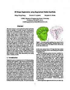
Fig.1: Microwell array with different shapes in the size of a singe cell (left), close up of a triangular microwell (middle), stained cell in a triangular shaped well with a blue nucleus and a green actin cytoskeleton (right). RESULTS: Endothelial cells constrained within microwells were viable, although cell death was increased in very constrained microwells as has been reported for cells on flat substrates. In contrast to studies on 2D surfaces, actin stress fibers were present even within cells cultured in very constrained microwells, and in addition the
cytoskeleton exhibited 3D arrangement and was not only limited to the cell-substrate interface. 3 DISCUSSION & CONCLUSIONS: These observations demonstrate that microwells can be used to produce microenvironments for large numbers of single cells with 3D shape control and can be added to a repertoire of tools which are ever more sought after for both fundamental biological studies as well as cell-based assays for drug development and screening. REFERENCES: 1
M.R. Dusseiller, M.L. Smith, V. Vogel, M. Textor; Biointerphases (2006), 1, 1
2
M.R. Dusseiller, D. Schlaepfer, M. Koch, R. Kroschewski, M. Textor, Biomaterials (2005), 26, 5917-5925
3
M. Ochsner, M.R. Dusseiller, H. M. Grandin, S. Luna-Morris, M. Textor, V. Vogel, M.L. Smith; Lab on a Chip (2007), In Press ACKNOWLEDGEMENTS: We gratefully acknowledge the Swiss National Science Fundation (FN 205321-112323/1).