ARTICLE Development and Validation of a Computational Method for Assessment of Missense Variants in Hypertrophic Cardiomyopathy Daniel M. Jordan,1,2,8 Adam Kiezun,1,8 Samantha M. Baxter,3,8 Vineeta Agarwala,2,3 Robert C. Green,4,5,6 Michael F. Murray,1 Trevor Pugh,3,6 Matthew S. Lebo,3,6 Heidi L. Rehm,3,7 Birgit H. Funke,3,7,* and Shamil R. Sunyaev1,* Assessing the significance of novel genetic variants revealed by DNA sequencing is a major challenge to the integration of genomic techniques with medical practice. Many variants remain difficult to classify by traditional genetic methods. Computational methods have been developed that could contribute to classifying these variants, but they have not been properly validated and are generally not considered mature enough to be used effectively in a clinical setting. We developed a computational method for predicting the effects of missense variants detected in patients with hypertrophic cardiomyopathy (HCM). We used a curated clinical data set of 74 missense variants in six genes associated with HCM to train and validate an automated predictor. The predictor is based on support vector regression and uses phylogenetic and structural features specific to genes involved in HCM. Ten-fold cross validation estimated our predictor’s sensitivity at 94% (95% confidence interval: 83%–98%) and specificity at 89% (95% confidence interval: 72%–100%). This corresponds to an odds ratio of 10 for a prediction of pathogenic (95% confidence interval: 4.0–infinity), or an odds ratio of 9.9 for a prediction of benign (95% confidence interval: 4.6–21). Coverage (proportion of variants for which a prediction was made) was 57% (95% confidence interval: 49%–64%). This performance exceeds that of existing methods that are not specifically designed for HCM. The accuracy of this predictor provides support for the clinical use of automated predictions alongside family segregation and population frequency data in the interpretation of new missense variants and suggests future development of similar tools for other diseases.
Introduction DNA sequencing is quickly becoming the method of choice for clinical genetic diagnostics. The improvement in clinical sensitivity that sequencing provides over genotyping platforms is invaluable, especially in disorders that show locus and allelic heterogeneity. However, there are also important challenges presented by the use of DNA sequencing, including the difficulty of interpreting novel sequence variants. There is currently little standardization of variant classification in the genetics community. Most clinics use a combination of traditional genetic methods relying on segregation with the disease in families, frequency in controls, biochemical characterization, and evolutionary conservation at the variant position.1 This manual classification process is time consuming and requires significant expert knowledge. More frustratingly, it often fails to produce a classification at all: variants with incomplete or conflicting data are routinely classified as variants of unknown significance (VUSs), and no confident classification is reported to the patient or the referring physician. In some genes, these VUSs comprise as many as one-quarter to one-half of all reported variants.2 This problem is only getting worse. As next-generation sequencing technologies begin to enter widespread clinical
use, the volume of novel variants is expected to expand by several orders of magnitude. The genetics community must therefore begin to develop robust automated methods to classify novel variants accurately. There currently exist several computational tools for predicting the functional effects of genetic variants.3–5 However, these tools in general were not designed for clinical use, have not been rigorously tested on individual genes or diseases, and have not undergone any kind of validation against well-curated data sets. Therefore, the sensitivities and specificities of these predictors are in general ill-defined. This lack of proper validation has created the perception among medical professionals that automated predictors cannot be trusted.6 Consequently, although most geneticists are familiar with these tools, the predictions they produce are typically not formally included in clinical variant classification methods and are therefore not communicated to physicians via clinical reports. Several studies have attempted to address this problem by validating existing predictors against known diseasecausing variants, largely arriving at the conclusion that these methods are not yet mature enough for clinical use.6–8 Variant classification pipelines that are considered mature enough for clinical use are generally designed
1
Division of Genetics, Department of Medicine, Brigham and Women’s Hospital and Harvard Medical School, Boston, MA 02115, USA; 2Program in Biophysics, Harvard University, Cambridge, MA 02138, USA; 3Laboratory for Molecular Medicine, Partners HealthCare Center for Personalized Genetic Medicine, Cambridge, MA 02139, USA; 4Boston University School of Medicine, Boston, MA 02118, USA; 5Boston University School of Public Health, Boston, MA 02118, USA; 6Harvard Medical School Genetics Training Program, Boston, MA 02115, USA; 7Department of Pathology, Harvard Medical School, Boston, MA 02115, USA 8 These authors contributed equally to this work *Correspondence:
[email protected] (B.H.F.),
[email protected] (S.R.S.) DOI 10.1016/j.ajhg.2011.01.011. Ó2011 by The American Society of Human Genetics. All rights reserved.
The American Journal of Human Genetics 88, 183–192, February 11, 2011 183
from the ground up with clinical use in mind and are designed, demonstrated, and validated using variants classified according to clinical criteria. Examples of such pipelines include the classification procedure currently in use at the Laboratory for Molecular Medicine (LMM), a clinical diagnostic laboratory in the U.S., and the integrated evaluation of BRCA gene variants that developed from the work of Goldgar et al.9 However, fully automated computational predictors are not currently designed in this way. We therefore set out to test whether this methodology could successfully create an automated predictor that would be useful to medical professionals as a tool for classifying novel missense variants. We chose to target one specific disease and a limited number of genes in which diseasecausing variants might be found so that we would be able to generate a high-quality set of manually classified missense variants to use as the gold standard for training and validating our predictions. We also hoped that focusing on a limited number of functionally related genes would allow us to identify common features of these genes and common mechanisms of disease in these genes, which would help us to make our predictor more accurate. The disease we chose was hypertrophic cardiomyopathy (HCM [MIM 192600]), an autosomal dominant disease of the myocardium (heart muscle) with an incidence of roughly one in 500 individuals and a largely genetic basis.10 Variants in over 20 genes are associated with HCM, with over 900 unique variants reported in the literature, and sequencing of many of these genes can be ordered for clinical testing in CLIA-approved laboratories. The vast majority of pathogenic variants are found in eight genes that encode for units of the cardiac sarcomere, a contractile protein complex in the heart: b-cardiac myosin heavy chain (MYH7 [MIM 160760]), cardiac actin (ACTC1 [MIM 102540]), cardiac troponin T (TNNT2 [MIM 191045]), a-tropomyosin (TPM1 [MIM 191010]), cardiac troponin I (TNNI3 [MIM 191044]), cardiac myosin-binding protein C (MYBPC3 [MIM 600958]), and the myosin light chains (MYL2 [MIM 160781] and MYL3 [MIM 160790]). Sequencing of these genes yields a high number of novel variants, mainly because of the high prevalence of private familial variants. Roughly 50% of probands tested have a disease-causing variant in one of these genes, and approximately 80% of those are in MYH7 and MYBPC3 (H.L.R., unpublished data).11 Missense variants represent nearly all such variants detected in MYH7 and 35% of those in MYBPC3. Missense variants exerting dominant negative effects on the sarcomere structure represent the vast majority of all variants. The notable exception is MYBPC3, where missense variants constitute only 35% of all variants, the remainder being splice, nonsense, or frameshift variants leading to loss of function. At the time of this study, the LMM had identified over 700 variants in HCM-related genes over 5 years of testing, over half of which were novel at the time of reporting and over half of which were missense changes. We performed a systematic manual classification
of these variants, producing a final data set of 74 missense variants with extremely confident manual classifications. Using these 74 variants as our gold standard, we then set out to develop and validate a computational method that could predict the pathogenicity of any variant in these six genes.
Material and Methods We created a computational method to predict the pathogenicity of a novel variant in any of the six genes we chose to screen for HCM mutations. Our method, like other existing methods12–16 and, particularly, the recently developed algorithm PolyPhen2,17 integrates phylogenetic and structural information from several heterogeneous sources with a probabilistic classifier. However, unlike these methods, it exploits the narrow focus on six specific genes known to contain variants that cause the disease to improve the prediction strategy significantly. Also unlike these methods, it uses variants classified according to clinical criteria of pathogenicity to train the probabilistic classifier. The selection and classification of these variants, the features used for classification, and the training and validation of the classifier are all described below. This study was performed under an institutional-reviewboard-approved protocol through Partners Healthcare System.
Selection of Target Genes HCM is caused primarily by variants in eight genes encoding protein subunits of the cardiac sarcomere. We initially attempted to use all eight genes to develop our predictor. However, after constructing our data set (see Manual Classification of HCM Variants below), we examined the distribution of variants and found that the final data set contained no variants in ACTC1 and only one in MYL3. We discarded these two genes and built our classifier around the remaining six (MYH7, TNNT2, TPM1, TNNI3, MYPC3, and MYL2).
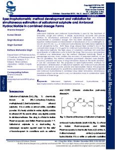
Manual Classification of HCM Variants We relied on LMM’s standard variant-assessment pipeline to create our data set of manually classified variants. To ensure unbiased training and testing of our computational method, we excluded from manual classification information that was accessible to the method such as evolutionary conservation or structural data, even though this information is currently used in the pipeline. Each variant recieved a classification of pathogenic, likely pathogenic benign, likely benign, or VUS. The basic decision process we used is described below and shown in Figure 1. Pathogenic Variants with a minimum of five informative meioses supporting familial cosegregation with HCM, absent in healthy controls, and/ or having strong functional data are classified as pathogenic. In HCM, informative meioses typically only include individuals who are positive for both phenotype and genotype. This level of stringency is required because of the highly variable expressivity and reduced penetrance, which make individuals without the phenotype largely uninformative, regardless of their genotype. Likely Pathogenic The minimum requirement to classify a variant as likely pathogenic is absence from race-matched controls or a large cohort of race-matched probands. The LMM has previously sequenced sarcomere genes in over 1000 HCM probands of European
184 The American Journal of Human Genetics 88, 183–192, February 11, 2011
VARIANT NOT PREVIOUSLY IDENTIFIED IN TESTING LABORATORY
PREVIOUSLY REPORTED?
NO
YES
PRESENT IN CONTROLS OR dbSNP?
NO
YES SUFFICIENT # OF RACE MATCHED PROBANDS OR CONTROLS TESTED? HIGH FREQ 3%
LOW FREQ A, which affects the last base of exon 6. This position is known to be part of the splice consensus and five different splice predictors (SpliceSiteFinder-like, MatEntScan, NNSPLICE, GeneSplicer, and Human Splice Finder; see Figure S6) predict an impact on splicing. This is supported by evidence showing that this may result in skipping of exon 6.28,29 Therefore, the conservation of the nucleotide and not the amino acid at this position is essential, possibly explaining a misprediction by our predictor. This is a limitation of this method and clearly lends itself to future improvement and generation of tools that incorporate a splice assessment. It is also important to point out that clinical laboratories are typically aware of this limitation. Novel variant assessment is a lengthy and complex process that relies on a large collection of different computer tools in combination with traditional genetic evidence such as familial segregation with disease and absence from race-matched healthy controls. All evidence is taken into account to synthesize a final probability for pathogenicity. In our laboratory, a splice assessment is performed for every variant, regardless of whether it changes an amino acid or not, and a benign prediction by this predictor would not lead to a final classification of benign, particularly not for genes for which pathogenic splice variants are known to be common. This example illustrates that this predictor or any other predictor developed with this methodology should not be used as a sole foundation for a diagnosis but rather be used in combination with other lines of evidence in agreement with recommendations from the American College of Medical Genetics and the International Agency for Research on Cancer.1,2 We envision future development of a single probabilistic classifier that would automatically combine heterogeneous factors such as familial segregation, frequency in controls, functional evidence, and computational predictions following early work in this area.
Conclusion We have addressed the problems that prevent automated predictors from being widely used in genomic medicine by developing a custom-tailored predictor specifically designed for clinical use. Our analysis suggests several important considerations that can increase the accuracy of computational methods. Manual adjustment of multiple sequence alignments and time-consuming computational methods of molecular evolution are feasible when focusing on a small set of genes and may improve predictions that use comparative sequence analysis. Exploitation of specific structural properties of proteins also becomes feasible when focusing on a specific disease. Most importantly,
190 The American Journal of Human Genetics 88, 183–192, February 11, 2011
a highly accurate manually curated data set is necessary to train and validate an accurate predictor, and this level of validation enables clinical laboratories to include it as part of their variant assessment processes. Where previous studies have concluded that existing tools are not mature enough for clinical use, we believe that our tool is ready for clinical use now, in combination with other sources of information. Our collaborating clinical laboratory, the LMM, has already begun to use our predictor as a source of information about HCM variants, and we look forward to helping additional laboratories do the same. Our study focused on HCM, but we believe that our approach is general and that analogous methods can be constructed for many other diseases where genetic testing is an important part of the diagnosis. In the future, we expect to work with additional laboratories and on additional diseases to expand the use of automated predictors in genomic medicine and simplify the problem of interpreting novel variants.
3.
4.
5.
6.
7.
8.
Supplemental Data Supplemental Data includes six figures and six tables and can be found with this article online at http://www.cell.com/AJHG/.
9.
Acknowledgments This research was supported by grants from the National Institutes of Health (R01 GM078598, T32 GM008313). We would like to thank Steve DePalma, Dan Hermann, and Jon and Christine Seidman for their help with the CardioGenomics database and insights into HCM.
10.
11. Received: August 19, 2010 Revised: January 17, 2011 Accepted: January 19, 2011 Published online: February 10, 2011 12.
Web Resources The URLs for data presented herein are as follows:
13.
Online Mendelian Inheritance in Man (OMIM), http://www.ncbi. nlm.nih.gov/Omim PolyPhen-HCM method, http://genetics.bwh.harvard.edu/hcm
14.
15.
References
16.
1. Richards, C.S., Bale, S., Bellissimo, D.B., Das, S., Grody, W.W., Hegde, M.R., Lyon, E., and Ward, B.E.; Molecular Subcommittee of the ACMG Laboratory Quality Assurance Committee. (2008). ACMG recommendations for standards for interpretation and reporting of sequence variations: Revisions 2007. Genet. Med. 10, 294–300. 2. Plon, S.E., Eccles, D.M., Easton, D., Foulkes, W.D., Genuardi, M., Greenblatt, M.S., Hogervorst, F.B., Hoogerbrugge, N., Spurdle, A.B., and Tavtigian, S.V.; IARC Unclassified Genetic Variants Working Group. (2008). Sequence variant classification and reporting: recommendations for improving the inter-
17.
18.
19.
pretation of cancer susceptibility genetic test results. Hum. Mutat. 29, 1282–1291. Ng, P.C., and Henikoff, S. (2006). Predicting the effects of amino acid substitutions on protein function. Annu. Rev. Genomics Hum. Genet. 7, 61–80. Thusberg, J., and Vihinen, M. (2009). Pathogenic or not? And if so, then how? Studying the effects of missense mutations using bioinformatics methods. Hum. Mutat. 30, 703–714. Jordan, D.M., Ramensky, V.E., and Sunyaev, S.R. (2010). Human allelic variation: perspective from protein function, structure, and evolution. Curr. Opin. Struct. Biol. 20, 342–350. Tchernitchko, D., Goossens, M., and Wajcman, H. (2004). In silico prediction of the deleterious effect of a mutation: proceed with caution in clinical genetics. Clin. Chem. 50, 1974–1978. Dorfman, R., Nalpathamkalam, T., Taylor, C., Gonska, T., Keenan, K., Yuan, X.W., Corey, M., Tsui, L.C., Zielenski, J., and Durie, P. (2010). Do common in silico tools predict the clinical consequences of amino-acid substitutions in the CFTR gene? Clin. Genet. 77, 464–473. Tavtigian, S.V., Greenblatt, M.S., Lesueur, F., and Byrnes, G.B.; IARC Unclassified Genetic Variants Working Group. (2008). In silico analysis of missense substitutions using sequence-alignment based methods. Hum. Mutat. 29, 1327–1336. Goldgar, D.E., Easton, D.F., Deffenbaugh, A.M., Monteiro, A.N.A., Tavtigian, S.V., and Couch, F.J.; Breast Cancer Information Core (BIC) Steering Committee. (2004). Integrated evaluation of DNA sequence variants of unknown clinical significance: application to BRCA1 and BRCA2. Am. J. Hum. Genet. 75, 535–544. Wang, L., Seidman, J.G., and Seidman, C.E. (2010). Narrative review: harnessing molecular genetics for the diagnosis and management of hypertrophic cardiomyopathy. Ann. Intern. Med. 152, 513–520, W181. Richard, P., Charron, P., Carrier, L., Ledeuil, C., Cheav, T., Pichereau, C., Benaiche, A., Isnard, R., Dubourg, O., Burban, M., et al; EUROGENE Heart Failure Project. (2003). Hypertrophic cardiomyopathy: distribution of disease genes, spectrum of mutations, and implications for a molecular diagnosis strategy. Circulation 107, 2227–2232. Ng, P.C., and Henikoff, S. (2001). Predicting deleterious amino acid substitutions. Genome Res. 11, 863–874. Ng, P.C., and Henikoff, S. (2003). SIFT: Predicting amino acid changes that affect protein function. Nucleic Acids Res. 31, 3812–3814. Bromberg, Y., and Rost, B. (2007). SNAP: predict effect of nonsynonymous polymorphisms on function. Nucleic Acids Res. 35, 3823–3835. Yue, P., and Moult, J. (2006). Identification and analysis of deleterious human SNPs. J. Mol. Biol. 356, 1263–1274. Yue, P., Melamud, E., and Moult, J. (2006). SNPs3D: candidate gene and SNP selection for association studies. BMC Bioinformatics 7, 166. Adzhubei, I.A., Schmidt, S., Peshkin, L., Ramensky, V.E., Gerasimova, A., Bork, P., Kondrashov, A.S., and Sunyaev, S.R. (2010). A method and server for predicting damaging missense mutations. Nat. Methods 7, 248–249. Ronquist, F., and Huelsenbeck, J.P. (2003). MrBayes 3: Bayesian phylogenetic inference under mixed models. Bioinformatics 19, 1572–1574. Lupas, A., van Dyke, M., and Stock, J. (1991). Predicting coiled coils from protein sequences. Science 252, 1162–1164.
The American Journal of Human Genetics 88, 183–192, February 11, 2011 191
20. Lupas, A. (1996). Prediction and analysis of coiled-coil structures. Methods Enzymol. 266, 513–525. 21. Letunic, I., Doerks, T., and Bork, P. (2009). SMART 6: recent updates and new developments. Nucleic Acids Res. 37 (Database issue), D229–D232. ¨ rgyi, 22. Houdusse, A., Kalabokis, V.N., Himmel, D., Szent-Gyo A.G., and Cohen, C. (1999). Atomic structure of scallop myosin subfragment S1 complexed with MgADP: a novel conformation of the myosin head. Cell 97, 459–470. 23. Himmel, D.M., Gourinath, S., Reshetnikova, L., Shen, Y., ¨ rgyi, A.G., and Cohen, C. (2002). Crystallographic Szent-Gyo findings on the internally uncoupled and near-rigor states of myosin: further insights into the mechanics of the motor. Proc. Natl. Acad. Sci. USA 99, 12645–12650. 24. Vinogradova, M.V., Stone, D.B., Malanina, G.G., Karatzaferi, C., Cooke, R., Mendelson, R.A., and Fletterick, R.J. (2005). Ca(2þ)-regulated structural changes in troponin. Proc. Natl. Acad. Sci. USA 102, 5038–5043. 25. Martı´nez, L., Andreani, R., and Martı´nez, J.M. (2007). Convergent algorithms for protein structural alignment. BMC Bioinformatics 8, 306.
26. Thomas, P.D., Campbell, M.J., Kejariwal, A., Mi, H., Karlak, B., Daverman, R., Diemer, K., Muruganujan, A., and Narechania, A. (2003). PANTHER: a library of protein families and subfamilies indexed by function. Genome Res. 13, 2129–2141. 27. Thomas, P.D., Kejariwal, A., Guo, N., Mi, H., Campbell, M.J., Muruganujan, A., and Lazareva-Ulitsky, B. (2006). Applications for protein sequence-function evolution data: mRNA/ protein expression analysis and coding SNP scoring tools. Nucleic Acids Res. 34 (Web Server issue), W645–50. 28. Andersen, P.S., Havndrup, O., Bundgaard, H., Larsen, L.A., Vuust, J., Pedersen, A.K., Kjeldsen, K., and Christiansen, M. (2004). Genetic and phenotypic characterization of mutations in myosin-binding protein C (MYBPC3) in 81 families with familial hypertrophic cardiomyopathy: total or partial haploinsufficiency. Eur. J. Hum. Genet. 12, 673–677. 29. Marston, S., Copeland, O., Jacques, A., Livesey, K., Tsang, V., McKenna, W.J., Jalilzadeh, S., Carballo, S., Redwood, C., and Watkins, H. (2009). Evidence from human myectomy samples that MYBPC3 mutations cause hypertrophic cardiomyopathy through haploinsufficiency. Circ. Res. 105, 219–222.
192 The American Journal of Human Genetics 88, 183–192, February 11, 2011