www.nature.com/scientificreports
OPEN
Received: 18 September 2017 Accepted: 11 May 2018 Published: xx xx xxxx
Development and validation of a LC-MS/MS assay for pharmacokinetic studies of complement C5a receptor antagonists PMX53 and PMX205 in mice Vinod Kumar1, John D. Lee1,2, Richard J. Clark1 & Trent M. Woodruff 1 PMX53 and PMX205 are cyclic hexapeptide inhibitors of complement C5a receptors (C5aR1), that are widely used to study C5aR1 pathobiology in mouse models of disease. Despite their widespread use, limited information regarding their pharmacokinetics have been reported. Here, a bioanalytical method for the quantitative determination of PMX53 and PMX205 in plasma, brain and spinal cord of mice was developed using liquid chromatography-tandem mass spectrometry (LC-MS/MS) techniques. The LC-MS/MS method was validated in all three matrices according to regulatory guidelines and successfully applied to pharmacokinetic studies of PMX53 and PMX205 in C57BL/6 J mice following intravenous administration. The developed method was highly sensitive and sufficiently accurate with a lower limit of quantification within the range of 3–6 ng/ml in extracted plasma samples and 3–6 ng/g in processed tissue samples, which outperforms previously published LC-MS/MS methods. The results thus support the suitability, reliability, reproducibility and sensitivity of this validated technique. This method can therefore be applied to perform a complete pre-clinical investigation of PMX53 and PMX205 pharmacokinetics in mice. An enzymatic cascade, the complement system is a vital component of the immune system creating a bridge between the innate and adaptive immune systems. This signalling pathway is omnipresent throughout the animal kingdom including invertebrates lacking a circulatory system1. Activation of the complement system results in terminal activation of an extremely potent complement fragment, C5a, that exhibits various immuno-regulatory and pro-inflammatory biological activities2. C5a binds to two known receptors, termed C5a receptor 1 (C5aR or CD88 – now referred to as C5aR1) and C5a receptor-like 2 (C5L2 or GPR77 – now referred to as C5aR2)3. C5aR1 is generally expressed at higher levels than C5aR2, and activation of C5aR1 enhances disease pathology, including diseases affecting the brain2,4–7. As such, there has been much interest in developing inhibitors to C5aR1 as therapeutic treatments for a wide range of diseases8–10. The most well-studied inhibitors of C5aR1 are Ac-Phe-[Orn-Pro-dCha-Trp-Arg] (3D53 or PMX53)11 and hydrocinnamate-[Orn-Pro-dCha-Trp-Arg] (PMX205)12. These small cyclic peptidic molecules specifically target C5aR1 at nanomolar concentrations and act in a pseudo-irreversible and insurmountable manner13,14. PMX205 is a lipophilic analogue of PMX53 that demonstrates improved in vivo stability and efficacy5,15,16, and has been suggested as a more ideal drug candidate, particularly for neurological diseases. For example, this drug has shown beneficial effects in models of Huntington’s disease5, amyotrophic lateral sclerosis4,16, spinal cord injury6,17, and in reduction of memory loss in mice with Alzheimer’s disease18,19. Both antagonists have been used in numerous
1
School of Biomedical Sciences, the University of Queensland, Brisbane, QLD, 4072, Australia. 2University of Queensland Centre for Clinical Research, the University of Queensland, Brisbane, QLD, 4029, Australia. Correspondence and requests for materials should be addressed to T.M.W. (email:
[email protected])
Scientific REPOrTS | (2018) 8:8101 | DOI:10.1038/s41598-018-26387-4
1
www.nature.com/scientificreports/
Analyte
PMX53
PMX205
Q1 (Da)
Q3 (Da)
Dwell time (msec)
DP (V)
EP (V)
CEP (V)
CE (V)
CXP (V)
Fragment 1 (Quantifier)
448.6
120.2
150
46
4
20
53
4
Fragment 2 (Qualifier 1)
448.6
70.0
150
46
4
20
73
14
Fragment 3 (Qualifier 2)
448.6
162.1
150
30
7
20
29
4
Fragment 1 (Quantifier)
420.2
70.0
150
30
4
20
53
6
Fragment 2 (Qualifier 1)
420.2
105.2
150
26
2.5
20
55
12
Fragment 3 (Qualifier 2)
420.2
126.0
150
26
7
20
53
4
ID
Table 1. Optimized MS parameters for PMX53 and PMX205. Q1, mass of analyte in (+2) ionization state; Q3, fragment mass; DP, declustring potential; EP, entrance potential; CEP, collision cell entrance potential; CE, collision energy; CXP, collision cell exit potential. experimental inflammatory conditions for over 15 years, and oral and topical PMX53 has also been tested in early Phase I human clinical trials20. Despite this extensive usage of these C5aR1 inhibitors, relatively few studies have reported the quantitative pharmacokinetic determination of these antagonists7,13,15,21,22. Further, none of these prior studies have reported validated LC-MS/MS methods for the quantitative determination of PMX53 and PMX205 in mice, the major species in which these compounds are used. The present research describes the development and validation of a simple, rapid, specific and sensitive LC-MS/MS method with high accuracy and precision, allowing for the quantitative determination of drug levels in plasma, brain and spinal cord of mice. This method was successfully utilised for pharmacokinetic studies of PMX53 and PMX205 in mice following the intravenous (i.v.) route of administration, and may be useful for determining the complete comparative preclinical pharmacokinetics of these drugs in mice.
Results
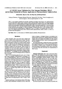
LC-MS/MS conditions and optimization. The mass spectrometer was operated in multiple reaction monitoring (MRM) scan mode for identification of an ideal ionization state for fragmentation. Fragmentation was not achieved for both PMX53 and PMX205 in +1, −1 and −2 charge ionization mode. In the +2 ionization state, PMX53 and PMX205 underwent fragmentation resulting in formation of three main fragments. Compound-dependent and source-dependent mass spectrometer conditions were optimized using automatic optimization by infusion and flow injection analysis (FIA). The optimized compound dependent instrument parameters for PMX53 and PMX205 with their MRM transitions are summarized in Table 1. Fragmentation pattern and relative intensities of fragment ions are shown in Fig. 1A,B. For quantitative purposes, one mass/ charge (m/z) transition per analyte, i.e. 448.6 → 120.2 for PMX53 and 420.2 → 70.0 for PMX205, were monitored. Additional transitions were monitored for qualification purposes, i.e. for PMX53 m/z transition of 448.6 → 70.0 and 448.6 → 162.1 and for PMX205 420.2 → 105.2 and 420.2 → 105. For both analytes, the ion spray voltage, collision cell entrance potential and source temperature/auxiliary gas temperature were set at 5000 V, 20 V and 550 °C respectively. Nebulizer gas (ion source gas 1, GS1) and auxiliary gas (ion source gas 2, GS2) were set at 70 psi. The curtain and collision-activated dissociation gases were set at 20 and 5 on an arbitrary scale. LC conditions were optimized to obtain a short run and better chromatographic separation of PMX53 and PMX205 by using various mobile phase concentrations. After several LC-MS/MS method trials, a mobile phase under binary gradient condition (Fig. 1C) of mobile phase A (milliQ water containing 0.1% formic acid) and mobile phase B (acetonitrile containing 0.1% formic acid) with variable flow rate was selected for simultaneous determination of PMX53 and PMX205 without interference from each other resulting in better resolution. Using these optimized LC-MS/MS parameters, the retention time of PMX53 and PMX205 was approximately 6.5 min and 6.7 min respectively with a total run time of 18 min including the washing phase and equilibrium phase (Fig. 1D). Due to unavailability of radiolabelled analytes, it was difficult to find an ideal internal standard to be used in validation and analytical experiments. The chemical structure of PMX53 and PMX205 are closely related to one another and with both analytes having an adjacent retention time, this makes them ideal candidates to act as an internal standard for each other. Consequently, PMX53 was used as the internal standard for PMX205 experiments and vice versa. Selectivity and specificity. Chromatograms of processed blank matrices, matrices spiked with analytes
and samples from mice administered with drug candidates are shown in Supplementary Figs 1 and 2. Lack of additional specific peaks of endogenous substances was observed in blank matrices indicating an absence of interference from endogenous components for the quantification of PMX53 and PMX205. The unchanged peak shape and retention time of PMX53 and PMX205 in matrices spiked with low quality control (LQC) compared to samples obtained from mice i.v. administered with drug candidates, indicates the reliability of LC-MS/MS method in terms of selectivity and specificity.
Scientific REPOrTS | (2018) 8:8101 | DOI:10.1038/s41598-018-26387-4
2
www.nature.com/scientificreports/
Figure 1. HPLC-MS/MS of PMX53 and PMX205: Product ion MS/MS scan spectra and relative intensities of fragment ions following positive (+2) electrospray ionization of PMX53 (m/z 448.6) (A) and PMX205 (m/z 420.2) (B). Developed MS method under multiple reaction monitoring mode of mass spectrometer combined with liquid chromatography gradient and flow rate conditions (C) for fragment ions resulted HPLC-MS/ MS chromatograms of analytes as represented by overlayed chromatograms of complement C5a receptor 1 antagonists PMX53 and PMX205 (D).
Linearity of calibration curves and sensitivity. For the bioanalytical method validation, the calibration curve parameters for PMX53 and PMX205 in different matrices were determined on two consecutive days as summarised in Supplementary Table T1. Calibration curves were linear over the selected concentration range in different matrices. All curves with an average correlation coefficient r > 0.99 were considered for this analysis. The mean of six standard curves in plasma, brain homogenate and spinal cord homogenates were used in the validation of the bio-analytical method. Following validation guidelines, deviation of ±15% for all quality controls (QCs) and ±20% for lower limit of quantification (LLOQ) from the theoretical value was used as an acceptance criterion. Additionally, Supplementary Table T1 summarizes the lower limit of detection (LLOD) and LLOQ values for determination of PMX53 and PMX205 in various matrices. The results support that the developed method was sufficiently accurate and sensitive with LLOD and LLOQ within the range of 1.76–5.35 ng/ml for PMX53 and 1.23–3.73 ng/ml for PMX205 in extracted plasma samples. Additionally, lower limits of detections and quantifications of developed method in processed tissue samples are within the range of 2.75–6.31 ng/g for PMX53 and 1.95–5.9 ng/g for PMX205.
Accuracy and precision. Intra-day and inter-day % relative standard deviation and % relative error for both analytes in various matrices were within acceptable limits (i.e. ±15%) (Table 2) for the validation of bioanalytical methods as per The USA Food and Drug Administration (FDA) guidelines23. These results favour the accuracy and precision of this analytical method over the range of the assay. Process recovery, efficiency and matrix effects. The process efficiency, extraction efficiency and recovery values as summarized in Table 3 suggested that the sample preparation results in exceptional extraction of analytes from various biological matrices with minimum matrix effect. It should be noted however, that the matrix effect was compensated for by the use of the structurally similar analogue (i.e. PMX53 or PMX205) as the internal quantitative standards during sample preparation and analysis.
Stability studies. Stability studies were performed in the biological matrices, plasma, brain and spinal cord,
to reflect and identify any degradation of PMX53 and PMX205 during the entire period of sample collection, storage, processing and analysis. The main aim of this method development was to apply a validated protocol for pharmacokinetic studies in mice not only by the i.v. route, but through various other routes of drug administration that involve an array of biological fluids such as the serum, gastric and intestinal environments. For these cyclic peptidic drugs (PMX53 and PMX205) it was important to identify in vivo metabolic stability responsible for duration of action in circulation, absorption from gut, and gastric stability, which may reflect oral activity.
Scientific REPOrTS | (2018) 8:8101 | DOI:10.1038/s41598-018-26387-4
3
www.nature.com/scientificreports/ Intra-day (n = 6)
PMX53
Accuracy (RE, %)
Measured conc. Precision (mean ± SD) (RSD, %)
Accuracy (RE, %)
Matrix
Spiked conc. LQC (6.25 ng/ml)
6.24 ± 0.17
2.75
−0.06
6.20 ± 0.28
4.56
−0.87
Plasma
MQC (25 ng/ml)
24.81 ± 0.46
1.85
−0.74
24.96 ± 1.46
5.84
−0.17 −0.59
Brain
Spinal Cord
Plasma
PMX205
Inter-day (n = 6 × 3)
Measured conc. Precision (mean ± SD) (RSD, %)
Brain
Spinal Cord
HQC (200 ng/ml) 199.9 ± 1.23
0.62
−0.03
198.83 ± 1.9
0.96
LQC (6.25 ng/g)
6.30 ± 0.46
7.36
0.73
6.24 ± 0.3
2.70
0.46
MQC (25 ng/g)
24.7 ± 0.78
3.17
−1.20
25 ± 1.62
2.06
−0.68
HQC (200 ng/g)
199.3 ± 5.48
2.75
−0.35
198.4 ± 1.82
0.58
0.11
LQC (6.25 ng/g)
6.27 ± 0.17
2.70
0.46
6.11 ± 0.13
2.18
−2.18
MQC (25 ng/g)
24.82 ± 0.51
2.06
−0.68
24.32 ± 0.65
2.68
−2.71
HQC (200 ng/g)
200.2 ± 1.15
0.58
0.11
197.6 ± 0.76
0.38
−1.17
LQC (6.25 ng/ml)
6.2 ± 0.10
1.72
−0.80
6.3 ± 0.11
1.72
1.46
MQC (25 ng/ml)
24.7 ± 1.06
4.31
−0.93
25.2 ± 1.09
4.31
0.96
HQC (200 ng/ml) 198.7 ± 3.01
1.52
−0.64
202.6 ± 3.07
1.52
1.29
LQC (6.25 ng/g)
1.29
1.98
6.5 ± 0.25
3.85
4.12
6.37 ± 0.08
MQC (25 ng/g)
25.1 ± 1.15
4.57
0.37
24.7 ± 0.94
3.78
−1.07
HQC (200 ng/g)
203.2 ± 3.04
1.50
1.58
198.1 ± 1.84
0.93
−0.94
LQC (6.25 ng/g)
6.23 ± 0.08
1.29
−0.29
6.40 ± 0.06
1.00
2.41
MQC (25 ng/g)
24.6 ± 1.12
4.57
−1.51
25.3 ± 1.1
4.33
1.51
1.50
−0.35
202.9 ± 3.5
1.71
1.50
HQC (200 ng/g)
199.3 ± 2.3
Table 2. LC-MS/MS method’s accuracy and precision for PMX53 and PMX205 determination. Intra-day and inter-day precision and accuracy of the LC-MS/MS method for PMX53 and PMX205 determination in mice plasma, brain and spinal cord (n = 3 days, 6 imitates per day). RSD: relative standard deviation; RE: relative error.
Hence, in addition to storage and post-preparative stability, metabolic stability of both analytes was analysed in serum, gastric and intestinal environments. Storage and post-preparative stability. Results, as expressed in Table 4, represent the storage stability of analytes in biological matrices. The stability of PMX53 and PMX205 in plasma, brain and spinal cord matrices stored for four hours at room temperature, in −20 ± 5 °C storage conditions for up to twelve months and after three freeze-thaw cycles were within an acceptable range of guidelines (i.e. ±15% for medium QC (MQC) and high QC (HQC) samples. For LQC samples, ±25% criteria with a minimum of three values within the range of ±20% was used as per regulatory guidelines). Further stability could potentially be improved by reducing storage conditions from −20 ± 5 °C to −80 ± 5 °C. Supplementary Table 2, represents the high stock solution stability of both analytes up to six months in the current storage conditions. Results of post-preparative stability of PMX53 and PMX205 as determined by performing auto-sampler stability, auto-sampler reproducibility and comparing the results of processed samples with unprocessed standard samples, support the reliability of developed method. In summary, the combined results reflect the stability of PMX53 and PMX205 under post-preparative conditions and the reliability of conditions for analyte quantification. Plasma and serum stability. The results illustrate the stability of PMX53 and PMX205 in plasma (88 ± 1.2% and 91.4 ± 3.1%) and serum (88 ± 1.5% and 91.4 ± 1.4%) respectively when incubated at 37 °C for up to 60 min, and were comparable with stability of analytes in control solution (i.e. PBS) when stored and analysed using similar conditions (Fig. 2A,B). In conclusion, during pharmacokinetic studies for up to 60 min, the amount of analytes detected reflects the unchanged form of PMX53 and PMX205 in plasma and serum. Gastric environment stability. Figure 2C,D illustrates the gastric stability of PMX53 and PMX205. The stability of the antagonists was expressed as a percentage of concentration detected at different time points in comparison to concentration detected immediately following addition of antagonists, i.e. at t = 0 min. For PMX53 and PMX205, the proteinaceous nature of analytes resulted in some degradation in the gastric environment. PMX53 was more stable in gastric lavage fluids with more than 60% of unchanged form compared to PMX205 (