Journal of Chromatographic Science, Vol. 49, February 2011
Development and Validation of New Assay Method for the Simultaneous Analysis of Diltiazem, Metformin, Pioglitazone and Rosiglitazone by RP-HPLC and its Applications in Pharmaceuticals and Human Serum Najma Sultana1, M. Saeed Arayne2, Nighat Shafi1, and Farhan Ahmed Siddiqui2,* 1Research
Institute of Pharmaceutical Sciences, Faculty of Pharmacy, University of Karachi-75270, Pakistan and of Chemistry, University of Karachi.
2Department
Abstract Simple, sensitive, rapid, and accurate high-performance liquid chromatographic (HPLC) method has been developed and validated for the simultaneous determination of diltiazem, metformin, pioglitazone, and rosiglitazone hydrochloride in raw materials, their pharmaceutical formulations, and human serum. In HPLC, all the above drugs were chromatographed using acetonitrile–methanol–water (30:20:50 v/v, pH 2.59 ± 0.02) as the mobile phase at a flow rate of 1.0 mL/min at ambient temperature. The separation is carried out on a Hiber, 250-4.6 RP-18 column, equipped with a UV–vis detector at 230 nm. All the antidiabetic drugs eluted at different retention time and each showed a good resolution from diltiazem. The method has been successfully applied to pharmaceutical formulations because no chromatographic interferences from the tablet excipients are found. The method was found to be linear, accurate, and precise with apposite detection and quantification limit. Suitability of the method for the quantitative determination of the drugs is proved by validation in accordance with the requirements laid down by International Conference on Harmonization (ICH) guidelines. The validation results, together with statistical treatment of the data, demonstrated the reliability of this method.
Introduction Hypertension in diabetics represents an important health problem as the combination of the two diseases is common, carries significant morbidity and mortality, and is frequently difficult to treat. The prevalence of hypertension in diabetic people is probably 1.5–2 times higher than in the general population (1). Reduction of cardiovascular risk is therefore a high priority in the management of diabetes. Microalbuminuria is an important predictor of cardiovascular events and forms one of the components of insulin resistance/metabolic syndrome, which confers a particularly high risk of cardiovascular death (2). Diverse classes *Author to whom correspondence should be addressed: email
[email protected]
of antihypertensive prescription may be used for blood pressure management in diabetes among these calcium channel blockers, angiotensin-II type 1 receptor blockers (ARBs), thiazide diuretics, and ACE inhibitors are common (3). Cheung demonstrated that the calcium antagonists have been extensively used in hypertensive patients with diabetes (4). Use of verapamil, a calcium channel blocker, significantly reduced the risk of developing diabetes (5). Similarly diabetic patients often take anti-hypertensive medications and are co-administered with antidiabetic drugs (6). Treatment of patients with hypertension and diabetes with ARBs improved both macrovascular and microvascular alterations (7). Diltiazem is a peripheral and coronary vasodilator with limited negative inotropic activity inhibiting cardiac conduction, particularly at the sino-atrial and atrioventricular nodes (8,9). The currently available glucose-lowering agents or hypoglycaemic drugs for treating type II (non-insulin-dependent) diabetes mellitus are sulphonylurea derivatives, α-glucosidase inhibitors (metformin) and thiazolidinediones (pioglitazone and roziglitazone). Metformin appears to have little effect on microalbuminuria expressed as urinary albumin/creatinine ratio, while the thiazolidinediones have unique effects on this risk factor, in parallel with their effects on insulin resistance (10). Literature survey revealed that number of assay methods have been used for analysis of diltiazem in bulk drug, pharmaceutical preparations, and serum using different techniques including Raman spectroscopy (11), capillary electrophoresis (12), spectrophotometry (13), and RP-HPLC (14–16). However, several analytical procedures have also been described for simultaneous separation and enantio-separation of diltiazem, its analogs, possible degradation products and metabolites (17–19). Similarly, a number of assays have been reported for the quantitative determination of antidiabetic drugs using spectrophotometry and RPLC (20–24) in pharmaceutical dosage forms, bulk material and human plasma or serum. Kolte et al., established simultaneous HPLC methods for the determination of pioglitazone (25) and rosiglitazone (26) in a combined pharmaceutical dosage form with metformin.
Reproduction (photocopying) of editorial content of this journal is prohibited without publisher’s permission.
1
Journal of Chromatographic Science, Vol. 49, February 2011
Since diltiazem can be used for cardiovascular dealings in diabetes and co-administered with antidiabetic drugs, there is a need for the simultaneous determination of metformin, rosiglitazone, and pioglitazone hydrochloride with diltiazem. There was no HPLC method reported for the simultaneous determination of all the above co-administrated drugs. The projected method was effectively applied to the determination of these drugs in finished product and in human serum. The established method was validated with respect to linearity, limit of detection and quantification, precision, accuracy, specificity and robustness.
Experimental Instrumentation
A Shimadzu HPLC system equipped with LC-10 AT VP pump and SPD-10 A VP UV–vis detector was utilized. The chromatographic system was integrated via Shimadzu model CBM-102 to P-IV computer loaded with Shimadzu CLASS-VP software (Version 5.03) for data acquisition and mathematical calculations. Rheodyne manual injector fitted with a 20 μL loop, a Hiber, RT 250-4.6 Purospher STAR RP-18 column and DGU-14 AM on-line degasser. In addition, Mettler Toledo electronic balance, microliter syringe, and micropore filtration assembly were used in this study. Materials and reagents
Diltiazem, was kind gift from Hilton Pharma (Private) Limited. Metformin (Neodipar 250 mg), pioglitazone (Poze 45 mg), and rosiglitazone (Rosita 8mg) were of Aventis pharma (Pvt.) Ltd., Ali Gohar pharmaceuticals (Pvt.) Ltd., and Pharm Evo (Pvt.) Ltd., and respectively and were purchased from the market. Reference standards of all these statins were supplied by lab-9 of Department of Chemistry, University of Karachi. Each product was labeled and had an expiry of not less than 365 days at the time of study. All reagents used were of HPLC grade. Acetonitrile, methanol, and phosphoric acid 85% (Merk, Germany) and HPLC-grade deionized filtered water was used to prepare the mobile phase. Stock solutions of all drugs were prepared in the mobile phase. Fresh working solutions were prepared daily. All solutions were filtered (0.45 μm) and degassed using sonicator. Preparation of solutions
Standard solutions of diltiazem and each antidiabetic drugs were prepared by dissolving appropriate amounts of drug in mobile phase acetonitrile–methanol–water (30:20:50, v/v) to yield final concentrations of 100 μg/mL. Seven calibrators of each drug were prepared by making serial dilutions from stock solutions. For the assay preparation, the content of 20 tablets were powdered and a weighed portion of the powder equivalent to suitable amount of drug (according to the labeled claimed) was transferred into a 50 mL volumetric flask. The drug was fully dissolved in mobile phase and then diluted with this solvent up to the mark, seven dilutions of each drug were prepared, portion of this solution was filtered through a disposable 0.45 μm filter and then injected.
2
Serum drug analysis
Blood samples were collected from healthy volunteers and then centrifuged at 3000 rpm for 10 min. The supernatant obtained was stored at –20°C. After thawing, serum was deprotinated by acetonitrile and spiked daily with working solutions to produce desired concentrations of diltiazem and antidiabetic drugs. 20 µL volume of each sample was injected and chromatographed under the described conditions. Chromatographic conditions
The chromatographic analysis was performed at ambient temperature with isocratic elution. The mobile phase consisted of acetonitrile–methanol–water (30:20:50 v/v) with pH adjusted to 2.59 ± 0.02 with phosphoric acid (85%) and pump at a flow rate of 1 mL/min, using a column Hiber, 250-4.6 RP-18 column, sample volume of 20 μL was injected in triplicate onto the HPLC column and effluents were screened over the dative UV region at 230 nm.
Results and Discussion Method development
Method development and validation are very momentous for pharmaceuticals and pathological laboratories such as drug quality control, stability, metabolism, pharmacokinetics, and toxicity studies. This is always desirable to select and develop a simple, efficient, validated, and economical analytical methods are very critical requirements for all these investigation (27). Method optimization and isocratic scouting An HPLC–UV method was developed and successfully validated for the simultaneous quantitation of diltiazem, metformin, pioglitazone, and rosiglitazone. Chromatographic parameters were isocratically optimized on a set of different C18 stationary phases with different lengths and particle sizes. Isocratic scouting does not involve re-equilibration of column (28). Firstly a Hypersil, ODS, C18 (150 × 4.6 mm, 5 µ) column was tested with a variety of mobile phases, but the results were still not adequate. Analyte peaks were not efficient or reproducible with short analyses time (< 10 min) and acceptable resolution was not reached because of peak tailing and placebo obstruction in the formulations. The best choice of the stationary phase that provided satisfactory peak shape, resolution, and run time was based on a Hiber, RT 250-4.6 Purospher STAR RP-18 endcapped (5 μm). A variety of mobile phases were investigated for optimization of chromatographic conditions with respect to the quantity and different rates of organic modifier. In order to select an appropriate organic modifier, different compositions of acetonitrile and methanol were tested. Beneficial partition was established with acetonitrile. Various mobile phase compositions were attempted to separate diltiazem and given antidiabetic drugs and the amount of acetonitrile varied when it was less than 20% of the total volume of mobile phase, diltiazem was retained on the column. Mobile phase between 20–27% of acetonitrile showed that antidiabetic drugs could not be separated, but each showed a good resolution from diltiazem. The optimum results [capacity
Journal of Chromatographic Science, Vol. 49, February 2011
factor (k'), specificity, good resolution and short omega peak] were obtained when the compositions of acetonitrile in the mobile phase was 30%. The fitness of the mobile phase was achieved on the foundation of the sensitivity, suitability for stability studies, run time required for the investigation, simplicity of preparation, and utilization of readily available cost-effective solvents. Variation in pH of the mobile phase had a great influence to the retention time, chromatographic peak, and efficiency of the chromatographic system. The mobile phase pH varies between 2.5 to 3.5, and best results were attained at pH 2.59 ± 0.2; if the pH is less than 2.59, peak tailing was observed while, at a higher pH peak of diltiazem and antidiabetic drugs merged as well as retention time were higher. For detection wavelength, UV spectra of both diltiazem and antidiabetic drugs overlap to each other. It was found that both the drugs exhibited the highest response (maximum UV absorption) at 230 nm and offered a greatest intensity with the smallest interference. In RP Chromatography any analyte will elute earlier when it is in the ionized form as oppose to the neutral form. Metformin is more basic than any other drugs present in the sample, so it was more ionized at this pH of mobile phase and eluted earlier than other drugs. Rosiglitazone and pioglitazone is less basic than metformin and these were the less ionized form, so these eluted latter than metformin; rosiglitazone is little less basic than pioglitazone, and diltiazem is more acid than any other drug so it might be its neutral form or less ionize form, so it eluted last. Peak identification At the selected chromatographic conditions, the peaks of diltiazem, metformin, pioglitazone, and rosiglitazone were identified using retention times by matching their values with those of standard solutions. The method was established to be a rapid and universal approach for the identification as diltiazem, metformin, pioglitazone, and rosiglitazone eluted out at 2.34, 3.1, 3.8, and 5.3 min respectively, which is important for routine analysis (total run time < 10 min). All the antidiabetic drugs eluted with a good resolution.
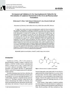
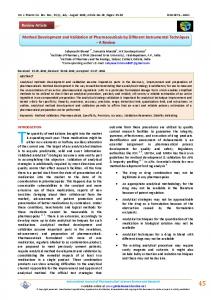
performed (29). The developed method was validated according to the ICH guidelines (30) under such parameters as system suitability, selectivity, specificity, linearity (concentration–detector response relationship), accuracy, precision, sensitivity, detection, and quantification limit. System suitability parameters System suitability test parameters is evaluated by analyzing the repeatability, peaks symmetry (symmetry factor), and theoretical plates of the column, resolution, mass distribution ratio (capacity factor) and relative retention. All the outcomes for the system suitability were within acceptable boundary (31). The system suitability studies were carried out by analyzing the symmetry of the peaks (symmetry factor < 2), theoretical plates of the column (> 3,500), retention factor was found from 2–7, and resolution between the peaks was greater than 2. Selectivity/specificity The selectivity and specificity of the projected method was appraised for the intervention of impurities caused by excipients existing in the finished product. Specificity was evaluated by matching up to the chromatograms attained from the drug and the most frequently used excipients mixture with those attained from blank. Figure 1 represents chromatograms of plasma containing all drugs. Four distinct, sharp, and symmetrical peaks were obtained with good baseline resolution. Column elutions were obtained as 2.34, 3.1, 3.8, and 5.3 for metformin, rosiglitazone, pioglitazone, and diltiazem respectively. Figure 2 represents a typical chromatogram of human serum containing all drugs respectively. A typical chromatogram of blank serum is shown in Figure 3.
Method validation
Method validation has received considerable attention in the literature and from industrial committees and regulatory agencies. Although there is general agreement about what type of studies should be done, there is great diversity in how they are Figure 2. Representative chromatogram showing resolution between diltiazem and NIDDMS drugs in human serum (Concentration 3.12 µg/mL).
Figure 1. Representative chromatogram showing resolution between diltiazem and NIDDMS drugs (Concentration 3.12 µg/mL).
Figure 3. Representative chromatogram of blank human serum.
3
Journal of Chromatographic Science, Vol. 49, February 2011
isfactory for all the analytes as confirmed by the correlation coefficients (r2) of greater than 0.9992 and characteristic parameters for regression equation (y = a + bx) obtained by least squares treatment of the results. LOD and LOQ for each compound, along with linearity data, are reported in Table I–II. In order to estimate the limit of detection and limit of quantification, mobile phase was Table I. Linearity and accuracy of the proposed method injected six times and the noise level was determined. The limit of detection was calFormulations (%) Raw Materials (%) Conc. Formulations (%) Raw Materials (%) Conc. culated to be three times the noise value and (µg/mL) Recovery RSD Recovery RSD (µg/mL) Recovery RSD Recovery RSD ten times the noise which gave limit of quanMET DLZ tification and also cross checked by formulas given as under. 0.39 98.8 1.31 100.27 1.17 1.56 99.37 0.54 101.25 0.57
Linearity and sensitivity
In order to evaluate the linearity of the proposed method calibration curves were constructed in the range of investigated concentrations and were found to be linear relationships within the quantification ranges for all the assayed drugs. Linearity was sat-
0.78 1.56 3.12 6.25 12.5 25.0
99.32 100.61 99.52 100.33 99.28 100.97
0.95 0.74 0.93 1.28 0.79 0.43
99.22 100.72 99.16 101.29 99.34 99.77
1.36 0.84 0.39 0.58 0.66 0.84
3.12 6.25 12.5 25.0 50.0 100.0
98.47 100.47 99.17 98.85 101.52 99.84
ROZI 0.78 1.56 3.12 6.25 12.5 25.0 50.0
100.32 99.44 100.62 98.74 100.36 99.47 100.28
1.2 0.84 0.92 0.84 0.71 1.07 0.95
Metformin Rosiglitazone Pioglitazone Diltiazem
Concentration range (µg/mL) 0.39–25 0.78–50 1.56–100 1.56–100
99.47 101.27 99.37 101.44 100.63 98.44 100.69
0.88 1.4 0.94 0.84 0.98 1.26 0.75
1.56 3.12 6.25 12.5 25.0 50.0 100.0
100.29 99.14 98.74 99.24 100.6 99.25 98.67
Regression equation A = 20736x + 5043 A = 13663x + 6115.6 A = 9885.8x + 9489.3 A = 9213.4x + 38905
r2
LOD (µg/mL)
LOQ (µg/mL)
0.9999 0.9998 0.9999 0.9992
0.011 0.033 0.033 0.043
0.034 0.102 0.101 0.130
Table III. Intraday and Inter-Day Precision
Analyte Metformin
Rosiglitazone
Pioglitazone
Diltiazem
4
100.69 100.77 98.66 99.37 100.84 101.34
1.07 0.85 1.08 1.47 0.94 0.78
LOD = 3.3 σ/S LOQ = 10 σ/S
100.34 99.47 98.47 99.57 100.47 101.33 101.27
0.86 1.34 0.59 0.88 0.57 1.18 0.94
Repeatability, intra-day, and inter-day precision and accuracy
Where σ is the standard deviation of the lowest standard concentration and S is the slope of the standard curve.
PIO
Table II. Regression Statistics and Sensitivity of this Method
Drug
1.06 0.84 1.11 0.97 0.77 0.92
% Working Range
Conc. (µg/mL)
% Day 1
CV Day 2
80 100 120 80 100 120 80 100 120 80 100 120
12 15 18 24 30 36 20 25 30 20 25 30
0.98 1.21 0.86 1.08 1.02 0.59 1.04 1.28 1.31 0.99 1.23 1.06
0.86 0.99 0.73 1.06 1.1 0.86 1.11 1.39 1.29 1.01 1.18 0.99
0.51 1.24 0.85 0.58 1.22 1.55 0.41
Method precision, articulated as repeatability and inter- and intra-day precision, was determined for each of the analyte. Repeatability was made sure by scheming the relative standard deviation (% RSD) of six replicate determinations. The same trial was completed on two different days for evaluating intermediate precision. The relative standard deviations ranged from 0.56 to 1.31% and 0.56 to 1.39% for intraday and inter-day repeatability, respectively. The results are shown in Table III. Accuracy/Recovery Accuracy tests were conducted at three different concentration levels (80, 100, and 120%) within the operational range, for each compound by measuring the recovery of known amounts of each drug which were spiked into their corresponding formulation, and these spiked matrices were expressed as percentage. For this purpose placebos were prepared of all the drugs and known quantities of drugs were added. The percentage of recovery was calculated by comparing the determined amount of these standards with the added amount. The consequences obtained for the measured concentration and the accepted recoveries indicate that the method is accurate; results are shown as in Table IV). Ruggedness Degree of reproducibility or ruggedness of the proposed method was established by determining diltiazem, metformin, pioglitazon, and roziglitazone using same chromatographic conditions by two analysts on two different days with two different HPLC systems (same model). Results of coefficient of variation (%CV) of two different days ranged from 0.59–1.39% verified the ruggedness of the projected method (Table III). Additionally, the elution order and the resolution factors of the compounds were not affected.
Journal of Chromatographic Science, Vol. 49, February 2011
Robustness The robustness of the method was investigated by deliberate modifications are made to the method operational conditions (32,33) for instance flow rate, pH, and composition of the mobile phase. Composition of solvent have a considerable effect of retention of analytes and flow rate as well but variation of pH up to ± 0.2 do not have any great influence on chromatographic behavior but any further change will affect the chromatographic behaviors. Serum drug analysis
Recovery of all the drugs from human serum was determined under the established chromatographic conditions. Blood samples were collected from 10 healthy 22–25-year-old volunteers. They were non-smokers, were not involved in any exhausting activity, and did not accept any other medications. Multiple Table IV. Accuracy of Drugs by Proposed Method
Drugs
Spiked conc. (µg/mL)
Accuracy %
12 15 18 24 30 36 20 25 30 20 25 30
97.3 101.0 101.8 99.4 100.9 98.4 100.2 101.9 100.6 97.9 99.5 101.7
MET
ROSI
PIO
DLZ
Recovered conc. (µg/mL) 11.676 15.150 18.324 23.856 30.270 35.424 20.040 25.475 30.180 19.580 24.875 30.510
Table V. Recovery from Human Serum by Proposed Method Conc. (µg/mL)
% Recovery
Conc. (µg/mL)
MET 0.39 0.78 1.56 3.12 6.25 12.5 25.0
DLZ 100.66 98.71 98.4 99.34 100.44 98.52
1.56 3.12 6.25 12.5 25.0 50.0 100.0
ROZI 0.78 1.56 3.12 6.25 12.5 25.0 50.0
% Recovery
98.27 99.1 100.26 98.37 98.15 99.08 100.52 PIO
98.44 99.37 98.41 100.45 101.35 98.32 99.18
1.56 3.12 6.25 12.5 25.0 50.0 100.0
99.67 98.24 100.45 99.35 98.27 100.28 101.33
blood samples (10 mL) were collected in evacuated glass tubes directly from a vein or using an indwelling forearm vein cannula. After collecting, the blood was slightly shaken, centrifuged at 3,000 rpm for 10 min, and the plasma was separated. To 1.0 mL of the plasma, 10.0 mL of acetonitrile were added. The mixture was vortexed for 1 min, and then centrifuged for 10 min at 10,000 rpm. The supernatant was filtered through a 0.45-µm pore size membrane filter. The obtained serum was mixed with drug solutions in a ratio of 1:1 to obtain the required drug concentration. The obtained solutions were stored at 20°C before the analysis. They were injected into the HPLC system and chromatographed. The results are given in Table V.
Conclusion The projected RP–HPLC method allows simultaneous quantitation of diltiazem, metformin, pioglitazone and rosiglitazone permitting excellent separation and resolution of the chromatographic peaks. First time, this method provides rapid, simple, specific, sensitive, accurate and reproducible quantitative analysis of these drugs. There was no interference from excipients in the tablets. The consequences found are in a first-class agreement with the declared contents. Statistical analysis showed the calculated value was established to be less than critical value.
References 1. J.C. Pickup and G. Williams. “Hypertension and diabetes mellitus”. Textbook of Diabetes. Oxford: Blackwells, 719-720, 1991. 2. E. Erdmann. Microalbuminuria as a marker of cardiovascular risk in patients with type 2 diabetes. Int. J. Cardiol. 107(2): 152–158 (2006). 3. Curtis Triplitt. Drug interactions of medications commonly used in diabetes. Diabetes Spectrum 19(4): 202–211 (2006). 4. B.M.Y. Cheung. Blockade of the renin-angiotensin system. HKMJ. 8(3): 185–191 (2002). 5. Certain Blood Pressure-lowering Drugs Reduce Diabetes Risk In Hispanic Patients. Science Daily (May 22, 2006). 6. B. Steven. Leichter and stephanie thomas combination medications in diabetes care: An opportunity that merits more attention. Clinical Diabetes 21: 175–178 (2003). 7. L.H. Lindholm, H Ibsen B. Dahlof R.B. Devereux, G. Beevers, U. de Faire, F. Fyhrquist, S. Julis, S.E. Kjeldsen, K. Kristiansson, O. Lederballe-Pedersen, M.S. Nieminem, P. Omvik, S. Oparil, H. Wedel, P. Aurup, J. Edelman, and S. Snapinn. The LIFE Study Group. Cardiovascular morbidity and mortality in patients with diabetes in the Losartan Intervention for Endpoint reduction in hypertension study (LIFE): a randomized trial against atenolol. Lancet. 359: 1004–1010 (2002). 8. D.J. Abraham. Burger’s Medicinal Chemistry and Drug Discovery, A John Wiley and Sons, Inc., Publication Hoboken, New Jersey, 6th 3: 15–17 (2003). 9. A.R. Gennaro. Remington: The Science and Practice of Pharmacy. University of the Sciences in Philadelphia 21st Ed, Philadelphia, PA 1364–1365 (2005). 10. E. Erdmann. Microalbuminuria as a marker of cardiovascular risk in patients with type 2 diabetes. Internat. J. Cardiology 15:107(2) 152–158 (2005). 11. G.J. Vergote, C. Vervaet, J.P. Remon, T. Haemers, and F. Verpoort.
5
Journal of Chromatographic Science, Vol. 49, February 2011
Near-infrared FT-Raman spectroscopy as a rapid analytical tool for the determination of diltiazem hydrochloride in tablets. Eur. J. Pharm. Sci. 16: 63–67 (2002). 12. B. Chankvetadze, M. Saito, E. Yashima, and Y. Okamoto. Enantioseparation using selected polysaccharides as chiral buffer additives in capillary electrophoresis. J. Chromatogr. A 773: 331–338 (1997). 13. M. Akram El-Didamony. Indirect spectrophotometric determination of diltiazem hydrochloride in pure form and pharmaceutical formulations. Central European Sci. J. 3(3): 520–536 (2005). 14. P.M. Lacroix, N. Beaulieu, T.D. Cyr, and E.G. Lovering. High performance liquid chromatography methods for assay of diltiazem hydrochloride and its compounds in bulk drug and finish tablet. J. Pharm. Sci. 78: 243–246 (1989). 15. Z. Dragica, S. Traje, and S. Marina. High-performance liquid chromatographic determination of diltiazem in human plasma after solid-phase and liquid–liquid extraction. Anal Bioanal. Chem. 376: 843–853 (2003). 16. L.X. Zhang and Z. Feilang. HPLC determination of diltiazem in human plasma and its application to pharmacokinetics in humans. Biomed. Chromatogr. 17(8): 522–525 (2003). 17. R. Shimizu, K. Ishii, N. Tsumagari, M. Tanigawa, and M. Matsumoto. Determination of optical isomers in diltiazem hydrochloride by high-performance liquid chromatography. J. Chromatogr A. 253: 101–108 (1982). 18. R. Shimizu, T. Kakimoto, K. Ishii, Y. Fujimoto, H. Kishi and N. Tsumagari. New derivatization reagent for the resolution of optical isomers in diltiazem hydrochloride by high-performance liquid chromatography. J. Chromatogr A. 357: 119–125 (1986). 19. K. Ishii, K. Minato, N. Nishimura, T. Miyamoto, and T. Sato. Direct chromatographic resolution of four optical isomers of diltiazem hydrochloride on a Chiralcel OF column. J. Chromatogr. A 686(1): 93–100 (1994). 20. B.S. Madhira, D.M. Vaibhav, A.S. Dimal, K.B. Kashyap, S.M. Rajendra, G. Madhira, J.P. Binita. Estimation of pioglitazone hydrochloride and metformin hydrochloride in tablets by derivative spectrophotometry and liquid chromatographic methods. J. AOAC Internat. 88(4): 1167–1172 (2005). 21. T. Radhakrishna, R.D. Sreenivas, and R.G. Om. Determination of pioglitazone hydrochloride in bulk and pharmaceutical formulations by HPLC and MEKC methods. J. Pharmaceut. Biomed. Anal. 29(4): 593–607 (2002). 22. P. Venkatesh, T. Harisudhan, H. Choudhury, R. Mullangi, N.R. Srinivas. Simultaneous estimation of six anti-diabetic drugs- gliben-
6
23. 24.
25.
26.
27. 28.
29. 30. 31. 32. 33.
clamide, gliclazide, glipizide, pioglitazone, repaglinide, and rosiglitazone: development of a novel HPLC method for use in the analysis of pharmaceutical formulations and its application to human plasma assay. Biomed. Chromatogr. 20(10): 1043–1048 (2006). D.G. Sankar, J.M.R. Kumar, and M.V.V.N. Reddy. UV spectrophotometric methods for the determination of anti-diabetic drugs. Asian J. Chem. 16(1): 537–539 (2004). R. Bhushan, D. Gupta, and A. Jain. TLC supplemented by UV spectrophotometry compared with HPLC for separation and determination of some antidiabetic drugs in pharmaceutical preparations. J. Planar Chromatogr. Modern TLC, 19:(110) 288–296 (2006). B.L. Kolte, B.B. Raut, A.A. Deo, M.A. Bagool, and D.B. Shinde. Simultaneous high-performance liquid chromatographic determination of pioglitazone and metformin in pharmaceutical-dosage form. J. Chromatogr. Sci. 42(1): 27–31 (2004). B.L. Kolte, B.B. Raut, A.A. Deo, M.A. Bagool, and D.B. Shinde. Simultaneous determination of metformin in combination with rosiglitazone by reversed-phase liquid chromatography. J. Chromatogr. Sci. 42(2): 70–73 (2004). N. Zaltýne, E. Ucakturk. Simultaneous determination of ezetimibe and simvastatin in pharmaceutical formulations by dual-mode gradient LC. Chromatographia. 66: S87–S91 (2007). K. Suenami, L.W. Lim, T. Takeuchi, Y. Sasajima, K. Sato, Y. Takekoshi, and S. Kanno. Rapid and simultaneous determination of nonsteroidal anti-inflammatory drugs in human plasma by LC–MS with solid-phase extraction. Anal Bioanal Chem 384: 1501–1505 (2006). G.S. Clarke. The validation of analytical methods for drug substances and drug products in UK pharmaceutical laboratories. J. Pharm. Biomed. Anal. 12: 643 (1994). ICH guideline Q2B: Validation of Analytical Procedures: Methodology (2003). I. Krull and M. Swartz. Validation viewpoint, introduction: National and international guidelines. LC-GC 15(6): 534–539 (1997). R.L. Plackett and J.P. Burman. The design of optimum multi-factorial experiments. Biometrika. 33: 305–325 (1943–1946). E. ASTM. American Society for Testing and Materials. Standard Guide for Conducting Ruggedness Tests (Plackett-Burman Design). 100 Barr Harbor Drive, West Conshohcken PA 19428–2959.
Manuscript received revision received