maltotetraose, and phosphate (see "Materials and Methods"), and ali- quots (2 pl) ..... Brick, P., Carter, P., Waye, M., and Winter, G. (1985) Nature 314.235-238.
Vol. 269, No. 4, Issue of January 28, pp. 2485-2490, 1994 Printed in U S A .
Tm JOURNAL OF B I O ~ I CCHEMISTRY AL 0 1994 by The American Society for Biochemistry and Molecular Biology, Inc.
Dissecting Differential Binding in the Forward and Reverse Reaction of Escherichia coli Maltodextrin Phosphorylase Using 2-Deoxyglucosyl Substrates* (Received forpublication, May 14, 1993, and in revised form, August 18, 1993)
Stefan BeckerS, DieterPalm, and Reinhard Schinzelg From the Theodor-Boveri-Institut fur Biowissenschaften (Biozentrum), Physiologische Chemie I, A m Hubland, 0-97074 Wiirzburg, Germany
It is assumed thatglycogen or linear polysaccharide chains Substrate analogs are used in combination with sitedirected mutagenesis to probe specific interactions be- can bind to phosphorylase in two distinct modes: one in which the terminalglucosyl residue is bound in a positionsuitable for tween substrate and enzyme in the forward and reverse direction of the Escherichia coli maltodextrin phospho- phosphorolysis(phosphorolysis mode, P-mode) and one in which the terminalglucosyl residue isbound in a position that rylasereaction. In thephosphorolysis(degradation) allows it to act as a glucosyl acceptor (synthesis mode, S-mode) mode, removal of the 2-OH group of the terminal glucose (French and Wild, 1953; Chao et al. 1969; Gold et al. 1974; of the polysaccharide results in a 30-fold reduction ofK , Segel, 1975). This together with thelong held conception that while similar changes were of no influence when the same polysaccharide was used for priming the synthe- synthesis of polysaccharide from Glc-1-P by phosphorylase acor T y l d s E does tion requires the presence of a “primer” molecule composed of sis. Mutation ofactive site residues Gluss7 of not change apparent affinity of substrates during deg- at least four consecutiveglucose residues raises the question radation. In the synthesis mode, 2-deoxyglucose-1-Pas whether there are distinguishable glycosyl binding subsites. Despite the outstanding contributions of studies with phossubstratecausesa2-foldreductionofthewild-type k,,dK, while for theYS38F mutant a %fold reduction phorylases for the solution of major enzymological problems is observed. In contrast, the mutation of Gluss7 to Asp like allosteric control, interconversion of covalently modified causes a 10-fold increase kcaJK,. in Therefore, different forms of an enzyme, and finally to thenonclassical catalysis by oli- pyridoxal-P, the problem of primer requirement is still unbinding sites for the terminal glucose residue of the gosaccharide and glucose-1-P exist. GluSS7 and solved (for review, see Madsen and Withers(19861, Palm et al. are part of the glucose-1-P binding site and do not inter- (1990), andJohnson (1992)). Theanswertothequestion act with the terminal glucose residue. A 2-fold increase whetherbinding of oligosaccharides inthe phosphorolysis in rate was observed in both directions using the 2-de-mode is differentfrombinding in the synthesis mode on a oxy derivatives. This confirms the role of intrinsic elecstructural level presumably will provide another clue to the tronic effects in stabilizing the transition state. Uncom- enigma of the primer requirement. petitive substrate inhibition at high concentrations of Present knowledge about substratebinding sites of glucosyl maltoheptaoseinthephosphorolysisdirection is ex- residues inphosphorylases is stillincomplete. From x-ray crysplained by inhibitory binding of the sugar in the synthetallography, molecular recognition and site directed mutagensis mode. esis studies the binding of Glc-1-P and its derivatives in ground state and transition state is well characterized (Street et al., 1986,1989; Schinzel and Palm,1990; Johnson, 1992). SubstanGlycogen phosphorylases (EC 2.4.1.1) constitute a well char- tially less known is about productive bindingof the polysacchaacterized class of enzymes that catalyze the phosphorolytic ride awaiting degradation. So far no binding of oligosaccharides cleavage of a-1,4-linked glucose units to produce a-D-glucose at the active siteof rabbit muscle glycogen phosphorylase has 1-phosphate (Glc-l-P)l: been observed in crystals (Spranget al., 1991; Johnson, 1992). In contrast, there isat least cross-structural information on a (Eq. 1) Glucose, + Pi glucose,-, + Glc-1-P second carbohydrate binding site responsible for the attachAlthough at equilibrium (Keq= Pi/Glc-1-P = 3.6 at pH 6.8) ment of carbohydrates toglycogen particles, the“glycogen stor“synthesis” isfavored, the physiological role of phosphorylase is age site” (Kasvinskyet al., 1978, Philip et al., 1982; Goldsmith the energy conserving mobilization of storage polysaccharides et al., 1989, Johnson, 1992). Beside the direct structural approach, molecular recognition through phosphorolysis. studies were used to map groups of substrates important for * This work was supported by DeutscheForschungsgemeinschaft binding. Withers and co-workers (1989)used deoxy and fluoriGrant DFG Pd19-3. The costs of publication of this article were de- nated analogs of Glc-1-P to analyze the contribution of the frayed in part by the payment of page charges. This article mustthere- different hydroxyl groups of the sugarto binding and catalysis fore be hereby marked “advertisement”in accordance with 18 U.S.C. of phosphorylases (Street et al., 1986, 1989). This strategy, Section 1734 solely to indicate this fact. however, was not applicable to assess the substrate requireDedicated to Prof. E. J. M. Helmreich on the occasion of his 70th ments of the P-mode, because analogs of the nonreducing terbirthday. minal residue werenot available. The synthesis of 2-deoxygluf Present address:TAD PharmazeutischesWerk D-27472 Cuxhaven. 8 To whom reprint requests should be addressed. cose-(Glc), and 2-deoxyglucose 1-phosphate provide the means The abbreviations usedare: Glc-1-P, a-D-glucopyranosyl phosphate; to discriminate positions of the terminalglucose residue when phosphate;2-deoxy-Glc2-deoxy-Glc-1-P, 2-deoxy-cr-~-glucopyranosyl used together with Glc-1-P and (Glc),. Complementary siteGlc,, a-l,4-(2-deoxyglucosyl)-(glucose)4; Glc,, maltoheptaose; Glc,, maltotetraose; MES, 2-(N-morpholino)ethanesulfonic acid; P-mode, directed mutagenesis can be used to analyze interactions of phosphorolysis mode; S-mode,synthesis mode. specific amino acid residues with the substrates. Acombination
-
wsE
2485
2486
Carbohydrate Binding to Maltodextrin Phosphorylase
of both methods should allowthe identification of interactions RF = 0.17). The composition was tested by hydrolysis at acidic pH and between specific amino acid residues and essential functional subsequent determination of 2-deoxyglucose and phosphate by the glugroups of the substrate molecule. Sierks and Svensson (1992) cose dehydrogenase assay (see above) and a colorimetric assay, respectively (Saheki et al., 1985). Since 2-deoxy-Glc-1-Pis thermolabile and used this approach to identify a hydrogen bonding pair in the instable at acidic pH, it was prepared freshly and used immediately. glucoamylase-maltose transition state complex. For enzymes Enzyme Purification-Wild-type and mutant enzyme were purified catalyzing reversible reactions, this approach should allowthe as described (Schinzel and Palm, 1990) with an additional fast protein detection of differences in binding of substrates in the forward liquid chromatography purification step on a Mono-Q anion exchange and reverse reaction. Such differences would be a strong indi- column (Pharmacia LKB Biotechnology Inc.). The enzyme was eluted cation for structural rearrangements at the active site during from the MonoQ column by a gradient (0-0,3 M NaCl in 10 m Trisacetate, 1m EDTA, pH 6.9) and dialyzed against 10 m Tris-acetate, catalysis. 1 m EDTA, 50 m KCl, pH 6.9. Purification oflow activity mutant From the crystal structure of the rabbit skeletal muscle en- proteins was followed up by SDS-polyacrylamide gel electrophoresis. zyme and its complexes with glucose and Glc-1-P derivatives, Protein concentration was determined by the method of Bradford (1971) ~ ' )been assigned or from the absorbance at 280 nm, using EA;"S = 1.36. two active site residues (lj76'73 and G ~ u ~had Kinetic Measurements-Enzyme activity was assayed a t 30 "C in the which are involved in interactions between the 2-OH group of the glucose residue and theenzyme (Johnson et al., 1990; Mar- direction of phosphorolysis in a coupled assay as described (Schinzel and Palm, 1990). Initial velocity studies were performed at 30"Cby tin et al. 1990).In aparallel approach, we mapped functionally varying the concentration of the substrates within a 10-fold range. The important amino acid residues of the Escherichia coli enzyme kinetic parameters were determined by non-linear regression using the by site-directed mutagenesis and kinetic studies. We found that GFtAFIT-program (Leatherbarrow, 1992). Activity using 2-deoxy-Glc-Glc4 as substrate was determined in an Ty1"538 and G ~ uwhich ~ ~ occupy ~ , the equivalent positions in the m highly conserved sequences of the catalytic site of phosphory- assay containing in 250pl of 50 m Tris-acetate, pH6.9,20 M for the mutant lases (Newgard et al., 19891, participate in ground state and KHzP04, lo-' M maltodextrin phosphorylase enzymes) and 0.5-7.5 m 2-deoxy-Glc-Glc4. At preset times aliquots (50 transition state binding of the glucosyl residue. When the ki- pl) were drawn and the 2-deoxy-Glc-1-P content was determined in a netic properties of the Y538F mutant enzyme and the wild-type coupled enzymeassay (25 m Tris-acetate, pH 8.0, 1m NAD, 20 pg of enzyme were compared using substrate analogs lacking either glucose dehydrogenase (Merck),2 units of alkaline phosphatase (Boehthe 2-OH group of the terminal glucose residue of linear oligo- ringer Mannheim)). In the direction of synthesis the enzyme activity saccharide (degradation) or the 2-OH group of Glc-1-P (synthe- was assayed by measuring the release of Pi from either Glc-1-Por 2-deoxy-Glc-1-P. sis), we found that this mutation had significant effects on RESULTS overall binding energy of Glc-1-Pin the synthesis direction, but was without effects in the degradation direction. On the other Synthesis of Z-Deoxy-Glc-Glc4 and 2-Deoxy-Glc-1-P-Reachand, removal of hydrogen bondingcapacity of the substrate at tion of maltodextrin phosphorylase with D-glucal in the presthe 2-position weakens overall apparent binding in theforward ence of maltotetraose and Pi yields 2-deoxyglucosyl oligosacand the reverse direction. charides and 2-deoxy-Glc-1-P (Fig. lA). Both products were In the present work differential binding of substrate analogs isolated in pure form. The number of 2-deoxyglucosylresidues and different properties of active site amino acid residues in the added to maltotetraose vaned from 1 to 50, depending on the P- or S-mode were used to derive a model for substrate versus D-glucallmaltotetraose ratio. Deoxyglucosyl oligosaccharides product binding. with a high deoxyglucose content ("deoxyamylose") are insoluble. Due to precipitation of the product, the reaction beMATERIALS AND METHODS came virtually irreversible. The soluble fraction of (2-deoxy-Glc),-Glc4 was isolated unIsolation and characterization of the substrate analogs was perder appropriate conditions (ratio of maltotetraoseh-glucal, 1:lformed as follows. a - 1 , 4 - ( 2 - D e o ~ y g l u c o s y l ) - f g l w o s e ) ~ M ~ ~ t e t(33 r a o sm, e Boeh- 1:3). Phosphorolysisof the soluble form of the 2-deoxyglucosyloligosaccharide by maltodextrin phosphorylaseyields 2-deoxyringer Mannheim) was incubated in 25 m KHzPO, with 100 m DGlc-1-P and D-glucal as analyzed by thin layer chromatography glucal and M maltodextrin phosphorylasein 25 m Tris-acetate, pH 6.9, for 30 min at 30 "C. The enzyme was removed by ultrafiltration (Fig. 1B). 2 units of alkaline (Amicon Centricon 30). To hydrolyze 2-deoxy-Glc-1-P, Substrate Znhibition during Phosphorolysis-Initial velocity phosphatase (Boehringer Mannheim) were added and incubated at studies at saturating concentrations of phosphate showed sig30 "C for 30 min. After a second ultracentrifugation step the solution was applied to a Fractogel TSK HW40 S gel filtration column (Merck) nificantly reduced rates at maltoheptaose concentrations and eluted with degassed bidistilled water (1d m i n ) . Peaks were de- higher than 2lll~(Fig. 2A 1. It was ascertained that theactivity tected by absorption a t 190 nm, 2-deoxy-Glc-Glc4 was foundin the first of the coupled assay was notlimited by concentrations up to 20 peak. The compound was homogeneous as tested by thin-layer chroma- lll~maltoheptaose. Corresponding kinetic studies were pertography (RF= 0.43). To prove its composition the compound was hy- formed with two mutants of maltodextrin phosphorylase, drolyzed in 1 N HzS04. f i r neutralization total monosaccharide was E637D and Y538F. Like the wild-type enzyme the E637D mumeasured by using glucose dehydrogenase (Merck). In addition, the glucose content was measured by a coupled enzyme assay using hexo- tant enzyme was subject to substrate inhibition, while for the kinase/glucose-6-P dehydrogenase(Boehringer Mannheim). Since 2-de- Y538F mutant enzyme substrate inhibition was not observed oxyglucose-6-P is not a substrate of glucose-6-phosphatedehydrogenase for concentrations up to 20 nm maltoheptaose (Fig. 2 4 ) . SubfromLeuconostocmesenteroides in the presence ofNAD instead of strate binding in the inhibitory mode (EG*) was treated as NADP (Bergmeyer et al., 1983),this assay allows the determination of uncompetitive inhibition according to Scheme I. the monosaccharide:glucoseratio. The ratio was found to be 5:4, confirming 1residue of 2-deoxyglucose per moleculeof maltotetraose. Compared to the 'H NMR spectra of maltotetraose, the 'H spectra of 2-deoxy-Glc-Glc, exhibit signals of the two protons at the 2-position at 6 1.7 (dt, J = 7.5 MHz, 3 Hz) and 6 2.18 (dd, J = 9 Hz, 7 Hz). a-2-Deoryglucose-l-phosphate-Maltotetraose(3 m) was incubated with inorganic phosphate and D-glucal as described above.The enzyme was removed by ultrafiltration and the filtrate was lyophilized. The lyophilisate was dissolved in 1-butanoYpyriWwater 6:43 (v/v), applied to a silica gel column (Lobar Kieselgel 60; Merck) and subsequently eluted with the same solvent (2 d m i n ) . Fractions were screened by thin-layer chromatography (1-butanollpyridirdwater 6:4:3,
E+G=EG=E+P
11 EG*
SCHEME 1 Equation 2 applies if [I1 = [GI: u = V-[GYKm
+ [GI + [GI2/Ki
(Eq. 2)
The data where fitted to this equation by non-linear regression to estimate the apparent binding constant for the inhibitory
Carbohydrate Binding to Maltodextrin Phosphorylase A
2487
1w
A
80
-
EW
z 4 0
20 0
0
B 0
1'
2'
5'
10
20
2'
5'
10
20
6
8 1 10 2 4 6 Gk7 (mM)
I"'"''''''''''T1
0
2deOxyGlcGlc, Glc, 2deoxYGlc-1-p
1'
4
30 M
--
0
2
30 M
Rc. 1. Time couree of formation of 2-deoxy-Glc-Glc4 (A)and of the degradation of 2-deoxy-Glc-Glc4 ( B ) .A, E. coli maltodextrin phosphorylase was incubated with D-glucal, maltotetraose, and phosphate (see "Materials and Methods") and aliquots (2 pl) were applied on TLC at the given times. The lacking 2-OH group causes a RF value for 2-deoxy-Glc-Glc4 largerthan the RF value for maltotetraose.B , E. coli maltodextrinphosphorylasewasincubatedwith 2-deoxy-Glc-G1c4, maltotetraose, and phosphate (see "Materials and Methods"), and aliquots (2 pl) were applied on TLC at the given times. Markers (right lane) are maltotetraose, glucose, and D-glucal.
binding mode of maltoheptaose (Ki).Values of about 8 m were determined for the wild-type enzyme and the E637D mutant enzyme, while for the Y538F mutant enzyme a value >lo0 mM was calculated. Substrate inhibition was notobserved with 2-deoxy-Glc-Glc4 at concentrations up to 5 m. Determination of inhibition constants at higher substrate concentrations appeared impracticable due to thehigh K , values (see below) and the low solubility of this compound. In contrast to phosphorolysis, no substrate inhibition was observed when the reaction was studied in theopposite direction. Maltoheptaose was used as primer in the presence of saturating concentrations of Glc-1-P and the initialvelocities were determined by measuring the phosphate release from Glc1-P.Up to of 50 mM maltoheptaose the reaction followed hyperbolic saturation kinetics withno detectable substrate inhibition (Fig. 2B). An apparent K , value of 3 mM was determinedfor the oligosaccharide primer. For both mutant enzymes K , values comparable to the wild-type enzyme values were observed. Initial Velocity Studies with Substrate Analogs-The results of a comparative kinetic study substituting 2-deoxy-Glc-Glc4 for Glc7 in the phosphorolysis direction are shown in Table I. About a 2-fold increase of kcatwas observed for the wild-type as well as for the mutant enzymes, E637D and Y538F. The ratio kat D.dsoq.O,~,d/k,t ~ 1 remained ~ 7 unaffected (Table I). The apparent binding of 2-deoxy-Glc-Glc4was lowered by a factor of 30 for the wild-type enzyme and by a factor of about 10 for both
2
4
6
8 10 1 12 4 6 Glc7 (d)
Rc.2. Substrate inhibition of E. coli maltodextrin phosphorylase by maltoheptaose in the direction of degradation ( A )and synthesis ( B ) for the wild-type enzyme (0).the E637D mutant (O), and the Y538F mutant enzyme (0).The rates are normalized (V,, = 100%). mutant enzymes. Quantitative binding data are restricted by the low solubility of 2-deoxy-Glc-Glc4at the concentrations required. Further, thereliability of the resultsmay sufferfrom a release of glucose caused by an increased hydrolytic error of mutant enzymes (Schinzel and Palm, 1990). Despite these difficulties in interpretation,a significant increase of the K , values compared to maltotetraoseis apparent. Theobserved about 15-fold decrease of k,,/K,,, for both the wild-type enzyme and the two mutant enzymes caused by the removal of the 2-OH group mainly reflects the decrease in apparent binding. Initial velocity studies in the direction of synthesis were performed with eitherof the two substrate pairs,2-deoxy-Glc1-P and 2-deoxy-Glc-Glc4in thepresence of saturating concentrations of maltotetraose andGlc-1-P, respectively. In contrast to the degradation reactionthe steadystate kinetic parameters for 2-deoxy-Glc-1-P determined for the wild-type and mutant enzymes differ considerably. For the wild-type enzyme a twofold increase in rate using 2-deoxy-Glc-1-P as substrate was observed when compared to that of Glc-1-P. This increase was accompanied by a threefold increase of the K, value for 2-deoxy-Glc-1-P. The k, of the E637D mutant enzyme also increased 2-fold, but surprisingly the E637D mutant enzyme binds 2-deoxy-Glc-1-P 10-foldbetter than the physiological substrate Glc-1-P. Therefore, the E637D mutant enzyme is theonly case observed where the modified substrate caused an increase in k,,/K, (Table 11). The kinetic parameters of the Y538F mutant enzyme differ from the wild-type and theE637D mutant enzyme. The Y538F mutant enzyme exhibits K , similar values for Glc-1-P and 2-deoxy-Glc-1-P. A decrease in rate by a factor of 7 rather than the 2-fold increase was found for the Y538F mutant enzyme with 2-deoxy-Glc-1-P as substrate. This is in contrast to the other forms of maltodextrin phosphorylase. Consequently, the deletion of the 2-OH group of Glc-1-P caused a relatively large decrease in k,,IK, compared to the moderatechanges in k&Km observed for the wild-type enzyme. Using 2-deoxy-Glc-Glc4as primer in the synthesisdirection no or only minor changes of the kinetic parameters were observed when compared to physiological primers in mutant and
Carbohydrate Binding
2488
Maltodextrin Phosphorylase to
TABLEI Kinetic parameters of wild-type and mutantE. coli maltodextrin phosphorylase with2-deozyglucosyl substrates in the phosphorolysis direction in the presence of saturating concentrations of inorganic phosphate Standard errors are 5% for wild-type and the Y538F enzyme and 15% for the E637D enzyme. GIc7
Wild-type 0.5 Y538F0.7 E637D
2-Deoxy-Glc-Glc4
kcat
Km
kc,JK,
kcat
K,
S-1
mM
S-l/mM
S"
mM
28 3.3 0.05
45
3 4.7 0.13
0.4
5.4 0.08
>15 6-9 5-6
kc,JKm" S"/mM
0.3 0.01
Directly determined dueto the low accuracy of the K , values.
TABLEI1 Kinetic parameters of wild-type and mutantE. coli maltodextrin phosphorylase with 2-deoxy-Glc-1-P as substrate in the direction of glycogen synthesis in the presence of saturating concentrations of maltotetraose Standard errors are 5% for wild-type and the Y538F enzyme and 15% for the E637D enzyme. Glc-1-P
73
Wild-type Y538F E637D
38
2-Deoxy-Glc-I-P
kcat
K,"
kc.JKm
kcat
K,
L J L
S-1
mM
s-l/rnM
S-1
mM
S"/mM
1.7 0.12
3.0
38 12.5 0,03 0.06
0.4
1.0 2.9 2.3
22 493 0.3
0.6
wild-type enzymes (Table 111). However, the data have to be interpreted cautiously, since the products of the reaction may themselves act as primer if the reaction proceeds, especiallyat low primer concentrations. These primers will behave like the physiological primers when more than 4 glucose units have been added. To circumvent this problem, rates were determined only in the initial phase of the reaction. At the corresponding time points the reaction was assayed for the presence of elongated primer by thin layer chromatography. No elongated primers were detectable under those conditions (data not shown).
P-mode and S-mode binding sites by purely kinetic methods, especially ifone is restricted to glucosyl derivatives. Those limitations can be overcome with a combination of alternate substrates and site-directed mutagenesis. An essential prerequisite of the latter approach is, however, that there will be no structural or conformational changes in ligands or the mutagenized enzyme other than the targeted groups. Such prerequisites were met by 2-deoxyglucosederivatives of Glc-1-Por the terminal glucose residue of oligosaccharide chains and by some mutagenized residues of the active site in E. coli maltodextrinphosphorylase. DISCUSSION Street et al. (1989)predicted that the2-deoxyglucosylresidue Early concepts of the action of phosphorylase on the polysac- would contribute to the stabilization of the hypothetical oxocharide substrate are based on a complementary substrate- carbonium ion-like transition state and thus lower activation enzyme relation. A series of binding positions is present which free energy. We observed incorporation of 2-deoxyglucosein the hold the substrates in appropriate steric arrangement so that terminal residue of Glc,-acceptor (primer) molecules when Dother functional groups present at the enzyme surface are able glucal was utilized by phosphorylases in thepresence of Pi and to complete the reaction. Five or more glucosebinding subsites a primer (Kleinet al., 1982). Phosphorolysisof 2-deoxy-Glc-Glcn are lined up complementary to an amylose chain or to Glc-1-P yields 2-deoxy-Glc-1-Pin a reversible reaction. Full reversibiland an amylose chain fragment of 4 ormoreglucose units ity of all stepsdemonstrates that substrates andproducts meet (French and Wild, 1953). Based on a complete analysis of the the above stated requirements of minimal structural changes kinetic consequences, Segel (Goldet al. 1974; Segel 1975) pro- in the ligands (Palm et al., 1991). Replacement of residue T y ? 3 8 by Phe or G ~ by uAsp ~in ~ posed two kinds of central complexes where maltooligosaccharides can bind in two different modes, onein which the termi- maltodextrin phosphorylase elicited distinct changes in kinetic nal glucosyl residue is bound ina position suitable for parameters leaving all measurable physical parameters intact phosphorolysis (P-mode)and one in which the terminalresidue (Schinzel and Palm, 1990). Overall changes in kinetic behavior can be assessed by is bound in a position that allows it to act as a glucosyl acceptor in particular. This value proved to be quite during synthesis (S-mode).In the phosphorolysis direction the changes in kCatlKm, presence of inorganic phosphate will allowboth, otherwise mu- informative for comparison of wild-type and mutant enzymes tually exclusive, modes of binding for the carbohydrate sub- or alternativesubstrates. Changes in performance are exstrate. In contrast, in the synthesis direction, the presence of pressed by AAG$ = RT In ( k c a ~ l K ~ ) ~ ( k ~ ~ t l K ~ ) l . Binding of the oligosaccharide in the degradation direction Glc-1-P should exclude binding of the polysaccharide in the P-mode (Fig. 3). The model would predict substrate inhibition was defined as P-mode binding (Fig. 3). The replacement of by a significant loss in the degradation direction, but not in the synthesis direction. Glc7by 2-deoxy-Glc-Glc4 was accompanied While we observed substrate inhibition, the data were best of binding affinity to wild-type and mutantenzymes. Fromthe difference a contribution of 2 kcal/mol by the 2-hydroxyl group fitted by uncompetitive inhibition assuming [I] = [GI and Ki 8 mM Glc7. Uncompetitivesubstrate inhibition is based on the to binding is calculated. This corresponds to the binding energy assumption that product binds to an independent binding site, provided by a hydrogen bond between uncharged groups which by similarity of product and substrate, will be occupied (Fersht et al., 1985; Street et al., 1986).On the other hand, the by the substrate athigh substrate concentration (Bisswanger, terminal 2-deoxyglucose residue causes a rate increase as an1979).Mutation of T y ? 3 8 to a Phe relieves substrate inhibition ticipated by Withers et al. (1989) on the basis of intrinsic elecpresumably by loss of inhibitory binding in theS-mode (Fig. 31, tronic effects. The similarity of kinetic performance shows that and hyperbolic binding in the P-mode is observed. There are in the direction of degradation the deoxyglucosyl residue limitations to obtain a more detailed structural inventory of the caused the same changes in wild-type and mutant phospho-
~
Carbohydrate Binding to
Maltodextrin Phosphorylase
2489
TABLE I11 Kinetic parameters of wild-type and mutant E. coli maltodextrin phosphorylasewith 2-deozy-Glc-Glc4as primer in the direction of glycogen synthesis under saturating Glc-I-Pconcentrations Standard errors are 5%for wild-type and the Y538F enzyme and 15%for the E637D enzyme. 2-Deoxy-Glc-G1c4
GIc4
Wild-type Y538F E637D
kcat
K,
kcad&
kcat
K,
kcadK,
s-1
mM
S-l/mM
S"
mM
S"lmM
38 12.5 0.07
3.0 2.9 3.6
2.5 3.9 2.6
14.4 3.8
12.7 4.3 0.02
n
0.03
A
36 15 0.08
WT
WT
At1.4
+lS\
Y598F 2dG-04
E6370 2dffi
B
WT
WT 0 1P
0 1P
2 r . Dogmdatlon
+
E6370 2dG1P
WT 2dG1 P
\4.3
y
WT
Y S 0 1P
2dG1 P
t1.2\
C n
Y53BF E6370 Glu a2\
/*.l
E6370 2dGlP
Y!i38F 2dGlP
WT Glu
WT Glu
WT 2dGe /+a4 E6370 2doc.
wr 2dG-G4
o.o\
2dG4 A . 8 Y S 2dG-G4
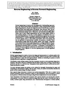
FIG.4. Overall activation free energy differencesbetween wild-type and mutant E. coli maltodertrinphosphorylases. Substrate analogs used are (A) 2-deoxy-Glc-Glc, (2dG-GJ in the degradation (phosphorolysis)direction, ( B ) 2-deoxy-Glc-1-P (2dGIP), and (C) 2-deoxy-Glc-Glc4(asprimer analog) in the synthesis direction. The energy differences (in kcal/mol) are calculated from the values given in Tables 1-111 using AAG$ = RT ln(k,,JK, )J(k.,JK,
1.
FIG.3. Model of oligosaccharide binding in the synthesis direction (1)and the degradation direction (2a).The inhibitory binding of the oligosaccharide in the S-mode during degradation is shown in 26.The numbers refer to the proposed subsites.
fects in performance can be attributed to changes in binding of the second substrate and not to changes in binding of the primer. rylases (Fig. 4). A difference of about 1.5 kcal/molwas found for Effects of alternative substrates and replacement of or wild-type and mutant enzymes. Since replacement of or G ~ were u observed ~ ~ when ~ the kinetics of binding for Glc-1-P G ~ didunot ~affect ~ the~relative values, these residues appear during synthesis were studied. This clearly distinguishes this not to be engaged in P-mode binding of the terminal glucose glucosyl binding subsite from the foregoing sites. In the wildresidue. type enzyme 2-deoxy-Glc-1-P caused a rate increase, indicating Binding of the oligosaccharide forelongation was defined as that the reverse reaction goes through a transition state comS-mode binding (Fig. 3). Differing from the preceding case, no parable with the forward reaction. This state isnot influenced changes in kinetic performance were observed whenthe termi- by the mutation of Glu637,but the mutation '&?38 Phe nal glucose residue is replaced by the 2-deoxyglucosyl analog impairs the capability of the catalyst to support the formation (Fig. 4). Therefore, the 2-OH group of the terminal glucose or stabilization of the transition state complex. From the kiresidue of the primer is not involved in binding of the primer netic data it can be calculated that the 2-OH-hydrogen bond molecule. Since the mutations Y538F or E637D do not influ- may contribute not only to binding in the ground state, as ence the overall binding energy of the oligosaccharide, the ef- proposed earlier (Schinzel and Palm, 1990), but contributes
w3'
w38
"-f
2490
Carbohydrate Binding to Maltodextrin Phosphorylase
about 1.2 kcaYmol to the stabilization of the transition state. The kinetic performance showsclearly that residues ‘13@3s and are in contact with the 2-hydroxylgroup of Glc-1-P (however, in case of E637D with 2-deoxy-Glc-l-P loss of the proposed 2-OH-phosphate bond can indirectly cause the observed effects). “he binding site for the glucosyl moiety of Glc-1-P is therefore different from the binding site for the terminal glucose residue of the oligosaccharide substrate in the degradation direction. Our present comprehension of substrate binding at the active site of phosphorylases is still dictated by the basic concept of French and Wild (19531,but now the functional differences of subsites can be described at a molecular level.In the direction of synthesis, subsite 1 is occupied by the glucose residue of Glc-1-P. Subsite 2 is occupied by the terminal residue of the oligosaccharide primer. From kinetic studies with oligosaccharides of different length (Glc3-Glc7)as primers in E. coli maltodextrin phosphorylase2 and potato phosphorylase (Suganuma et al., 1991) it can be concluded that the fifth subsite plays a critical role in primer binding and controlling the functional state of the catalytic site. Monosaccharides and short oligosaccharides like glucose, maltose, and maltotriose are only weak inhibitors of unregulated E. coli phosphorylase3and thus corroborate that sites 2 3 should be occupiedto allow the reaction to proceed. On the other hand,subsites 2 through 4 contribute only little to the affinity and specificity of polysaccharide binding. In contrast, the 2-OH group of the glucose moietyof Glc-1-P together with ‘13@3s and Gluss7 in position 1 of the subsite array are essential for the kinetic performance of the ternary enzyme substrate complex. This is in accordance with the crystal structure of the rabbit muscle phosphorylase b-heptulose2-P complex (Johnson et al., 19901, where the 2-OH group of heptulose-2-P is in hydrogen bond contact with the corresponding The removal of the hydroxyl group of “ y P 3 8 inthe bacterial enzyme causes similar effects on binding as the removal of substrate hydroxyl group. Although Glu672(rabbit muscle phosphorylase) is located in hydrogen bonddistance to the substrate, there isno evidence from kinetic data that hydrogen bonds betweenthe 2-hydroxyl group of Glc-1-P and the glutamate exist in ground state binding. The 2-OH group of the terminal glucose residue of the oligosaccharide substrate in the degradation direction participates in binding. However, the active site residue %53s, in contact to the 2-hydroxyl group in Glc-1-P, seemed not to be involved in binding of the oligosaccharide in the degradation direction. A second subsite 1(subsite 1* in Fig. 3) for the terminal glucose residue of the oligosaccharide different from the binding site of Glc-1-P is proposed. In thecourse of the reaction during transglycosylation there should be a rearrangement of the binding site of the glycosyl moiety of Glc-1-P to allow the formation of the a-1,4 bond. Indeed sugar-induced local changes at the acK. Rusch and D. Palm, unpublishedresults. D. Palm and R. Schinzel, unpublishedresults.
tive site of phosphorylase b upon binding of the inhibitor heptulose-2-P were observed (Johnson et al., 1990). Further evidence for a structural rearrangement during catalysis comes from the observation that different dianion binding sites for the substrate and the product phosphate exist at the active site (Barford and Johnson, 1989; Sprang et al., 1992; Leonidas et al., 1992). Future work will concentrate on the characterization of the second subsite l*. Amino acid residues potentially involved in oligosaccharide binding in the degradation direction willbe identified by site-directed mutagenesis. Further, subsite 5 will be identified, and the proposed contribution of this subsite to binding and catalysis will be characterized. Acknowledgments-We thank
Dr. K. Schnacken for help with the
N M R measurements and for critical reading of the manuscript andS. Spahr for technical assistance. REFERENCES Barford, D., and Johnson, L. N. (1989) Nature 218.233-236 Bergmeyer, H., Grassl, M., and Walter, H.-E. (1983) in Methods of Enzymatic Annlysis (Bergmeyer, H. U.,ed) 3rd Ed., Vol. 2, pp. 204-205, VCH, Weinheim, Germany Bisswanger, H. (1979) Theorie der Enzymkinetik, pp. 101-102, VCH, Weinheim, Germany Bradford, M. M. (1976) A d . Biochem. 72,248-254 Chao J., Johnson, G . F., and Graves, D. J. (1969) Biochemistry 8,1459-1466 Fersht, A. R, Shi, J. P., KnillJones, J., Lawe, D. M., Wilkinson, A. J., Blow, D. M., Brick, P., Carter, P., Waye, M., and Winter, G . (1985) Nature 314.235-238 French, D., and Wild, G. M. (1953) J. Am. Chem. Soc. 76,4490-4492 Gold, M. H., Farrand, R. J., Livoni, J. P., and Segel, I. H. (1974) Arch. Biochem. Biophys. 181,515-527 Goldsmith, E.J., Sprang, S. R., Hamlin, R., Xoung, N.-H., and Fletterick, R. J. (1989) Science 246,528-532 Johnson, L. N. (1992) FMEB J. 6,2274-2282 Johnson, L. N., Acharya, K, Jordan, M. D., and McLaughlin, P. (1990) J. Mol. Biol. 211,645-661
Kasvinsky, P. J., Madsen, N. B., Fletterick, R. J., and Syguach, J. (1978) J. Bwl. Chem. 263,1290-1296 Klein, H. W., Palm, D., and Helmreich, E. J. M. (1982) Biochemistry 21.6675-6684 Leatherbarmw, R. J. (1992) GraFit Version 3.0, Erithacus Software, Staines, UK Leonidas, D. D., Oikonomakos, N. G., Papageorgiou, A., Acharya, K , Barford, D., and Johnson, L. N. (1992) Protein Sei. 1, 1112-1122 Madsen, N. B., and Withers, S.G. (1986) in Coenzymes and Cofactors (Dolphin D., Poulsen, R., and Awamovic, O.,eds) Vol. I, pp. 355-389, John Wiley & Sons, New York Martin, J. L., Johnson, L. N., and Withers, S.G. (1990) Biochemistry 29, 1074510756
Newgard, C. B.,Hwang, P. K, and Fletterick, R. J. (1989) Crit. Rev. Biochem Mol. Biol. 24, 69-99 Palm D., Klein, H. W., sehinzel, R., Buehner, M., and Helmreich, E. J. M. (1990) Biochemistry 29, 1099-1107 Palm, D., Becker, S., and sehinzel, R. (1991) in Enzymes Dependent on Pyrida+al Phosphate and Other Carbonyl Compounds as Cofactors (Fukui, T., yama, H., Soda, K , and Wada, H., eds) pp. 377-385, Pergamon Press, oxford Philip G., Gringel G., and Palm, D. (1982) Biochemistry 21,3043-3050 Saheki, S.,Takeda, A,, and Shimazu, T.(1985) A M I . Biochem. 148,277-281 Schinzel R., and Palm, D. (1990) Biochemistry 29,9956-9962 Segel, I. (1975) Enzyme Kinetics, pp. 316-320, John Wiley & Sons, New York Sierks, M.R., and Svensson, B. (1992) Protein Eng. 6, 185-188 Sprang, S.R., Withers, S. G., Goldsmith, E., Fletterick, R. J., and Madsen, N.B. (1991) Science 264,1367-1371 Sprang, S. R., Withers, S. G., and Madsen, N. B. (1992) Protein Sci. 1, 1100-1111 Street, I. P., Armstrong, C. R., and Withers, S.G. (1986) Biochemistry 26,6021-
mami-
6027
Street, I. P., Rupitz, K , and Withers, S.G. (1989) Biochemistry 26,1581-1587 Suganuma, T.,Kitazone, J.-I., Yoshinaga, K, Fujimoto, S., and Nagahma, T. (1991) Carbohydrate Res. 217,213-220 Withers, S. G., MacLennan, D. J., and Street, I. P. (1989) carbohydrate Res. 164, 127-144