International Journal of
Molecular Sciences Article
Effects of Gelatin Hydrogel Containing Anti-Transforming Growth Factor-β Antibody in a Canine Filtration Surgery Model Michiko Maeda 1 , Shota Kojima 1, *, Tetsuya Sugiyama 1,2 , Denan Jin 3 , Shinji Takai 3 , Hidehiro Oku 1 , Ryohsuke Kohmoto 1 , Mari Ueki 1 and Tsunehiko Ikeda 1 1
2 3
*
Department of Ophthalmology, Osaka Medical College, Takatsuki-City, Osaka 569-8686, Japan;
[email protected] (M.M.);
[email protected] (T.S.);
[email protected] (H.O.);
[email protected] (R.K.);
[email protected] (M.U.);
[email protected] (T.I.) Nakano Eye Clinic of Kyoto Medical Cooperative, Kyoto 604-8404, Japan Department of Innovative Medicine, Osaka Medical College, Takatsuki-City, Osaka 569-8686, Japan;
[email protected] (D.J.);
[email protected] (S.T.) Correspondence:
[email protected]; Tel.: +81-072-683-1221; Fax: +81-072-681-8195
Academic Editor: Rei Ogawa Received: 3 April 2017; Accepted: 2 May 2017; Published: 5 May 2017
Abstract: In this present study, we investigated the effect of a controlled release of anti-transforming growth factor β (TGF-β) antibody on intraocular pressure (IOP), bleb formation, and conjunctival scarring in a canine glaucoma filtration surgery model using gelatin hydrogel (GH). Glaucoma surgery models were made in 14 eyes of 14 beagles and divided into the following two groups: (1) subconjunctival implantation of anti-TGF-β antibody-loaded GH (GH-TGF-β group, n = 7), and (2) subconjunctival implantation of GH alone (GH group, n = 7). IOP and bleb features were then assessed in each eye at 2- and 4-weeks postoperative, followed by histological evaluation. We found that IOP was significantly reduced at 4-weeks postoperative in the two groups (p < 0.05) and that IOP in the GH-TGF-β-group eyes was significantly lower than that in the GH-group eyes (p = 0.006). In addition, the bleb score at 4-weeks postoperative was significantly higher in the GH-TGF-β group than in the GH group (p < 0.05), and the densities of fibroblasts, proliferative-cell nuclear antigen (PCNA)-positive cells, mast cells, and TGF-β-positive cells were significantly lower in the GH-TGF-β group than in the GH group. The findings of this study suggest that, compared with the GH-group eyes, implantation of anti-TGF-β antibody-loaded GH maintains IOP reduction and bleb formation by suppressing conjunctival scarring due to the proliferation of fibroblasts for a longer time period via a sustained release of anti-TGF-β antibody from GH. Keywords: trabeculectomy; glaucoma; gelatin hydrogel; transforming growth factor-β; beagles
1. Introduction Glaucoma filtration surgery (i.e., trabeculectomy) is a primary treatment for glaucoma that results in decreased intraocular pressure (IOP) by draining the aqueous humor to the subconjunctival space and forming a bleb. Reportedly, the most common cause of unsuccessful trabeculectomy surgery is subconjunctival scarring of the filtration bleb, which leads to subconjunctival fibrosis [1,2]. The findings of a large prospective randomized trial showed that a single application of mitomycin C (MMC) or 5-flurouracil (5-FU) during trabeculectomy surgery greatly improves the surgical results; i.e., the prolonged bleb persistence and IOP reduction via strong suppression of fibroblast proliferation [3]. However, their application also increases the risk of complications such as a thin bleb, bleb infection, and infectious endophthalmitis at the late phase [4–6].
Int. J. Mol. Sci. 2017, 18, 985; doi:10.3390/ijms18050985
www.mdpi.com/journal/ijms
Int. J. Mol. Sci. 2017, 18, 985
2 of 11
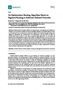
In this present study, we investigated transforming growth factor β (TGF-β), which is known to have three isoform types in humans; i.e., β1, β2, and β3. Isoforms β1 and β2 are known to greatly stimulate the dermal scarring response [7,8]. β2 is the mainly expressed ocular isoform, and is identified in both normal and diseased eyes [9,10]. The conjunctival scarring response in trabeculectomy surgery is thought to be affected by the passage of the aqueous humor including growth factors such as TGF-β, and subconjunctival scarring post glaucoma surgery is strongly affected by cytokines (especially TGF-β in the aqueous humor) [11,12]. Compared with other growth factors, TGF-β2 is reportedly dominant in the aqueous humor of glaucoma patients [13,14]. The TGF-β family is the main stimulator leading to conjunctival scarring post trabeculectomy, and various cells, such as fibroblasts and macrophages, can secrete them [15]. It was previously reported that TGF-β2 could increase α-smooth muscle actin (α-SMA) expression and the transdifferentiation of fibroblasts in conjunctiva to myofibroblasts [16]. In another previous study, the authors’ findings revealed that bleb failure post trabeculectomy primarily occurred due to the excessive accumulation of collagen in the subconjunctival space, and that high activity of TGF-β was associated with scarring [17]. Numerous studies have reported that the expression of TGF-β activates the proliferation by human Tenon’s fibrosis, and excessive production of granulation tissue constituents leading to scar formation [18–20]. In addition, several studies have reported that TGF-β inhibitors may effectively reduce scarring by reducing TGF-β activity via neutralization with antibodies [21,22]. Subconjunctival injections of anti-TGF-β antibody, as a drug substituting for MMC, were performed in a clinical trial for the suppression of fibroblast proliferation post trabeculectomy, however, the outcome was reportedly unsuccessful [23]. Various drug delivery systems (DDSs) have been tested for sustained drug release, since it is important to prevent scarring over an extended period following glaucoma surgery. Several previous studies have focused on subconjunctivally implanting DDSs to provide a sustained release of antiproliferative drugs over an extended time period post glaucoma surgery [24–26]. Most of those studies reported that these DDSs maintained IOP reduction and prolonged bleb persistence to the same degree as the conventional application of MMC and 5-FU, while significantly reducing their toxicity. However, most of those DDSs have yet to obtain successive results in the treatment of glaucoma patients [25]. Gelatin hydrogel (GH), a biodegradable material developed in Japan, has reportedly been used as a DDS for bioactive proteins in other medical fields [27]. Various growth factors gradually released from GH have been effective for therapy of various tissues [28,29]. In addition, GH has been applied to clinical therapies, such as for severe skin lesions complicating autoimmune vasculitis syndromes, peripheral arterial disease, and severe ischemic limb pain, and was found to be both safe and effective [30,31]. In the field of ophthalmology, GH impregnated with basic fibroblast growth factor has reportedly been used to induce experimental models of subretinal or corneal neovascularization [32,33]. We previously reported the possibility of using GH containing chymase inhibitor and GH containing MMC for longer-term maintenance of filtering blebs and IOP reduction by the prolonged suppression of subconjunctival scarring [34,35]. In this present study, we investigated the effect of a sustained release of anti-TGF-β antibody from GH in a canine glaucoma surgery model for IOP reduction and the effect on tissue in comparison with the application of GH alone. 2. Results 2.1. Verification of Anti-TGF-β Antibody in GH Goat anti-Chicken IgY (H + L) secondary antibody was utilized to detect anti-TGF-β1-2 antibody. GH soaked overnight in phosphate-buffered saline (PBS) did not show a positive staining image by immunostaining, however, we were able to verify a wide range of positive staining images at sections of sliced GH with anti-TGF-β antibody overnight (Figure 1).
Int. J. Mol. Sci. 2017, 18, 985
3 of 11
Int. J. Mol. Sci. 2017, 18, 985 Int. J. Mol. Sci. 2017, 18, 985
3 of 12 3 of 12
(A) (A)
(B)(B)
Figure 1.1.Gelatin Gelatin hydrogel (GH) containinganti-transforming anti-transforming growth factor (TGF-β) antibody. GH Figure1. Gelatinhydrogel hydrogel (GH) (GH) containing containing growth factor β (TGF-β) antibody. GHGH Figure anti-transforming growth factor ββ (TGF-β) antibody. soaked overnight in phosphate-buffered saline (PBS) (A) did not show a positive staining image by soakedovernight overnightin in phosphate-buffered phosphate-buffered saline a positive staining image by by soaked saline(PBS) (PBS)(A) (A)did didnot notshow show a positive staining image immunostaining, however, we were were able ableto toverify verifyaaawide widerange range of positive staining images (red) at immunostaining, however, however, we ofof positive staining images (red) at at immunostaining, were able to verify wide range positive staining images (red) sections of sliced GH with anti-TGF-β antibody (B) overnight. Scale bars: 500 μm. sections of sliced GH with anti-TGF-β antibody (B) overnight. Scale bars: 500 μm. sections of sliced GH with anti-TGF-β antibody (B) overnight. Scale bars: 500 µm.
USV Symbol Macro(s) Description 2.2.IOP IOPChange Change 2.2. 1EF9 ỹ \~{y} LATIN SMALL LETTER Y WITH TILDE 2.2. IOP Change 200C \textcompwordmark ZERO WIDTH NON-JOINER Theinitial initial IOP IOP values values (mean (mean ±± SD) GH containing anti-TGF-β The SD) were were15.9 15.9±±0.7 0.7mmHg mmHgininthethe GH containing anti-TGF-β The initial IOP \textthreequartersemdash values (mean ± SD) were 15.9 ± 0.7 mmHg inFIGURE theDASH GH containing anti-TGF-β 2012 ‒ antibodygroup group(GH-TGF-β (GH-TGF-β group) group) and 15.5 ±±0.8 mmHg ininthe GH alone group (GH group). TheThe antibody and 15.5 0.8 mmHg the GH alone group (GH group). 2013 group – (GH-TGF-β \textendash EN DASH group (GH group). The IOP antibody group) andwere 15.5 ± 0.8 mmHg in the GH GH-TGF-β alone IOP values at 2-weeks postoperative 8.1 ± 0.4 mmHg in the group and 8.0 ± IOP 2014 values at—2-weeks postoperative were 8.1 ± 0.4 mmHg in the EMGH-TGF-β group and 8.00.4 ± 0.4 \textemdash DASH values at 2-weeks postoperative were 8.1 ± 0.4 mmHg inpostoperative the GH-TGF-β group and 8.0mmHg ± 0.4 mmHg in mmHg in the GH group. The IOP values at 4-weeks were 9.4 ± 0.7 in the mmHg GH group. The IOP values at 4-weeks postoperative wereBAR9.4 ± 0.7 mmHg in the 2015in the ― \texttwelveudash HORIZONTAL the GH group. The IOP values at 4-weeks postoperative were 9.4 ± 0.7 mmHg in the GH-TGF-β group GH-TGF-β group and 12.9 ± 0.7 mmHg in the GH group. In the eyes in both groups, IOP was found 2016 ‖ \textbardbl VERTICAL LINE GH-TGF-β group and 12.9 ± 0.7 mmHg in the GH group. In the eyesDOUBLE in both groups, IOP was found and 12.9 ± 0.7 mmHg in the GH group. In the eyes in both groups, IOP was found to beFigure significantly to be significantly reduced at 2- and 4-weeks postoperative (p < 0.05, unpaired t-test, 2). \textdoublevertline to be significantly reduced at 2- and 4-weeks postoperative (p < 0.05, unpaired t-test, Figure 2). reduced at 2and 4-weeks postoperative (p < 0.05, unpaired t-test, Figure 2). Although there was no 2018 ‘ \textquoteleft LEFT SINGLE QUOTATION MARK Although there was no significant difference in IOP between the eyes in both groups at 2-weeks Although there was\textgrq no significant difference in IOP between the eyes in both groups at 2-weeks significant difference IOP between the eyeswas in both groups atlower 2-weeks postoperative, IOP at 4-weeks postoperative, IOP atin4-weeks postoperative significantly in the GH-TGF-β group than in 2019 ’ IOP at \textquoteright QUOTATION MARK group than in postoperative, 4-weeks postoperative was significantly lowerRIGHT in SINGLE the GH-TGF-β postoperative was lower in the group than IOP in the GHagain groupbegan (p < 0.05, unpaired the GH group (p