Wilber Sabiiti1 & Robin C May*2 Infection & Immunity, Clinical Sciences Division, St Georges’ University of London, London, SW17 0RE, UK Institute of Microbiology & Infection, School of Biosciences, University of Birmingham, Birmingham, B15 2TT, UK *Author for correspondence: Tel.: +44 121 41 45418 n Fax: +44 121 41 45925 n
[email protected]
1 2
Brain infection by the fungus Cryptococcus neoformans results in inflammation of the meninges and brain parenchyma, a condition known as meningoencephalitis. One million people are estimated to suffer cryptococcal meningitis globally and >60% of these cases die within 3 months of diagnosis. Humans are believed to contract infection by inhalation of spores or dried yeast cells, which subsequently colonize the lung tissue. In the lungs, cryptococci may be cleared by the lung phagocytes, stay latent, cause pulmonary infection and/or disseminate to other body parts, preferentially the brain, culminating in cryptococcal meningo encephalitis. In this review, we discuss the pathogenesis of C. neoformans from the environment to the brain, the current understanding of the mechanisms of cryptococcal transmission into the brain and cryptococcal meningitis. We also give an insight into future cryptococcosis research and the development of novel therapies.
Cryptococcosis is a life-threatening disease causing a significant global burden of disease. The CNS form of the disease, cryptococcal meningoencephalitis (CM; or meningitis) is the most severe and accounts for most mortality arising from cryptococcosis. One million cases of CM are estimated to occur globally per year, with >60% mortality within 3 months of infection. HIV-associated CM accounts for the majority of these cases, 80% of which occur in sub-Saharan Africa [1] . Despite increasing access to antiretroviral therapy, the incidence and mortality of cryptococcal meningitis remains high, with the diagnosis of HIV frequently being coincident with the diagnosis of meningitis [2–4] . Even when optimal treatment with amphotericin B and flucytosine is available, mortality is between 15–30% in both lowand high-income settings [5,6] and much higher in low-income countries where such antifungal regimens are not readily accessible. In addition, the incidence of non-HIV-associated CM is increasing in Pacific north-western America and far east Asia, caused by hypervirulent strains of Cryptococcus gattii and Cryptococcus neoformans, respectively [7–9] . Lastly, cryptococcosis is the third leading fungal infection in solid organ transplant patients [10,11] and, thus, advances in organ transplant application and the wide use of immunosuppressives is prospectively expected to account for an increased incidence of cryptococcosis in the medically developed world [10–12] . 10.2217/FMB.12.102 © 2012 Future Medicine Ltd
The genus Cryptococcus has been known for over 100 years [13] . First isolated from fruit juice in 1894 by an Italian scientist, Francesco Sanfelice, the fungus C. neoformans was later isolated from milk, humans, pigeon droppings and roosting places [13,14] . The nomenclature of C. neoformans evolved through the names Saccharomyces neoformans, Blastomyces neofor mans, Cryptococcus hominis and Torula histolyt ica, until the 1950s, when the current name was settled on [15] . C. neoformans is an encapsulated yeast and, based on the immunological reaction to the polysaccharide capsule, was classified into four serotypes: A, B, C and D. Serotypes A and D are characterized as C. neoformans var. gru bii and C. neoformans var. neoformans, while serotypes B and C were associated with C. neo formans var. gattii [13,16] . Molecular-based typing has led to the recognition of C. neoformans var. gattii as a distinct species, C. gattii [17,18] , restricting C. neoformans var. neoformans and C. neoformans var. grubii to serotypes D and A, respectively [13,19,20] . The varieties are further divided into molecular types VNI, VNII and VNB of C. neofor mans var. grubii; VNIV of C. neoformans var. neoformans; VNIII (hybrid, serotype AD); and VGI, VGII, VGIII and VGIV corresponding to C. gattii [20,21] . The serotype A C. neoformans grubii molecular types VNI and VNII have a global occurrence [22] and account for 95% of the worldwide cryptococcal infections and 98% among HIV/AIDS patients [23] . The VNB type Future Microbiol. (2012) 7(11), 1297–1313
Review
Future Microbiology
Mechanisms of infection by the human fungal pathogen Cryptococcus neoformans
Keywords n
blood–brain barrier n brain
n cryptococcal
meningoencephalitis neoformans n dissemination n infection n macrophages n Cryptococcus
part of
ISSN 1746-0913
1297
Review
Sabiiti & May
is geographically restricted to southern Africa [22] . Recent population and phylogenetic studies show that C. neoformans var. grubii VNI originated in Africa [21,24] . This finding follows the previous phylogenetic study, which suggested an African origin of the AD hybrid serotype [25] . The first account of human cryptococcosis is attributed to two German scientists, Busse and Buschke, who, in 1895, described a 31-year-old woman with a lesion on her tibia [23] . Although many clinical cases of cryptococcosis were reported in the 1900s, the prevalence remained generally low until 1947–1968, when high numbers of cases were reported in Africa. The increase in cases is presumed to have been due to the emergence of AIDS in the Congo river basin during that period [13,26,27] . The global HIV/AIDS epidemic of the 1980s to the present has seen a parallel rise in cases of human cryptococcosis, mainly due to the immunosuppressive nature of HIV/AIDS [15,28–30] . Cryptococcal infection can potentially occur in any part of the human body, with cases involving organs such as skin, bone, prostate gland, urinary tract, liver, spleen, lymph nodes, lungs and brain being reported [29–32] . Of all the cryptococcal sites of infection, pulmonary and CNS sites are the most commonly affected sites, with most mortality arising from CNS cryptococcosis [1] . The route of human infection is believed to be through inhalation of airborne propagules from an environmental source to the lungs. Colonization of the host can result in: no disease, where the fungus is cleared by the host immune response; asymptomatic infection, in which the fungus enters latency and may be reactivated when the host becomes immunocompromised; pulmonary disease, characterized by pulmonary nodules and inflammation of the lungs [33,34] ; and disseminated disease, which can potentially occur in all systemic organs, with the brain being the most preferred destination [29,35–38] . Interestingly, Ngamskulrungroj et al. recently demonstrated that, unlike C. gattii, C. neoformans prefers the brain to the lung as a site of infection [39] . Cryptococcal infection of the brain will depend on the successful maneuver by the fungus through the barriers involved in the early phase of infection. Host colonization & pulmonary infection
Greater than 50 basidiomycetous yeast Crypto coccus species live ubiquitously in the environment, but only C. neoformans and C. gattii are significant pathogens of humans. The infectious cryptococcal propagules are widely spread in the 1298
Future Microbiol. (2012) 7(11)
environment and, through inhalation, the spores or dried yeast cells gain access to the respiratory tract. Colonization and establishment of infection in the lungs is a process involving successful transmission of infectious propagules into the lungs, as well as a complex interplay of pathogen virulence factors and host immune response. Transmission into the alveolar space
Cryptococcal spores and/or dried yeast cells are very small in size and can deposit deep in the respiratory tract following inhalation [40] . The kinetic action of host cilia and mucus may serve to drive the infectious particles down into the alveolar space [40] . While spores are largely known to mediate transmission from the environment into the host by pathogenic fungi, such as Blastomyces dermatitidis, Histoplasma capsula tum, Coccidioides spp., Aspergillus fumigatus and Paracoccidioides brasiliensis [41,42] , the extent to which cryptococcal spores, rather than desiccated yeast cells, are infectious propagules is still debatable. Recent advances in isolating and characterizing C. neoformans spores has opened up opportunities to model infection using spores as infectious particles in order to determine their ability to infect and cause disease in the host [40,43] . Spores of serotype A C. neoformans var. grubii caused 100% lethal infection in a murine inhalation model of infection and in the invertebrate host, Galleria mellonella [40] . Both spores and yeast cells could be aerosolized by a stream of air, although there were more aerosolized spores than yeast cells [40,43] , suggesting a higher chance of spores being involved in environmental dispersal and host exposure. However, spores are more susceptible to killing by activated host alveolar macrophages and thus are speculated to depend on rapid germination into mature yeast cells in the host in order to establish infection [40,43] . Isolation of C. neoformans spores from the environment and characterization of a wide range of spores from both clinical and environmental strains are needed to further substantiate the potency of spores as C. neoformans infectious propagules. The lung alveolar space is lined by a tension-reducing fluid, surfactant, which promotes adherence of cryptococcal cells to lung epithelial cells [44] . SP-D binds to and promotes phagocytosis of both encapsulated and acapsular C. neoformans via macrophages [45] , an indication that cryptococcal opsonization with host surfactant is most likely one of the first innate factors mediating transition of cryptococci from extracellular to intracellular life. Interestingly, future science group
Mechanisms of infection by the human fungal pathogen Cryptococcus neoformans
SP-D -/- mice were partially protected from cryptococcal infection and had reduced lung fungal burden [45] . The presence of alveolar macrophages that scavenge microbes in the respiratory tract is also likely to drive cryptococci into deeper areas of the respiratory tract and lung tissue, since each alveolus contains at least one alveolar macrophage [46] . Pulmonary infection: barrier or stopover for disseminated cryptococcosis
Once in the alveolar space, cryptococcal yeast cells may survive extracellularly and/or transit into lung tissue either by direct internalization by lung epithelial cells or by resident alveolar macrophages [38,47] . At this stage, depending on the nature of host immune response, pulmonary colonization, in which cryptococci are either cleared or develop into a localized latent asymptomatic infection, occurs. Alternatively, pulmonary involvement can serve as a temporary stopover in which cryptococci go on to establish symptomatic infection with subsequent dissemination to other parts of the body. Alveolar macrophages comprise 95% of the bronchoalveolar cell population, which makes them the predominant first-line phagocytes in the lungs [46] . The interaction of cryptococci with alveolar macrophages most likely determines the establishment and fate of pulmonary infection and subsequent systemic dissemination. Increased fungal burden and extrapulmonary dissemination were evident in alveolar macrophage-depleted rats [48] . Absence of activated alveolar macrophages in T–NK-celldeficient mice (Tg26) resulted in no granuloma formation, conditions under which the attenuated C. neoformans glucosylceramide mutants (DgcS1) become able to proliferate and disseminate to the brain [49] . Cryptococcal phagocytosis and antigen presentation by alveolar macrophages induces a Th1 response, which in turn augments alveolar macrophages to kill intracellular cryptococci. Furthermore, immuno competent individuals produce a strong Th1 inflammatory response, resulting in the formation of granulomas, which contain cryptococci and prevent extrapulmonary dissemination [12] . C. neoformans’ ability to survive and proliferate inside macrophages and reside inside protective granulomas might explain latent cryptococcosis in immunocompetent hosts. Studies using the rat model of cryptococcosis have demonstrated that rats produce a strong immune response, mimicking that of immunocompetent humans. In this model, cryptococcal future science group
Review
infection is characterized by granuloma formation and pulmonary containment of infection [47] . This response is reversed by treating rats with an immunosuppressive dose of dexamethasone, resulting in the loss of granuloma formation and increased lung fungal burden [47] . This mirrors the weak immune response and susceptibility to cryptococcal infection in immunocompromised patients. For example, CD4 + lymphocyte deficiency in HIV/AIDS patients renders them more susceptible to cryptococcal infection and, once infected, the likelihood of disseminated cryptococcosis and involvement of the CNS is high. Localized pulmonary cryptococcosis is seen in HIV/AIDS patients with higher CD4 counts [50] , which further confirms the notion that the degree of brain involvement depends on the state of the pulmonary immune response. Survival & dissemination in the host
Host antimicrobial molecules are designed to destroy both extracellular and internalized microbes. C. neoformans has, however, evolved mechanisms to evade host antimicrobial agents and manipulate the immune response in a manner that promotes its survival, replication and persistence in the host. Antiphagocytic factors
Capsule expression is a major virulence determinant for C. neoformans as it is involved in both phagocytosis resistance and immune modulation. The Cryptococcus polysaccharide capsule is antiphagocytic and, consequently, highly encapsulated strains are less phagocytosed by macrophages [51,52] . Reduced phagocytosis translates into reduced T‑cell proliferation [52] and reduced antigen processing and presentation by macrophages, all resulting in an interference with the T‑cell response [53] . Large-sized and highly encapsulated cryptococci were found significantly more frequently in the lungs than the brain in histopathological sections of a heart transplant patient who died of cryptoc occal meningitis [54], implying that encapsulated cryptococci resist phagocytosis, hence reducing their rate of systemic dissemination. Recent studies have demonstrated that C. neoformans can deploy multiple antiphagocytic strategies, some of which are capsule independent and involve expression of antiphagocytic proteins, such as App1 [55] and Gat201 [56] . App1 inhibits phagocytosis through binding complement receptors CR2 and CR3, whereas deletion of Gat201 generates hypocapsulated crypotococci that are highly phagocytosed [56,57] . www.futuremedicine.com
1299
Review
Sabiiti & May
Phagocytosis avoidance may result in complex effects on dissemination. For instance, mating type a (MATa) strains of C. neoformans var. grubii are more likely to disseminate to the CNS during coinfection with congenic mating type a (MATa) strains via the pulmonary route of inoculation [58] . Interestingly, MATa and MATa cells form giant cells during pulmonary infection that are resistant to phagocytosis by macrophages [59,60] . However, in coinfection with both mating types, the proportion of giant MATa in the lungs was higher than the MATa cells, which is one reason why they had a reduced chance to disseminate to the brain [58] . Okagaki and Nielsen have further confirmed the role of these giant (titan) cells in resisting phagocytosis, but they note that titan cell size alone is not enough to induce antiphagocytosis [61] . Despite the elaborate ability of C. neoformans to resist internalization, the host overcomes this evasion through antibody [62–67] and complement [68,69] . However, once inside the phagocyte, C. neoformans shows a remarkable ability to survive and proliferate inside mature, acidic phagosomes [70–72] . Furthermore, the fungus is able to escape from macrophages through a nonlytic mechanism and spread from cell to cell [73–75] , an indication that cryptococci can potentially disseminate to the brain by hitchhiking inside phagocytes. Research into cryptococcal intracellular life has demonstrated that survival in this hostile environment stems from orchestrated expression of both protective and nutrient acquisition factors. Intracellular survival factors Capsule
C. neoformans produces a complex polysaccharide made of two sugars, glucuronoxylomannan and galactoxylomannan. The capsule forms a structural coating around the yeast cell surface and is secreted into the medium [51] . As well as reducing phagocytic uptake, capsule expression protects the fungus from oxidative stress when inside the host cell [76,77] . Capsular material accumulates in the phagolysosome and renders it leaky [78,79] , a process that may allow cryptococci to release factors to inactivate intracellular antimicrobial elements or allow for inflow of nutrients, as well as diluting lysosomal contents [57] . Melanin
The production of melanin provides further protection to the fungus. Melanin is a negatively charged hydrophobic pigment of high molecular weight produced by the oxidative polymerization 1300
Future Microbiol. (2012) 7(11)
of phenolic compounds [80]. Melanin production is catalyzed by the copper-dependent enzyme laccase, which is produced by C. neoformans when growing on substrates containing polyphenolic or polyaminobenzene compounds [81] . Melanin has antioxidant properties and protects C. neo formans from oxidative killing by phagocytes. Melanized cells have been shown to be resistant to killing by both oxidants [82] and antifungal drugs, such as amphotericin B and caspofungin [83] , although a recent in vitro susceptibility study shows that C. neoformans is generally not susceptible to caspofungin [84] , implying that the observed antifungal resistance may not be melanin dependent, but most likely due to low potency of the drug. High melanin production has been associated with chromosome copy number, in that C. neoformans strains with chromosome 13 monosomy produced more melanin and were more virulent than their non-melanized chromosome 13-disomic counterparts in a murine model of cryptococcosis [85] . Metabolic machinery
As a natural environmental inhabitant, C. neofor mans grows optimally under conditions of good oxygen supply. In addition to the ability to grow at the host body temperature of 37°C, cryptococci must adapt to growing under hypoxic conditions, particularly when in an intracellular environment. For instance, sre1D mutant strains, which are impaired in sterol biosynthesis, were unable to grow under low oxygen conditions and caused no disease in a mouse model of cryptococcosis [86] . A steady supply of energy is crucial for cryptococcal intracellular survival and thus glucose utilization mutants (lacking pyruvate kinase and/or hexokinase I and II) are less virulent in a murine inhalation model of cryptococcosis and demonstrate decreased persistence in rabbit cerebrospinal fluid [87] . On the other hand, genes involved in peroxosomal lipid metabolism were upregulated in macrophage-internalized cryptococci and cryptococci from mice in the pulmonary phase of infection [88,89] . A recent characterization of C. neoformans mitochondrial and peroxisomal b‑oxidation mutants has demonstrated that such mutants are unable to grow on fatty acids and have attenuated virulence in mice [90] , indicating that, similar to glucose, the metabolism of lipids is essential to C. neoformans survival and dissemination in the host. Nutrient acquisition
Nutrient supply is crucial for both intracellular and extracellular cryptococci in the host. future science group
Mechanisms of infection by the human fungal pathogen Cryptococcus neoformans
Biosynthesis of major virulence factors, melanin and capsule by Cryptococcus species require mineral ions, such as copper and iron. These minerals are equally important to the host physiology, implying that the pathogen competes with the host for the precious nutrients. An ability to acquire these minerals under conditions of limited availability is essential for cryptococcal survival and pathogenicity. Copper is a cofactor for the cryptococcal enzyme laccase, which is responsible for melanin synthesis [91] and is also involved in capsule biosynthesis through the copper transporter-encoding gene CTR2 [92] . Acquisition of copper by C. neoformans is believed to be via the copper sensing/transport (CFU1/CTR4) system, which, when genetically disrupted, reduces viability and virulence factor expression in the fungus. High expression of the copper transporter protein Ctr4 was observed in both macrophage-internalized and brain isolated cryptococci, implying that copper acquisition is essential for C. neoformans intracellular survival and dissemination to the brain [93] . Denying C. neoformans access to copper by the copper chelator microplusin reduces growth and completely inhibits melanization in vitro [94] . In addition to copper, iron is an essential micro nutrient required by many microbial organisms. However, high-affinity molecules, such as hemoglobin, transferrin and ferritin, tightly bind iron and make the availability of free iron very low in the human host [95] . For example, circulating transferrin binds ferric (Fe3+) iron with a stability constant of 10 -20 M [96] . To acquire this iron, C. neoformans deploys multiple strategies, including the high affinity iron transporters Cft1 (permease)/Cfo1 (ferroxidase), the siderophore transporter Sit1 and low-affinity uptake (heme utilization) systems [97] . The high-affinity uptake system can acquire Fe3+ iron from transferrin and is able to reduce ferrous (Fe2+) iron to the preferred Fe3+ iron for uptake using the metalloreductase (Fre) system [97,98] . Although C. neoformans cannot produce siderophores, it has the ability to use siderophores expressed by other microbes in its vicinity, an indication that coinfection may exacerbate cryptococcosis by making more iron available to the fungus [97] . The high-affinity iron permease cft1 mutants were unable to use transferrin as an iron source and showed less virulence and limited dissemination in the brains of infected mice [97] . Additionally, it has been shown that C. neofor mans can utilize heme as an iron source [98] , although the mechanism by which heme iron is accessed is still not clear. future science group
Review
Intracellular C. neoformans can also acquire and/or recycle nutrients using its own or the host’s autophagy system. C. neoformans autophagy genes ATG3 and ATG9 are upregulated within 2 and 24 h of intracellular life in J774 macrophages, respectively, and autophagy mutants are easily killed by macrophages and cause no disease in a mouse model of infection (reviewed in [12]). Equally, C. neoformans can exploit host autophagy mediators to maintain its intracellular survival. Using RNAi, Qin et al. have demonstrated that depletion of macrophage autophagy (Atg) proteins significantly reduces intracellular proliferation, as well as escape from macrophages [99] . However, a recent study suggests that macrophage auto phagy is a complex process that can promote or restrict pathogen growth, as well as modulate the immune response depending on the host cell type [100] . Immune modulation
The host immune response is a key determinant of cryptococcal pathogenesis. A strong Th1 response results in clearance or containment of pulmonary cryptococcal infection, but during early-stage infection, the fungus is thought to polarize the immune response towards Th2 to promote its survival and dissemination [101] . Cryptococcal Ure1 promotes accumulation of immature dendritic cells and biases immunity towards Th2 in the lungs, resulting in increased extrapulmonary dissemination of the fungus [102] . Through capsule expression, intracellular cryptococci ensure their survival by inhibiting lethal reactive nitrogen species, inducing production of anti-inf lammatory cytokines and dampening adaptive immune responses by blocking antigen presentation [103] . Furthermore, the capsule inhibits IL‑6 production by peripheral monuclear cells, a process that is thought to subvert the protective immune response against C. neoformans [104] . On the other hand, a recent study has shown that the C. neoformans PGE inhibits the production of IL‑17 by differentiating Th17 cells [105] . The C. neoformans enzyme laccase is responsible for PGE production and, consequently, laccase mutant (Dlac1) strains were less virulent in a murine model of infection [105–107] . Host cell disruption & barrier penetration
C. neoformans produces degradative enzymes, such as phospholipase, proteinase and urease, and increasing evidence shows that these enzymes act to degrade membranes, which www.futuremedicine.com
1301
Review
Sabiiti & May
subsequently compromises host intracellular and intercellular integrity and paves the way for dissemination of the fungus to the brain. Phospholipase
Phospholipases are a heterogeneous group of enzymes that are able to hydrolyze one or more ester linkages in glycerophospholipids. The C. neoformans phospholipase enzyme demonstrates PLB, lysophospholipase hydrolase and lysophospholipase transacetylase activities [108] . The action of phospholipases can result in the destabilization of membranes, cell lysis and release of lipid secondary messengers, promoting interstitial lung infection and the dissemination of cryptococci in lymph and blood [109,110] . Phospholipase B cleaves dipalmitoyl phosphatidylcholine, one of the main components of lung surfactant, enhancing adherence to lung epithelial cells and thus assisting fungal spread [44,110,111] . Furthermore, PLB enhances both cryptococcal phagocytosis by macrophages and intracellular survival [112,113] . Through PLB1 action, cryptococci take up macrophage arachidonic acid, which is subsequently used to generate eicosanoids [112,113] . The generated eicosanoids can be used to suppress the host immune response and hence promote intra cellular survival and dissemination of the fungus [112–114] . Interestingly, there is a correlation between phospholipase expression and virulence in a dose-dependent manner among strains used to infect mice [109,115] , and hence disruption of the PLB1 gene leads to reduced virulence in vivo and growth inhibition in a macrophage-like cell line [116] . PLB1 has also been shown to enhance penetration of the blood–brain barrier (BBB) through activation of host cell Rac1 (the small GTP-binding Rho family of proteins) and its association with STAT3 [117] . Proteinase
Possession of proteinase by both environmental and clinical isolates has been demonstrated to confer the ability to degrade host proteins including collagen, elastin, fibrinogen, immunoglobulins and complement factors [118] . It was further proposed that these proteinases, together with phospholipases, enable replication of C. neoformans inside the host macrophages by damaging phagosomal membranes and thus escaping killing by phagocytic enzymes [79] . Despite these advances a decade ago, no further work has been done to elucidate the mechanism by which proteinases enhance the virulence of C. neoformans in the host. 1302
Future Microbiol. (2012) 7(11)
Urease
Urease catalyzes the hydrolysis of urea to ammonia and carbamate and is an important pathogenesis factor in certain bacteria [111] . The cryptococcal urease Ure1 is an important virulence factor. Mice infected with Ure1D mutants live longer than mice infected with the wildtype strain H99 [119] . Although urease was not required for growth in the brain, the dissemination patterns to the brain, spleen and other organs after intravenous (iv.) inoculation differed from the wild-type strain, leading to the proposal that Ure1 is important for CNS invasion by enhancing yeast sequestration within microcapillary beds (such as within the brain) during hematogenous spread, thereby facilitating blood-to-brain transmission [120] . This sequestration affected cryptococcal adherence and/or toxicity to the brain endothelial cells, a view supported by Charlier and colleagues’ observation of brain lesions as early as 6 h after iv. inoculation of C. neoformans [121] . Indeed, Shi and colleagues, using intravital microscopy for real-time imaging of C. neoformans transmigration in a murine model, recently demonstrated that cryptococcal Ure1 was responsible for increased transmigration sites at the BBB [122] . Dissemination mechanisms: how the fungus gets into the brain
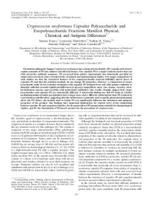
The BBB ensures that the brain is highly protected, with little access by circulating macromolecules and microorganisms. The human BBB is made up of microvascular endothelial cells supported by astrocytes, pericytes and neuronal feet [123,124] . Unlike the peripheral endothelial cells, the brain endothelial cells are joined together by tight junctions making the BBB a formidable barrier to many pathogens [124,125] . For C. neoformans to infect the brain, cryptococci must penetrate the normally impermeable BBB. The current understanding is that, once in the body, cryptococcal yeast cells may use multiple ways to enter the brain (Figure 1) . Trojan horse dissemination model
The Trojan horse form of dissemination involves parasitized phagocytes such as monocytes and/or macrophages smuggling pathogens across the BBB into the brain [126–128] . The intracellular parasitism of monocytes/macrophages has been demonstrated in both in vitro and in vivo models [70,72,129] . A study of infected mouse lungs demonstrated an 8-h peak of intracellular cryptococci in phagocytes after infection, and after day 7 of infection, intracellular cryptococci future science group
Mechanisms of infection by the human fungal pathogen Cryptococcus neoformans
Brain
Blood–brain barrier
Review
Brain capillary
Brain
Blood (systemic circulation)
Spores or dry cryptococci from the environment
Systemic circulation (infected monocytes, free cryptococci)
PMΦ
Lungs Heart
Alveoli
Extracellular cryptococci (Cap, App1, Gat201, MATa/α)
Phagolysosome Intracellular cryptococci (Cap, Mel, PLB1, PGE, Ctr4, Cft1/Cfo1)
AMΦ Granuloma
Future Microbiol. © Future Science Group (2012)
Figure 1. The dissemination model of Cryptococcus neoformans from the environment to the human brain. Through inhalation and with the help of mucociliary movement, spores or dry cryptococci colonize the alveolar space in the lungs [40] . In immunocompetent individuals, activated AMF phagocytose and kill cryptococci and surround cryptococci to form granulomas. Cryptococci in granulomas may stay latent or be reactivated and cause infection once the immune status of an individual changes. In immunodeficient individuals, PMF with intracellular cryptococci act as Trojan horses within the lungs, triggering dissemination into systemic circulation [12] . In order to survive in the hostile intracellular environment, Cryptococcus neoformans secretes or enlarges its Cap, forms Mel as an antioxidant against reactive oxygen and nitrogen species and PGE to downregulate antimicrobial activity, upregulates nutrient acquisition genes Ctr4 for copper and Cft1/Cfo1 for iron and secretes PLB1, which impairs intracellular antimicrobial responses [57,82,93,112,176] . Alternatively, cryptococci may resist phagocytosis by growing to a very large size (titan cells), generating a large Cap expressing App1, or through the activity of the antiphagocytic transcriptional regulator Gat201 [51,55,56,59–61] . Extracellular cryptococci may remain localized in the lungs or disseminate transcellularly into the blood circulation. In the systemic circulation, cryptococci associate with monocytes or are carried freely into the brain capillary bed. In the brain capillary, cryptococci may penetrate the blood–brain barrier by either: (A) paracellular traversal between endothelial cells, most likely permitted by damaged or weakened tight junctions [151] ; (B) transcytosis – binding to and being internalized by brain microvascular endothelial cells [122] ; or (C) riding inside infected monocytes/macrophages (Trojan horse) [128] . AMF: Alveolar macrophage; Cap: Capsule; Mel: Melanin; PMF: Parasitized alveolar macrophage.
were more abundant than extracellular ones [12] . Following phagocytosis, cryptococci survive and proliferate inside macrophages [70,72] . Furthermore, live cryptococci can be expelled in a novel nonlytic exoc ytosis (vomocytosis), leaving the host macrophage unharmed [70,72–75] . The exocytosed cryptococci then spread to other cells [70,72–75] . This direct cell-to-cell spread potentially explains how cryptococci can take advantage of phagocytes to penetrate the BBB in a Trojan horse manner [74] . By infecting mice with cryptococcal-laden monocytes, Charlier et al. demonstrated the occurrence of the Trojan horse traversal of C. neoformans across the BBB in vivo, since mice infected with cryptococcal-laden monocytes had high brain fungal burdens compared with mice future science group
infected with free cryptococci. The originally cryptococcal-laden monocytes (labeled) were found in the brain parenchyma, indicating that they had penetrated the BBB [128] . Reduction of disseminated cryptococcosis was observed in alveolar macrophage-depleted immunodeficient mice, which indirectly affirms the importance of the Trojan horse model in the dissemination of cryptococci [49] . Exploitation of host leukocytes to gain entry into the brain has also been demonstrated in other intracellular pathogens such as Listeria monocytogenes, Mycobacterium tuberculosis, HIV and the protozoan parasite Toxoplasma gondii [130–134] . T. gondii is a life-long intra c ellular parasite that infiltrates the BBB by hiding in phagocytes and lymphocytes – a process www.futuremedicine.com
1303
Review
Sabiiti & May
mediated by proinflammatory cytokines and involving high expression of endothelial cell adhesion proteins ICAM and VCAM [134,135] . The nonspecific lodging of cryptococci in brain capillaries observed by Shi et al., followed by transmigration (which is urease dependent) at 6 h, might be causal to localized toxicity and inflammation [122] . Inflammation can either result in the breakdown of intercellular junctions or attracting and promoting infiltration of inflammatory phagocytes, which, in the process, phagocytose and traffic cryptococcal yeast cells into the brain. Transcellular pathway
This pathway allows pathogens to cross the BBB by transcytosis through the brain microvascular endothelial cells (BMECs) by taking advantage of cellular endocytic processes [121,136,137] . A number of bacterial pathogens, such as Escherichia coli, group B Streptococcus, L. mono cytogenes, and the fungal pathogen Candida albi cans are reported to use the transcytosis pathway [121,127] . Cryptococcal transcellular traversal of the BBB has been widely studied using in vitro models of the BBB, which have demonstrated the ability of C. neoformans to adhere to, be internalized by and traverse BMECs [121,138] . Occurrence of transcytosis across the BBB depends on adherence and internalization of cryptococcal yeast cells by BMECs. Chen et al. showed that binding but not internalization could occur when cryptococci were incubated with human BMECs at 37°C and that binding induced cytoskeletal changes [139] . However, using transwell apparatus and electron microscopy, studies by Vu et al. and Chang et al. have demonstrated that coincubation of BMECs with encapsulated and acapsular strains of C. neoformans results in both adherence and internalization of crypotococci, with subsequent penetration of the BBB [138,140] . Crossing at the endothelial surface was further demonstrated in a murine model of cryptococcosis demonstrating that cryptococcal yeast cells were mostly localized in the brain parenchyma close to brain capillaries, and no association was observed with the choroid plexus at both early and late stages of infection [121,141] . A recent study, using real-time intravital imaging, has further elucidated the process of cryptococcal transcytosis across the brain endothelium. At 6 h after iv. inoculation, cryptococci were seen transmigrating across the BBB in a manner that involved transcytosis through brain endothelial cells [122] . 1304
Future Microbiol. (2012) 7(11)
The nature of interactions between Crypto coccus and BMECs remains poorly understood. While most bacterial pathogens are reported to use receptor-mediated endocytosis to traverse BMECs no receptor has been identified for adherence of C. neoformans to brain endothelial cells [126,138] . The nontethered movement and eventual trapping of cryptococci in the brain capillaries of similar diameter, described by Shi et al., implies that brain endothelium lacks specific surface molecules or receptors to capture cryptococci [122] . However, an independent in vitro study previously demonstrated the involvement of glycoprotein CD44 expressed by human BMECs in enhancing the binding of C. neoformans [142] . Binding of CD44 by cryptococcal hyaluronic acid induces downstream signaling mediated by PKA-a, which results in cytoskeleton reorganization and phagocytosis of cryptococci by BMECs [142] . In line with CD44 involvement, Huang et al. recently demonstrated that CD44 is exposed in lipid rafts, through which it interacts with C. neoformans hyaluronic acid, and that antibody blocking of CD44 inhibits cryptococcal adherence and uptake by human brain endothelial cells (F igur e 2) [143] . The glycoprotein CD44 is a widely expressed protein by many cell types [144,145] and thus Cryptococcus– CD44 interaction may not be specific to the brain endothelium alone. However, alternative splicing and post-translational modification make the multiexon CD44 gene functionally diversified in different cell types [144,145] , and hence investigating the nature of BMEC CD44 expression when exposed to cryptococci is needed to determine if the interaction is specific to brain endothelium. Apart from hyaluronic acid, a recent study has demonstrated that C. neoformans PLB1 interacts with lipid mediators in the brain endothelial cell membrane, thereby activating GDP–Rac1 to GTP–Rac1, which then associates with STAT3. The GTP–Rac1/STAT3 association induces cytoskeletal rearrangements resulting in phagocytosis of cryptococci (Figur e 2) [117] . Other studies have indicated the capsule to be important in Cryptococcus–brain endothelial cell interaction [121,141,146–149] . However, these studies demonstrated varying contributions of the capsule and/or its absence, leaving the role of capsule in C. neoformans transmission across the BBB unclear. By exposing both murine and human BMECs to encapsulated C. neoformans serotype A or D and their acapsular mutant derivatives, we have recently demonstrated future science group
Mechanisms of infection by the human fungal pathogen Cryptococcus neoformans
Review
Monocyte/macrophage
H
ya l ac uro id nic
Free budding cryptococci
Blood
Urea PLB1
Ure1
Ammonia >> TJd
Mannoproteins Plasminogen Plasmin >> TJd
Cytoplasm BBB
Endocytosis
Nucleus
Vacuole
TJd
Exocytosis
BMEC
Brain
GDP–Rac1
CD44/GM1
Cryptococci-infected brain macrophage
PKAα/DYRK3
GTP–Rac1 GTP–Rac1/STAT3
Cytoskeleton rearrangement
Cytoskeleton rearrangement
Transcytosis
Transcytosis
Future Microbiol. © Future Science Group (2012)
Figure 2. Cryptococcal interaction with brain microvascular endothelial cells and subsequent traversal of the blood–brain barrier. Cryptococcal yeast cells bind to and are encircled by microvilli-like protrusions on endothelial cells, resulting in internalization into intracellular vacuoles (endosomes) [141] . Cryptococcus neoformans interaction with BMECs induces lipid rafts (GM1) to expose cell adhesion glycoprotein CD44, which binds cryptococcal hyaluronic acid [143,177] . Hyaluronic acid-coated cryptococci bind to CD44 in lipid rafts (GM1) and the interaction induces cytoskeleton rearrangement mediated by PKAa or DYRK3 resulting in phagocytosis by and transcytosis across BMECs [143,177] . Alternatively, internalization/transcytosis may occur through GTP–Rac signaling induced by secreted C. neoformans PLB1 [117] . Cryptococcal Ure1 can convert urea to ammonia [122] , whereas mannoproteins bind and activate plasminogen to plasmin [151] . Both ammonia and plasmin have damaging effects on the extracellular matrix and may promote paracellular penetration of the BBB by cryptococci. While it is known that circulating monocytes are attracted to regions of inflammation through which they infiltrate into tissue, factors involved in the attraction of cryptococci-laden monocytes to brain endothelium and subsequent penetration of the BBB are still unknown. BBB: Blood–brain barrier; BMEC: Brain microvascular endothelial cell; TJ: Tight junction; TJd: Tight junction damage.
that the binding and uptake of cryptococci by BMECs is capsule independent [150] . Paracellular pathway
The paracellular pathway involves pathogens traversing the BBB through the intercellular spaces [127,141] . This mechanism involves the pathogen damaging and weakening tight junctions, which join the brain endothelial cells together, in order to gain entry. Trypanosoma species are thought to use the paracellular pathway [127] . Severe damage of the endothelium, characterized by neuropil edema (neuroaccumulation of fluid) and sequestration of yeast in the brain microvessels, was observed following cryptococcal penetration of the BBB [120,126] . Furthermore, Chen et al. showed that cryptococcal binding of the future science group
microvascular endothelium induced tight junction alteration [139] . In addition, a recent study has demonstrated that cryptococcal mannoproteins bind host plasminogen, which is activated to the effector form, plasmin. Plasmin binds and breaks down the extracellular matrix and membrane, thereby increasing the chance for paracellular penetration of the BBB to occur [151] . Taken together, these data indicate the possibility of cryptococci using a paracellular entry mechanism by inducing damage or exploiting host mechanisms that weaken the brain endothelial cell tight junctions (Figure 2) . The fact that >95% of cryptococcal meningitis cases are HIV/AIDS patients, often in the AIDS state in which the BBB is most likely inflamed and thus more permeable, makes the paracellular www.futuremedicine.com
1305
Review
Sabiiti & May
invasion of the brain quite plausible. During HIV, virus-infected monocytes are reported to infiltrate the BBB in the early stages of infection, resulting in inflammatory disease and damage to the BBB [152,153] . Moreover, the HIV-1 Tat protein has been implicated in damaging the BBB by inducing apoptosis of BMECs with subsequent increased permeability [154,155] . Whether HIV-infected monocytes or the HIV-1 Tat protein mediate damage of the BBB to promote cryptococcal penetration of the BBB remains unknown. Despite the occurrence of both diseases among HIV/AIDS patients, there are so far no clinical reports associating HIV-induced neuroinflammatory disorders (HIV-associated encephalitis and/or dementia) with the occurrence of cryptococcal meningitis, implying that the two conditions might occur independently. Interestingly, however, the HIV protein gp-41 has been shown to enhance cryptococcal binding to human brain endothelia [156] .
and severity of CM disease does not vary between the two etiological agents [164,165] . Treatment guidelines have been designed with consideration of the patient’s other underlying conditions, such as HIV status or the presence of an organ transplant [166] . For example, iv. amphotericin B deoxycholate 0.7–1.0 mg/kg plus flucytosine (100 mg/kg) for 4 days followed by 2 weeks of fluconazole 6 mg/kg is recommended as the primary induction and consolidation therapy for HIV-infected patients [166] . Despite the different regimens to manage this disease, new antifungal drug development is highly neglected, with the introduction of voriconazole in 2002 being the most recent new antifungal to come to market. The current most effective antifungal is amphotericin, which is unfortunately toxic and requires sophisticated administration procedures that are not easily accessible in resource-poor settings where cryptococcal meningitis is most prevalent. Conclusion
Cryptococcal meningoencephalitis
Once in the brain, C. neoformans causes meningoencephalitis, a severe form of the disease that is uniformly fatal if untreated [23] . Even with treatment, mortality can be as high as 30–100% in HIV/AIDS cohorts [3,157–160] . Patients present with headache, fever, malaise and altered mental status, and less often with signs such as meningism, papilledema, cranial nerve palsies, focal neurological deficits and depressed levels of consciousness [158] . Raised intracranial pressure is the most common complication, which results in visual and hearing loss [158] . Pathologically, nonHIV CM victims are found with more inflammation of the brain tissue and lower numbers of extracellular cryptococci than HIV-associated CM victims [161] . Post-mortems of HIVassociated CM victims showed large numbers of cryptococci in the arachnoid area, and this was associated with raised intracranial pressure, suggesting that the accumulation of cryptococci in the arachnoid and subsequent obstruction of cerebrospinal fluid outflow underlies raised intracranial pressure [162] . Cerebrospinal fluid fungal burden has been demonstrated to be associated with mortality in HIV-associated cryptococcal meningitis [163] . C. neoformans accounts for 99% of the crypto coccal meningitis cases and is the main cause of disease in immunocompromised people, while C. gattii is responsible for the remaining 1% of cases and primarily infects immunocompetent people. Although the underlying complications and clinical outcome can be different, the form 1306
Future Microbiol. (2012) 7(11)
Cryptococcal infection of the brain is the end result of a journey through hurdles imposed by the host. Entering the highly protected environment of the brain requires cryptococci to overcome barriers in the lung, circulatory system and, most significantly, the BBB. Alveolar macrophages are key in the pulmonary response to cryptococci, and the ability of cryptococci to survive and replicate inside these macrophages creates a path for transmission from the lung to systemic circulation. Cryptococci are endowed with protective strategies such as the production of capsule and melanin to downplay the antimicrobial activity of phagocytic cells, such as monocytes and neutrophils, as well as of complement proteins. At the brain gate – the BBB – cryptococci can gain entry by hiding inside phagocytes, by being engulfed directly by BMECs or by inducing damage to tight junctions to pave the way between brain endothelial cells. Once in the brain, the fungus proliferates, resulting in devastating inflammation of the meninges and brain parenchyma (meningoencaphalitis). The fact that the probability of survival from CM is less than 50%, even with treatment, makes improved diagnostic and therapeutic developments, as well as interventions focusing on the pulmonary and prebrain systemic life of C. neoformans, critical in order to reduce mortality from this disease. Future perspective
Our current understanding of the underlying mechanisms of the pathogenesis of future science group
Mechanisms of infection by the human fungal pathogen Cryptococcus neoformans
cryptococcosis largely stems from animal models of infection. Indeed, through mammalian/ non-mammalian and tissue culture models, various aspects of cryptococcosis, including pathogen virulence, manipulation of host immune response and penetration of the BBB, have been illustrated [167] . The challenge, however, is to what extent observations made in these models can explain what happens in the human host. For every infection, colonization, disease progression and clinical outcome are mutually modulated by the interplay between pathogen and host factors, which makes it challenging to decipher the human host factors by studying the pathogen
Review
in a different host. Fortunately, over the past decade, there has been an increase in clinical research on cryptococcosis and thus we envision that future research will see more clinical analysis, which will bridge the gap between laboratory models of infection and patient clinical parameters. Direct clinical analysis of samples and general underlying physiological parameters of patients will provide reference for observations made in laboratory models of infection. Del Poeta and Casadevall have recently contextualized a series of major unanswered questions in cryptococcosis, ranging from the nature of the infectious propagule to the possibility
Executive summary Cryptococcosis is a life-threatening fungal disease responsible for a significant global disease burden One million cases of cryptococcal meningitis are reported to occur globally per year. More than 60% of patients die within 3 months of diagnosis. Immunodeficiency, HIV/AIDS and immunosuppressive conditions such as solid organ transplant or steroid drug use are the main risks for cryptococcosis. Sub-Saharan Africa is mainly affected by HIV-associated cryptococcal meningitis, while non-HIV and immunocompetent cryptococcosis is on the rise in far-east Asia and north-west America.
The understanding of the fungus Cryptococcus neoformans has grown tremendously since its discovery over 100 years ago Cryptococcus neoformans was first described in fruit juice in 1894 by an Italian scientist and in a clinical sample in 1895 by German scientists. Over 50 species of the genus Cryptococcus live ubiquitously in the environment, but only two, C. neoformans and Cryptococcus gattii, are pathogenic to humans. By serotyping of their capsule, the two species are subdivided into variants, serotypes A, D, AD and B, corresponding to C. neoformans var. grubii, C. neoformans var. neoformans, diploid C. neoformans and C. gattii, respectively. By molecular typing, the varieties are now known by their molecular types, of which C. neoformans var. grubii molecular type I is responsible for the majority of cryptococcal infections worldwide.
Host colonization stems from inhalation of spores or dried yeast cells Light spores or dry yeast cells are swept down the respiratory tract by the help of mucociliarly movement. Host protein surfactant D aids cryptococci adherence to lung epithelial cells as well as phagocytosis by macrophages, which promotes transmission to lung alveoli.
Pulmonary involvement determines the fate of cryptococcosis In the lung, cryptococci can either be cleared by alveolar macrophages or be entangled by macrophages and lymphocytes, resulting in granuloma formation and/or localized benign pulmonary infection. Alternatively, cryptococci can cause pulmonary disease characterized by extracellular and intracellular proliferation of cryptococci, which subsequently disseminate to the rest of the body. Of all the body parts, there is a predilection of cryptococci for the brain, although the factors attracting C. neoformans to the brain are not yet known.
Transmission into the brain across the blood–brain barrier is a multimechanism process Cryptococci survive and multiply inside phagocytes, which they use as Trojan horses to penetrate the blood–brain barrier (BBB). Cryptococci can directly bind to – and be engulfed by – brain microvascular endothelial cells, resulting in transcytosis across the BBB. Cryptococci may weaken the brain microvascular endothelial cell tight junctions through the generation of ammonia or plasmin, creating intercellular spaces through which they penetrate the BBB.
Cryptococcal meningoencephalitis Once in the brain, cryptococci may form cysts within the brain parenchyma or proliferate in the arachnoid villi. High numbers of extracellular cryptococci and less inflammation is observed in immunocompromised cryptococcal meningitis than in immunocompetent cases. Treatment is difficult even in the presence of optimal antifungal regimens, with more than half of all cases resulting in death. Premeningitis diagnostic and therapeutic interventions may offer a better strategy for reducing disease burden and preventing the onset of fatal cryptococcal meningoencephlitis.
future science group
www.futuremedicine.com
1307
Review
Sabiiti & May
of predicting the likelihood of hematogenous dissemination to the brain [168] . In addition, there remain critical unanswered questions about the behavior of cryptococci in the environment. For instance, what are the nutritional requirements for cryptococcal growth in soil, pigeon excreta or on trees [23,169,170] , and what is the nature of the relationship between plants and Cryptococcus? For instance, Waikedre et al. have demonstrated that essential oils extracted from two species of heartwood tree, Callitris neocaledonica and Cookia sulcata, can inhibit growth of various human fungal pathogens, including C. neoformans [171] . Thus, research into understanding C. neoformans in its natural environment will be crucial to developing better control measures, as well as identifying new anticryptococcal compounds. In addition, understanding the molecular basis of the host–pathogen interactions will be crucial for effective management of cryptococcal disease, as well as discovering novel therapeutic targets. Despite having relatively similar levels of exposure to potentially Cryptococcus-infested environments, not all immunosuppressed people develop cryptococcosis [168] . Identifying host (genetic) factors that underlie differences in susceptibility to cryptococcal infection is thus a References Papers of special note have been highlighted as: n of interest nn of considerable interest 1.
2.
3.
4.
5.
Park BJ, Wannemuehler KA, Marston BJ, Govender N, Pappas PG, Chiller TM. Estimation of the current global burden of cryptococcal meningitis among persons living with HIV/AIDS. AIDS 23(4), 525–530 (2009).
major priority. Equally important will be pathogen transcriptomics and proteomics, especially of clinical isolates, to uncover virulence factors that are responsible for disease in different groups of patients. The damage caused by cryptococcal meningo encaphalitis is irreparable in most cases, with mortality reaching 100% in some cohorts, even with antifungal treatment [159] . While rabbits must be therapeutically immunosuppressed by repeated treatment with steroids for meningoencephalitis to take effect [172–174] , the effects of cryptococci in the human brain are equally devastating to both the immunodeficient and immunocompetent patients [175] . To this end, the ultimate prize for the future will be to prevent, rather than cure, cryptococcal infections.
6.
7.
Lortholary O. Management of cryptococcal meningitis in AIDS: the need for specific studies in developing countries. Clin. Infect. Dis. 45(1), 81–83 (2007). Byrnes EJ, Heitman J. Cryptococcus gattii outbreak expands into the northwestern United States with fatal consequences. F1000 Biol. Rep. 1, 62 (2009).
8.
Kambugu A, Meya DB, Rhein J et al. Outcomes of cryptococcal meningitis in Uganda before and after the availability of highly active antiretroviral therapy. Clin. Infect. Dis. 46(11), 1694–1701 (2008).
9.
Jarvis JN, Boulle A, Loyse A et al. High ongoing burden of cryptococcal disease in Africa despite antiretroviral roll out. AIDS 23(9), 1182–1183 (2009).
10. Singh N, Alexander BD, Lortholary O et al.
1308
The authors have no relevant affiliations or financial involvement with any organization or entity with a financial interest in or financial conflict with the sub ject matter or materials discussed in the manuscript. This includes employment, consultancies, honoraria, stock ownership or options, expert testimony, grants or patents received or pending, or royalties. No writing assistance was utilized in the production of this manuscript.
Society of America. Clin. Infect. Dis. 30(4), 710–718 (2000).
Meya DB, Manabe YC, Castelnuovo B et al. Cost–effectiveness of serum cryptococcal antigen screening to prevent deaths among HIV-infected persons with a CD4 + cell count