Oct 8, 1996 - hemangiosarcoma, 1 with congestive heart failure, 1 with severe, diffuse hepatic lipidosis, 1 with moderate, lymphocytic cholangiohepatitis, ...
JOURNAL OF CLINICAL MICROBIOLOGY, Mar. 1997, p. 673–675 0095-1137/97/$04.0010 Copyright q 1997, American Society for Microbiology
Vol. 35, No. 3
Development of a Nested PCR Assay for Detection of Feline Infectious Peritonitis Virus in Clinical Specimens DAVID A. GAMBLE,1 ANDREINA LOBBIANI,2 MAURIZIO GRAMEGNA,2 LISA E. MOORE,1 3 AND GIUSEPPE COLUCCI * The Animal Medical Center, New York, New York,1 and Clonit SpA2 and Fondazione Centro Studi di Patologia Molecolare Applicata alla Clinica,3 Milan, Italy Received 24 April 1996/Returned for modification 8 October 1996/Accepted 3 December 1996
A diagnostic test for feline infectious peritonitis virus (FIPV) infection based on a nested PCR (nPCR) assay was developed and tested with FIPV, feline enteric coronavirus (FECV), canine coronavirus (CCV), and transmissible gastroenteritis virus (TGEV) and clinical fluid samples from cats with effusive feline infectious peritonitis (FIP). The target sequence for the assay is in the S1 region of the peplomer protein E2 gene. A vaccine strain of FIPV and two wild-type FIPV strains tested positive, but FECV, TGEV, and CCV tested negative. Preliminary tests with 12 cats with clinical evidence of effusive FIP and 11 cats with an illness associated with effusions, but attributed to other causes, were performed. Eleven of the 12 cats with effusive FIP tested positive, while 1 was negative. Ten of the 11 cats ill from other causes tested negative, while 1 was positive. On the basis of clinical laboratory and histopathologic criteria, the preliminary sensitivity and specificity of the assay were 91.6 and 94%, respectively. Feline infectious peritonitis (FIP) is a fatal, immunologically mediated infectious disease of cats. The etiologic agent, feline infectious peritonitis virus (FIPV), is a single-stranded RNA virus of the family Coronaviridae (9, 12). The currently available serologic tests for anti-FIP antibody lack specificity and sensitivity for detection of active infection. Serologic tests for FIP cross-react with feline enteric coronavirus (FECV) (1, 13) and possibly other enteric coronaviruses which may infect cats. Antibody assays in general do not necessarily determine active infection. A more definitive diagnostic test for FIP infection would be one that detects components of the infectious agent. This report documents the development of a diagnostic test based on a nested PCR (nPCR) for the detection of viral RNA and evaluation of the test with several coronavirus isolates and clinical specimens. The intent of this test is that it be used in conjunction with fluid analysis for cats with illnesses which manifest effusions in celomic cavities. The target for amplification is a segment of the gene coding for peplomer protein E2. This gene was chosen because there is known antigenic difference in the E2 protein between FIPV and FECV (6) which may be due to differences in the RNA sequences of the E2 genes (not due to posttranslational modification). The E2 gene sequence is known (3) and available in a gene sequence database. The nPCR procedure was chosen because of its superior sensitivity and specificity over those from tests with a single target sequence (17).
TAATGCACGTGGTAAACC-39 (upstream; nucleotides 361 to 381) and 59-C ACTGGTTGGAGGTGAATTG-39 (downstream; nucleotides 530 to 510). Three microliters of each of the fluid specimens was directly processed for reverse transcription in 22 ml of a reverse transcription buffer containing 50 mM Tris-HCl (pH 8.2), 60 mM KCl, 10 mM MgCl2, and 4 mM dithiothreitol. The solution was heated to 928C for 30 s. Five units of avian myeloblastosis virus reverse transcriptase (5 U/ml) was added to the solution, and the mixture was incubated at 428C for 1 h. Twenty-five microliters of an amplification buffer containing 16.6 mM NH4SO4, 6.7 mM Tris-HCl (pH 8.8), 1.5 mM MgCl2, 10 mM b-mercaptoethanol, 0.1 mg of bovine serum albumin per ml, and 2 U of Taq I polymerase was added to the reverse transcription solution. The solution was heated to 948C, at which time primers were added. The solution was processed for 30 cycles of denaturation at 948C for 1 min, annealing at 538C for 1 min, and DNA polymerization at 728C for 3 min with a thermal cycler. One microliter of this amplification product was then subjected to a second round of amplification by using the same cycling procedure after adding the internal or nested primers. The final product was detected by gel electrophoresis, ethidium bromide staining, and UV light transillumination (11). Fifteen microliters of the final product was analyzed in each lane of the gel. The amplification product after the second, nested amplification appears as a band of 170 bp if the target sequence is present in the sample. No band is evident if the target sequence is not present in the sample. As a reaction control we used a synthetic FIPV E2 transcript that we developed by cloning the amplification product generated from an aliquot of the commercially available vaccine against FIP. This was blunt-end ligated at the SmaI site of the pGEM vector and was transformed into Escherichia coli HB101. Colonies were screened by the aforementioned nPCR protocol, and transcripts were obtained with T7 RNA polymerase (Promega Co., Madison, Wis.) following the instructions provided by the manufacturer. To rule out the possible presence of inhibitors of Taq polymerase, we performed for each sample a separate amplification in which the reaction mixture was spiked with an aliquot of the FIPV E2 transcript and looked for a reduction in the signal. Coronavirus strains. Commercially available modified live intranasal FIP vaccine was purchased from the manufacturer (SmithKline Beecham, Rixensart, Belgium). Two isolates of FIPV, strains DF-2 (NOR-15) (5) and NW-1 (UCD-1) (14), and one strain each of FECV (strain WSU 79-1683 [15]), canine coronavirus (CCV; strain 1-71 [10]), and transmissible gastroenteritis virus (TGEV; Miller strain [2]) were obtained from the American Type Culture Collection. Specimen selection and disease categorization. Fluid specimens were obtained from 21 cats presented to The Animal Medical Center as part of routine clinical evaluations for effusive disease of the peritoneal or the pleural cavity, or both. FIP was diagnosed in 12 animals, while the remaining animals were found to be affected by other, unrelated illnesses, including 1 with anaplastic sarcoma, 2 with hemangiosarcoma, 1 with congestive heart failure, 1 with severe, diffuse hepatic lipidosis, 1 with moderate, lymphocytic cholangiohepatitis, 1 with uterine adenocarcinoma, 1 with lymphosarcoma, 1 with pancreatitis, and 2 with idiopathic modified transudate. Fluid analysis, cytologic examination, and electrophoresis were performed. The cats were categorized as having clinical FIP or not having clinical FIP by
MATERIALS AND METHODS nPCR. Synthetic oligonucleotides were prepared with an automated oligonucleotide synthesis machine by standard techniques. The target sequence, chosen on the basis of mismatches with other related sequences, G1C content, and melting temperature, corresponds to nucleotides 251 to 601, which are 69 bp upstream from the transcription start codon of the peplomer E2 gene and which are defined by the following primers: outer primers 59-CTACAGAGGTGTGG TACAAC-39 (upstream; nucleotides 251 to 271) and 59-TTCCACTCAAGACC ATAGAT-39 (downstream; nucleotides 621 to 601) and internal primers 59-GG
* Corresponding author. Mailing address: Fondazione Centro Studi di Patologia Molecolare Applicata alla Clinica, via Pace 9, 20122 Milan, Italy. Phone: 39 2 4987020. Fax: 39 2 4815756. 673
674
J. CLIN. MICROBIOL.
GAMBLE ET AL.
TABLE 1. Categorization of cats without clinical evidence of FIP infection and results of PCR assay with fluid specimens Cat no.
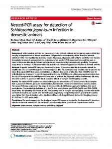
FIG. 1. Analytical sensitivity of nPCR for detection of FIPV. Decreasing concentrations of synthetic FIPV transcript were amplified as described in Materials and Methods. Lane 1, negative control; lanes 2 to 6, 500, 250, 50, 5, and 0.5 copies, respectively.
using established clinical laboratory (18) or histologic (19) criteria. Cats were not considered to have FIP infection when the fluid analysis did not show pyogranulomatous, plasma cellular inflammation and the albumin fraction was .50% and the gamma globulin fraction was ,30% of total protein, as determined by electrophoresis. Conversely, cats were categorized as having clinical FIP when the fluid analysis demonstrated pyogranulomatous, plasma cellular inflammation and the albumin fraction was ,50% and the gamma globulin fraction was .30% of the total protein, as determined by electrophoresis. Cats for which surgical biopsy specimens or necropsy specimens were available were considered to have FIP infection when multifocal pyogranulomatous lesions were found in a variety of parenchymal organs which included liver, kidney, spleen, and mesenteric lymph nodes and when fibrinosuppurative to pyogranulomatous inflammation in the visceral peritoneum or pleura but no evidence of bacterial infection was seen. The specificity and sensitivity (7) of the FIP nPCR test were determined by using the criteria established above.
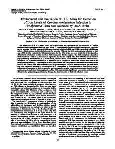
RESULTS Using serial dilutions of the FIPV synthetic E2 transcript, we assessed the analytical sensitivity of the assay, which corresponds to five copies per reaction (Fig. 1). The assay was then performed with commercially available modified live intranasal FIP vaccine, two isolates of FIPV, and one strain each of FECV, CCV, and TGEV (Fig. 2). The vaccine strain and the two FIPV isolates were positive, while the FECV, TGEV, and CCV isolates tested negative. When we selected the FIPV NW-1 strain for additional titration experiments, we could detect about 10 50% tissue culture infective doses (data not shown). Twenty-three cats with pleural or peritoneal effusions were tested, and sufficient clinical or histologic data were available to categorize their illnesses. Twelve cats fulfilled the established criteria for a clinical diagnosis of FIP, while 11 did not (Table 1). Of the 12 cats with clinical evidence of FIP infection, each had the effusive form of the disease. Eleven of these cats tested positive, and one tested negative. No evidence of inhibitory or interfering substances was found in this sample. Of the 11 cats that did not have evidence of FIP infection, 10 tested negative. The sensitivity of the nPCR assay is 91.6% and
FIG. 2. Nested amplification of different FIPV strains and related viruses, which were processed as described in Materials and Methods. Lane 1, FIPV DF-2; lane 2, FIP vaccine strains; lane 3, FECV; lane 4, TGEV; lane 5, negative control; lane 6, positive control (FIPV E2 synthetic transcript).
PCR result
1
Positive
2 3 4 5 8 7 8 9 10 11
Negative Negative Negative Negative Negative Negative Negative Negative Negative Negative
Final diagnosis or conclusion
Anaplastic sarcoma of spleen, liver, pancreas, and lung Hemangiosarcoma of spleen and pancreas Hemangiosarcoma of spleen, subcutis Congestive heart failure Hepatic lipidosis, diffuse, severe Cholangiohepatitis, lymphocytic, moderate Uterine adenocarcinoma Lymphosarcoma Pancreatitis Idiopathic modified transudate, recovered Idiopathic modified transudate, recovered
the specificity is 94% by comparison to clinical laboratory and histologic criteria. DISCUSSION The target sequence in the E2 gene chosen for this nPCR assay has sufficient sequence homology among strains to allow three different FIPV strains to be detected. The strains tested were a modified live, temperature-sensitive mutant FIP vaccine strain and two strains of FIPV with different cell culture characteristics: UCD-1 (NW-1), a low-titer virus, and DF-2 (NOR-15), a high-titer virus. The sequence from which the primers were synthesized was derived from a different hightiter strain, strain 79-1146. Although this strain was not tested, it is intuitively obvious that the assay would detect it as well. Although the assay was performed with only a single cytopathic strain of FECV, there was no cross-reactivity. Other strains of FECV or other enteric coronaviruses that have sufficient sequence homology to be detected in the assay could infect cats. This may not be a problem in the effusive form of the disease. FECV usually causes a mild transient enteric infection (13), and it is unlikely that FECV would be found in pleural or peritoneal effusions. Preliminary evaluation of the nPCR assay for the detection of viral nucleic acid in cats with the effusive form of FIP demonstrated that the assay has good sensitivity and specificity. The standard by which the nPCR assay was judged was established clinical and histopathologic criteria for effusive FIP. FIPV titers are not used in the routine clinical evaluation of cats suspected of having effusive FIP at The Animal Medical Center. Conventional indirect fluorescent-antibody assay or enzyme-linked immunosorbent assay titers do not consistently determine active infection, and fluid analysis has reasonable predictive value (18). Eleven of 12 fluid samples from cats with clinical FIP tested positive. There are several plausible explanations for the one apparent false-negative test result. There may be sufficient strain and nucleotide sequence variation such that the target sequence chosen for this assay may not detect all strains of FIPV. The assay requires reverse transcription of viral RNA to DNA prior to the amplification of DNA, and degradation of RNA could be a potential problem since RNases are virtually ubiquitous and are certainly found within inflammatory exudates. There could be inhibitors of reverse transcriptase and/or DNA polymerase in the fluid samples. However, this hypothesis was not confirmed by experiments carried out by spiking these samples with positive control RNA. One of 12 cats with illnesses not clinically compatible with
VOL. 35, 1997
DETECTION OF FIPV BY PCR
FIP tested positive. While this cat’s symptoms and clinical characteristics did not fulfill the established criteria for us to make a diagnosis of clinical FIP, there is no reliable means to prove that it did not have FIPV infection. This cat had systemic mesenchymal malignant neoplasia involving many viscera. It is possible that this cat was an infected carrier (16) without clinical evidence of FIPV infection and that the stress of chronic illness allowed for sufficient viral replication to be detected in the assay. The level of viremia or the titer of virus found in lymphoid tissue remains to be established in cats that are suspected of being persistently infected. The nPCR procedure can detect a single human immunodeficiency virus target sequence in approximately 70,000 peripheral leukocytes (4). If cats are sufficiently viremic such that virus approaching this level would be found in bodily fluids, the fluid may test positive, but the cat may not have evidence of clinical infection. The diagnostic test based on detection of viral nucleic acid by PCR is reasonably reliable for determining active FIPV infection. It is a valuable aid in making diagnostic decisions for cats with illnesses associated with pleural or peritoneal effusions. Recently, other investigators have described an nPCR system which is based on amplification of the feline coronavirus 39 untranslated region and which showed cross-reactivity with FECV (8). When it was used to detect FIPV in serum samples, this assay identified 78% of infected cats. In conclusion, the results of PCR tests must be interpreted in conjunction with other clinical findings and should not be used as the sole criterion for determining active FIPV infection. The potential problem of subclinical infection in cats with signs actually caused by a different illness is difficult to resolve. It remains to be determined if there is a target sequence of the FIPV genome with sufficient homology among all potential strains of the virus to be detected by PCR. ACKNOWLEDGMENT This work was supported in part by a grant provided by the Robert H. Winn Foundation. REFERENCES 1. Barlough, J. E. 1985. Cats, coronaviruses and coronavirus antibody tests. J. Small Anim. Pract. 26:353–362. 2. Bohl, E. H., R. K. Gupta, M. V. Olquin, and L. Saif. 1972. Antibody responses in serum, colostrum, and milk of swine after infection or vaccination
675
with transmissible gastroenteritis virus. Infect. Immun. 6:289–301. 3. de Groot, R. J., J. Maduro, J. A. Lenstra, M. C. Horzinek, B. A. van der Zeijst, and W. J. Spaan. 1987. cDNA cloning and sequence analysis of the gene encoding the peplomer protein of feline infectious peritonitis virus. J. Gen. Virol. 68:2639–2646. 4. Erlich, H. A., D. Gelfand, and J. J. Sninsky. 1991. Recent advances in the polymerase chain reaction. Science 252:1643–1651. 5. Evermann, J. F., L. Baumgartener, R. L. Ott, E. V. Davis, and A. J. McKeirman. 1981. Characterization of a feline infectious peritonitis virus isolate. Vet. Pathol. 18:256–265. 6. Fiscus, S. A., and Y. A. Teramoto. 1987. Antigenic comparison of feline coronavires isolates: evidence for markedly different peplomer glycoproteins. J. Virol. 61:2607–2613. 7. Gerstman, B. B., and D. T. Cappucci. 1986. Evaluating the reliability of diagnostic test results. J. Am. Vet. Med. Assoc. 88:248–251. 8. Herreweg, A. A. P. M., R. J. de Groot, A. Cepica, H. F. Egberink, M. C. Horzinek, and P. J. M. Rottier. 1995. Detection of feline coronavirus RNA in feces, tissue, and body fluid of naturally infected cats by reverse transcriptase PCR. J. Clin. Microbiol. 33:684–689. 9. Horzinek, M. C., and A. D. M. E. Osterhaus. 1979. The virology and pathogenesis of feline infectious peritonitis. Arch Virol. 59:1–15. 10. Keenan, K. P., H. R. Jervis, and R. H. Marchwicki. 1976. Intestinal infection of neonatal dogs with canine coronavirus 1-71: studies by virologic, histologic, histochemical, and immunofluorescent techniques. Am. J. Vet. Res. 37:247–256. 11. Maniatis, T., E. F. Fritsch, and J. Sambrook. 1982. Agarose gel electrophoresis, p. 150–163. In Molecular cloning: a laboratory manual. Cold Spring Harbor Laboratory, Cold Spring Harbor, N.Y. 12. Pedersen, N. C., J. Ward, and W. L. Mengeling. 1978. Antigenic relationship of the feline infections peritonitis virus to coronaviruses of other species. Arch. Virol. 58:45–53. 13. Pedersen, N. C., J. F. Boyle, K. Floyd, A. Fudge, and J. Barker. 1981. An enteric coronavirus infection of cats and its relationship to feline infectious peritonitis. Am. J. Vet. Res. 42:368–377. 14. Pedersen, N. C., J. F. Boyle, and K. Floyd. 1981. Infection studies in kittens utilizing feline infectious peritonitis virus propagated in cell culture. Am. J. Vet. Res. 42:2580–2585. 15. Pedersen, N. C., J. F. Everman, A. J. McKeirnan, and R. L. Ott. 1984. Pathogenicity studies of feline coronavirus isolates 79-1146 and 79-1683. Am. J. Vet. Res. 45:2580–2585. 16. Pedersen, N. C. 1987. Virologic and immunologic aspects of feline infectious peritonitis virus infection. Adv. Exp. Med. Biol. 218:529–550. 17. Porter-Jordan, K., E. I. Rosenberg, J. F. Keiser, J. D. Gross, A. M. Ross, S. Nasim, and T. Garrett. 1990. Nested polymerase chain reaction assay for the detection of cytomegalovirus overcomes false positives caused by contamination with fragmented DNA. J. Med. Virol. 30:85–91. 18. Shelly, S. M., J. Scarlett-Kranz, and J. T. Blue. 1988. Protein electrophoresis on effusions from cats as a diagnostic test for feline infectious peritonitis. JAAHA 24:495–500. 19. Weiss, R. C. 1989. Feline infectious peritonitis and other coronaviruses, p. 333–355. In R. G. Sherding (ed.), The cat: diseases and clinical managements. Churchill Livingstone, New York, N.Y.