cancer treatments. Thomas Krausz2 and Robert M.L.Winston2 ... Medical School, Hammersmith Hospital, Du Cane Road, London. Reimplantation .... St Louis, USA). Both media were ..... Newton, H., Aubard, Y., Rutherford, A. et al. (1996) Low ...
Human Reproduction vol.12 no.5 pp.1032–1036, 1997
Extracellular matrix improves survival of both stored and fresh human primordial and primary ovarian follicles in long-term culture
Outi Hovatta1,3, Rene Silye2, Ronit Abir2, Thomas Krausz2 and Robert M.L.Winston2 1Infertility
Clinic, The Family Federation of Finland, Kalevankatu 16, FIN-00100 Helsinki, Finland and 2Institute of Obstetrics and Gynaecology and Department of Pathology, Royal Postgraduate Medical School, Hammersmith Hospital, Du Cane Road, London W12 ONN, UK 3To
whom correspondence should be addressed
Ovarian cortical tissue was obtained during gynaecological operations by biopsy or after oophorectomy from 20 women aged 25–42 years. It was placed in organ culture, either fresh or following thawing after cryopreservation, for 1–4 months. The tissue was cut in slices 0.1–0.3 mm in diameter and transferred to 12 mm inserts in 24-well culture plates. These slices were cultured for 4–21 days in either α minimum essential medium (α-MEM) or Earle’s balanced salt solution with added pyruvate. Both media were supplemented with 10% human serum, insulin, gonadotrophins and antibiotics. Half of the inserts were precoated with extracellular matrix (Matrigel). Histological samples revealed that there were viable, non-atretic, primordial, primary and secondary follicles in all the cultures. Mitoses were seen in the granulosa cells of the secondary follicles. Although the proportion of atretic follicles increased during culture, non-atretic follicles were still present after 21 days. After 4–11 days the proportion of viable follicles was significantly higher when cultured in Earle’s solution supplemented with pyruvate, than when cultured in MEM (77 versus 38%, P , 0.001). In cultures with extracellular matrix the proportion of viable follicles was significantly higher after 10–15 days than it was without matrix (85 versus 19%, P , 0.001). Culture after thawing frozen ovarian tissue did not affect the density or the proportion of the viable follicles. Two-thirds of follicles in cryopreserved tissue were viable after 10–15 days in culture. The results indicate that it is possible to culture human primary and primordial follicles in vitro, and follicles in cryopreserved tissue are viable. Key words: culture/extracellular matrix/freezing/human ovary/ maturation in vitro
Introduction Maturation of oocytes in vitro from primordial and primary follicles would benefit many infertile patients. The risk of hyperstimulation after ovarian stimulation for clinical in-vitro fertilization (IVF) would be avoided and such follicles could 1032
be used for treatment involving oocyte donation and after cancer treatments. Cryopreservation of human ovarian tissue is already possible (Hovatta et al., 1996; Newton et al., 1996), but to utilize such tissue, it will be necessary to mature thawed follicles in vitro. Reimplantation of cryopreserved ovarian tissue has resulted in successful pregnancies in mice (Parrott, 1960; Carroll and Gosden, 1993) and sheep (Gosden et al., 1994b). Folliculogenesis has been shown in ovarian tissue transplanted under the kidney capsule of immunodeficient mice from several species (Gosden et al., 1994a; Candy et al., 1995). Small follicles in frozen–thawed human ovarian tissue have been shown to survive in the kidney of immunodeficient mice for 18 days (Newton et al., 1996). Fertilization and live offspring in mice have been obtained from fresh and frozen–thawed primary (Carroll et al., 1990; Spears et al., 1994) and recently also from primordial follicles (Eppig and O’Brien, 1996) matured in vitro. Initiation of follicle growth has recently been shown from cortical pieces of fetal bovine ovarian tissue (Wandji et al., 1996). It is more difficult to mature human follicles in vitro because of their large size and the long time needed for their development (Gougeon, 1986). Edwards (1965) matured oocytes from antral follicles of human, mouse, sheep, cow, pig and rhesus monkey ovarian tissue. Human oocytes have been aspirated from antral follicles of excised ovarian tissue and matured in vitro and pregnancy has been reported (Cha et al., 1991). Oocytes aspirated transvaginally from small antral follicles during unstimulated cycles have been fertilized by IVF, or intracytoplasmic sperm injection, after maturation in vitro (Trounson et al., 1994; Barnes et al., 1995). Healthy infants have been born. If small follicles could be cultured to the antral stage in vitro, similar maturation and fertilization methods could possibly be used. It is technically difficult to isolate follicles from the dense human ovarian stroma. Roy and Treacy (1993) developed a lengthy enzymatic isolation method, and pre-antral follicles were cultured for 4 days. Human pre-antral follicles can be grown to early antral stages after mechanical isolation (R.Abir et al., unpublished). However, it was seen that the majority of the early antral stage follicles had already lost their oocyte. Studies in our laboratory (Lass et al., 1997) show that human ovarian biopsy specimens contain low numbers of pre-antral follicles; the vast majority of follicles are either primordial or primary. Hence, maturing oocytes from primordial or primary follicles would be a better option. In order to optimize the conditions for maturation of fresh and frozen–thawed primordial and primary human follicles, we compared minimal essential medium (MEM) used for the culture of mouse pre-antral follicles (Spears et al., 1994), and © European Society for Human Reproduction and Embryology
Survival of ovarian follicles in long-term culture
Table I. Use of ovarian tissue from the 20 patients in the experimentsa Patients Patients Patients Patients Patients Patients
1–7 8–18 1–3, 12–17 4–10 4, 18 19, 20
MEM Earle’s balanced salt solution Extracellular matrix 1 Extracellular matrix – Frozen–thawed tissue, PROH Frozen–thawed tissue, DMSO
aControl
and cultured pieces of tissue were paired as far as possible, although tissue from every patient could not be used for both comparisons (medium and extracellular matrix). This was due to the small size of many of the laparoscopical biopsies. From open biopsies, tissue could be cultured in parallel in both experiments.
for pre-antral human follicles, with Earle’s balanced salt solution supplemented with pyruvate. The latter is known to be suitable for human oocytes and embryos. As extracellular matrix has been shown to modify granulosa cell function in culture (Amsterdam et al., 1989; Aten et al., 1995; Aston et al., 1996), we also evaluated its use in our cultures. Cryopreservation of oocytes or ovarian tissue followed by maturation of oocytes in vitro has also been recently discussed as a new treatment direction by Carroll (1996). Materials and methods Ovarian tissue was obtained during gynaecological operations by biopsy or from total oophorectomy specimens. In all, 20 women, aged 25–42 years (mean 35), donated tissue after informed consent. The ovarian tissue was placed in culture medium in the operating theatre and transferred to the laboratory within a few minutes. The tissue was either frozen in slices 0.3–1 mm in diameter (Hovatta et al., 1996), or placed immediately in organ cultures. Frozen tissue slices were thawed after 1–4 months and cultured further. Before culture, ovarian tissue was cut in slices of 0.1–0.3 mm thick, under a stereo microscope, using a scalpel and needles. The slices were transferred to Millicell CM inserts (12 mm diameter, 0.4 µm pore size; Millipore, Bedford, MA, USA) fitted into 24-well plates (Nunclon, Roskilde, Denmark). One to three slices were placed in each well. Half of the inserts had been coated with extracellular matrix (Matrigel, Becton Dickinson, MA, USA) consisting of laminin, collagen type IV and proteoglycans as major components. Matrigel had been diluted 1:3 with serum-free medium and 100 µl had been placed into each insert. The culture medium was either α-MEM (Gibco, Life Technologies Ltd, Paisley, UK) or Earle’s balanced salt solution (Gibco, Life Technologies Ltd) supplemented with 0.47 M pyruvate (Sigma, St Louis, USA). Both media were supplemented with 10% inactivated human serum (obtained from women undergoing pituitary desensitization for IVF), 0.3 IU/ml highly purified human follicle stimulating hormone (FSH) (Orgafol; Organon, Cambridge, UK), 0.3 IU/ml luteinizing hormone (LH; a gift from Dr S. Lynch, Birmingham and Midland Hospital for Women, West Midlands, UK), 33 ng/ml insulin (human recombinant insulin; Sigma) and antibiotics (50 U/ml penicillin G, 50 µg/ml streptomycin sulphate, 0.125 µg/ml amphotericin B, antibiotic antimycotic solution; Gibco). The medium was changed every second day. The tissue was cultured in a humidified incubator in 5% CO2 in air at 37°C. Tissue from each patient was used in either parallel cultures or in an individual experiment when the piece of tissue was so small that it could not be divided to all the various experiments, always with paired controls (Table I). Fresh tissue from 10 patients was cultured
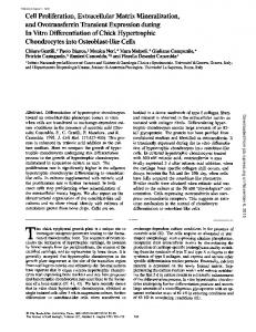
Figure 1. A cluster of follicles in ovarian tissue cultured for 7 days in Earle’s solution on extracellular matrix; original magnification 3400. in Earle’s medium and from seven patients in α-MEM. Tissue from nine patients was cultured with extracellular matrix and from eight without it. These cultures were continued for up to 21 days. Frozen– thawed tissue from four patients was cultured in Earle’s medium on extracellular matrix for up to 15 days. Tissue from two of these patients had been frozen using dimethylsulphoxide (DMSO; Sigma), and from the other two patients using propanediol (PROH; Sigma) as cryoprotectant. Pieces of each tissue specimen were fixed in Bouin’s solution for histology before and during the 3 week culture. The tissue was embedded in paraffin wax, cut into 2 µm sections and stained with haematoxylin and eosin. The numbers of viable and atretic follicles were counted per high power field (HPF, magnification 3400) and per patient. Eosinophilia of the ooplasm, contraction and clumping of the chromatin material and wrinkling of the nuclear membrane of the oocytes were regarded as signs of atresia (Gougeon, 1986). A pathologist received the coded samples from the culture laboratory, and carried out the follicle counts. The cultured pieces of ovarian cortical tissue were cut to sections at three levels. The whole area of the sections was counted as HPF, and the numbers of all the follicles seen in these sections were recorded. Fisher’s exact test and t-test for paired samples were used for statistical analyses.
Results Viable, non-atretic follicles were seen in freshly collected and frozen–thawed ovarian tissue cultured for up to 21 days (Figures 1–6), as were secondary follicles (Figures 5 and 6). Mitoses in granulosa cells (Figures 3 and 6) confirmed that the cells were dividing in culture. The proportion of atretic follicles increased significantly in all cultures with the duration of culture (Tables II, III and IV). Follicles were seen both in central and peripheral areas of the cultured pieces of tissue. After 4–11 days in culture, the percentage of primordial follicles was 20% and that of primary follicles 65%. There were 14% secondary follicles, and 2% tertiary follicles. After 14–21 days in culture the corresponding percentages were 21, 48, 28 and 3 respectively. The granulosa cells of the primary and the secondary follicles appeared enlarged in culture, especially when cultured on extracellular matrix. Viable follicles were seen in similar proportions in frozen–thawed tissue 1033
O.Hovatta et al.
Figure 2. One primordial and one primary follicle in ovarian tissue cultured for 11 days in Earle’s solution without extracellular matrix; original magnification 31000.
Figure 3. A cluster of follicles in ovarian tissue cultured for 5 days in Earle’s solution on extracellular matrix. A mitosis is seen in a secondary follicle; original magnification 31000.
Figure 5. A secondary follicle in ovarian tissue frozen with DMSO as a cryoprotectant, thawed after 4 months, and cultured for 11 days in Earle’s solution on extracellular matrix; original magnification 31000.
Figure 6. A secondary follicle with one of the granulosa cells in mitosis. The ovarian tissue had been frozen with DMSO as a cryoprotectant, thawed after 4 months and cultured for 11 days in Earle’s solution on extracellular matrix; original magnification 31000. Table II. Proportions of viable and atretic folliclesa in non-frozen ovarian tissue cultured for up to 21 days in either α minimum Eagle’s medium (αMEM) or Earle’s solution
Figure 4. A cluster of follicles in ovarian tissue frozen using PROH as a cryoprotectant, thawed after 3 months and cultured for 5 days in Earle’s solution on extracellular matrix. One follicle contains three oocytes; original magnification 3400.
after cryopreservation with either DMSO or PROH (Figures 5 and 6, Table V). There was no significant difference in the number of follicles per 10 HPF per patient before or after culture, or between any culture group (Table V). After 4–11 days in culture, the proportion of viable follicles was significantly higher (P , 1034
Control pieces before cultureb 4–11 days in culture 14–21 days in culture
α-MEM Earle’s α-MEM Earle’s α-MEM Earle’s
Number of patients
Viable follicles n (%)
Atretic follicles n (%)
7 10 7 10 7 5
13 (92) 90 (98) 24* (38) 105 (77) 9 (26) 4 (21)
1 2 39 31 26 15
(8) (2) (62) (23) (74) (79)
*P , 0.001 when compared with the pieces cultured in Earle’s solution. aDirect counts from the slides were made: numbers of follicles are low in these tissue pieces. bActual control pieces, taken from partially different patient populations (see Table I).
0.001, Fisher’s exact test) in the tissue cultured in Earle’s medium (Table II). After 14–21 days of culture in either Earle’s solution or MEM, the proportion of viable follicles was similar. The effect of extracellular matrix is shown in Table III.
Survival of ovarian follicles in long-term culture
Table III. Proportions of viable and atretic folliclesa in non-frozen ovarian tissue cultured with or without extracellular matrix (ECM) for up to 21 days
Control pieces before cultureb 4–9 days in culture 10–15 days in culture 18–21 days in culture
ECM1 ECM– ECM1 ECM– ECM1 ECM– ECM1 ECM–
Number of patients
Viable follicles n (%)
Atretic follicles n (%)
9 8 9 8 4 6 4 3
47 (100) 23 (96) 56 (49) 3 (43) 17* (85) 3 (19) 8 (26) 2 (17)
0 1 57 4 4 17 23 10
(0) (4) (49) (57) (15) (81) (74) (83)
*P , 0.001 when compared with the tissue cultured without ECM. aDirect counts from the slides were made: numbers of follicles are low in these tissue pieces. bActual control pieces, taken from partially different patient populations (see Table I).
Table IV. Proportions of viable and atretic folliclesa in frozen–thawed ovarian tissue cultured for up to 15 days
Frozen–thawed pieces before culture 4–9 days in culture 10–15 days in culture
Number of patients
Viable follicles n (%)
Atretic follicles n (%)
4
56 (97)
2 (3)
4 4
17 (40) 8 (67)
26 (60) 4 (33)
aDirect
counts from the slides were made: numbers of follicles are low in these tissue pieces.
Table V. Mean numbers of follicles per 10 high power fields per patient in the culturesa Number of Number of follicles patients n SD Control pieces before culture 4–11 days culture 14–21 days culture Control pieces before culture 4–9 days in culture 10–15 days in culture 18–21 days in culture Frozen–thawed pieces before culture 4–9 days in culture 10–15 days in culture
α-MEM Earle’s α-MEM Earle’s α-MEM Earle’s ECM1 ECM– ECM1 ECM– ECM1 ECM ECM1 ECM–
7 10 7 10 7 5 9 8 9 8 4 6 4 3
11.9 11.2 17.6 15.2 14.0 8.0 11.4 11.8 16.0 16.6 23.5 13.2 20.3 5.6
10.0 6.4 10.5 9.2 10.6 4.1 8.2 7.4 9.0 16.1 9.6 13.2 10.1 1.3
4 4 4
14.5 20.8 16.7
10.0 8.3 3.8
aAll
the follicles from all the sections from each patient were counted in order to make a fair comparison of follicular density in the different cultures. Nevertheless, the amount of tissue and sizes and shapes of the pieces and sections varied. ECM 5 extracellular matrix.
During the first days in culture there was no difference in the number or proportion of the follicles. After 10–15 days, the proportion of viable follicles was significantly higher
(P , 0.001, Fisher’s exact test) in tissue cultured on extracellular matrix. After 18–21 days the proportion of viable follicles and the number of the follicles still remaining in the tissue was still apparently higher when extracellular matrix was used, but this was not significant. In the cultures of frozen–thawed ovarian tissue, the density or proportion of follicles was similar to that in non-frozen tissue. Two-thirds of the follicles were viable after 10–15 days in culture (Table IV). Discussion The present study shows that human primordial or primary follicles can be maintained, and are obviously growing, in long-term organ cultures of ovarian tissue from adult women. The presence of secondary follicles and mitoses in granulosa cells can be regarded as evidence of follicular growth. In a quantitative analysis from the same patient population (Lass et al., 1997) it was shown that 88% of the follicles in the ovarian cortex are primordial, 8% primary and 4% secondary. In our cultured pieces the proportions of primary (48%) and secondary (28%) follicles had increased, hence suggesting activation of growth during the time in culture. The larger size of the granulosa cells in culture, especially on extracellular matrix, could be regarded as a sign of activation of growth. The small follicles were not isolated from the tissue. This approach maintains the structural integrity of the ovarian tissue. It allows cellular interactions between the cells of the follicle and the surrounding stroma. The same approach has been successfully used in culturing mouse primordial follicles (Eppig and O’Brien, 1996), bovine fetal follicles (Wandji et al., 1996) and for human fetal ovarian tissue culture (Zhang et al., 1995). After long enzymatic digestion, isolated human pre-antral follicles lose the theca layer and basement membrane surrounding the granulosa cells (Roy and Treacy, 1993). The loss of essential interactions may explain why they could only be cultured for 4 days. Mechanical isolation, although difficult from dense human ovarian tissue, retains the basement membrane and theca cells. The better maintenance and growth for up to 18 days of mechanically isolated human pre-antral follicles (R.Abir et al., unpublished) may be partly explained by the better integrity of the tissue components. However, for long-term culture, the primordial and primary human follicles will probably have to be isolated from the tissue at pre-antral stages to ensure nutrient and oxygen supply. Juvenile mice have been used for most successful mouse ovarian cultures (Spears et al., 1994; Eppig and O’Brien, 1996). However, adult infertile women have surprisingly few small follicles in their ovaries (Gougeon, 1986). The mean age of the women donating tissue for this study was 35 years. This explains why so few follicles were found, even in control pieces of ovarian tissue. The number of follicles and possibly the potential for growth might be improved in biopsies from young women. We have shown that frozen–thawed human follicles appear viable and capable of further development in culture. This confirms that it could be worthwhile to cryopreserve human ovarian tissue for possible future infertility treatments. This 1035
O.Hovatta et al.
approach may be valuable for young cancer patients, e.g. those about to have chemotherapy or radiotherapy in treatment of leukaemia. Extracellular matrix improves the growth of many cell types in culture. It contains growth factors (McGuire and Seeds, 1989) and is known as a survival factor (Meredith et al., 1993). Apoptosis is seen after disruption of epithelial cell-matrix interaction (Frish and Francis, 1994). In the present study, extracellular matrix improved the survival of ovarian follicles cultured for ù10 days. The proportions of viable follicles and the numbers of surviving follicles in ovarian tissue cultured for 18–21 days were higher in matrix cultures, although the difference was not significant. Because of the low numbers of the follicles in these cultures, the real physiological significance of these differences remains to be shown. The numbers of the tissue pieces cultured were rather low because of the difficulty in obtaining human ovarian tissue. Improvement in survival of follicles when using new culture media also shows that conditions for culture have still to be optimized. Further work will be needed to assess the effects of added nutrients, hormones and growth factors.
References Amsterdam, A., Rotmensch, S., Furman, A. et al. (1989) Synergistic effect of human chorionic gonadotropin and extracellular matrix on in vitro differentiation of human granulosa cells: progesterone production and gap junction formation. Endocrinology, 124, 1956–1964. Aston, K.E., O’Sullivan, M.J.B., Thomas, E.J. and Richardson, M.C. (1996) Effect of human chorionic gonadotrophin on the detachment of human granulosa cells from extracellular matrix layered onto glass or plastic. Hum. Reprod., 11, 336–340. Aten, R.F., Kolodecik, T.R. and Behrman, H.R. (1995) A cell adhesion receptor antiserum abolishes, whereas laminin and fibronectin glycoprotein components of extracellular matrix promote, luteinization of cultured rat granulosa cells. Endocrinology, 136, 1753–1758. Barnes, F.L., Crombie, A., Gardner, D.K et al. (1995) Blastocyst development and birth after in vitro maturation of human primary oocytes, intracytoplasmic sperm injection and assisted hatching. Hum. Reprod., 10, 3243– 3247. Candy, C.J., Wood, M.J. and Whittingham, D.G. (1995) Follicular development in cryopreserved marmoset ovarian tissue after transplantation. Hum. Reprod., 10, 2334–2338. Carroll, J. (1996) Development of oocyte banks and systems for the in-vitro development of oocytes: future directions for the treatment of infertility. Hum. Reprod., 11, 159–168. Carroll, J. and Gosden R. (1993) Transplantation of frozen-thawed mouse primordial follicles. Hum. Reprod., 8, 1163–1167. Cha, K.Y., Koo, J.J., Ko, J.J. et al. (1991) Pregnancy after in vitro fertilization of human follicular oocytes collected from non-stimulated cycles, their culture in vitro and their transfer in a donor oocyte program. Fertil. Steril., 55, 109–113. Edwards, R. (1965) Maturation in vitro of mouse, sheep, cow, pig, rhesus monkey and human ovarian oocytes. Nature, 208, 349–351. Eppig, J.J. and O’Brien M.J. (1996) Development of mouse oocytes from primordial follicles. Biol. Reprod., 54, 197–207. Frisch, S.M. and Francis, H. (1994) Disruption of epithelial cell-matrix interaction induces apoptosis. Cell Biol., 124, 619–626. Gougeon, A. (1986) Dynamics of follicular growth in the human: a model from preliminary results. Hum. Reprod., 1, 81–87. Gosden, R.G., Boulton, M.I., Grant, K. and Webb, R. (1994a) Follicular development from ovarian xenografts in SCID mice. J. Reprod. Fertil., 101, 619–623. Gosden, R., Baird, D.T., Wade, J.C. and Webb, R. (1994b) Restoration of fertility to oophorectomized sheep by ovarian autographs stored at –196°C. Hum. Reprod., 9, 597–603. Hovatta, O., Silye, R., Krausz, T. et al. (1996) Cryopreservation of human
1036
ovarian tissue using dimethylsulphoxide and propanediol-sucrose as cryoprotectants. Hum. Reprod., 11, 1268–1272. Lass, A., Silye, R., Abrams, D.-C. et al. (1997) Follicular density in ovarian biopsy of infertile women: a novel method to assess ovarian reserve. Hum. Reprod., 12, 1028–1031. McGuire, P.G. and Seeds, N.W. (1989) The interaction of plasminogen activator with a reconstituted basement membrane matrix and extracellular matrix macromolecules produced by cultured epithelial cells. J. Cell Biochem., 40, 215–227. Meredith, J.E., Fazeli, B. and Schwartz, M.A. (1993) The extracellular matrix as a cell survival actor. Mol. Biol. Cell, 4, 953–961. Newton, H., Aubard, Y., Rutherford, A. et al. (1996) Low temperature storage and grafting of human ovarian tissue. Hum. Reprod., 11, 1487–1491. Parrott, D.M.V. (1960) The fertility of mice with orthotopic ovarian grafts derived from frozen tissue. J. Reprod. Fertil., 1, 230–241. Roy, S.K. and Treacy, B.J. (1993) Isolation and long-term culture of human pre-antral follicles. Fertil. Steril., 59, 783–790. Spears, N., Boland, N.I., Murray, A.A. and Gosden R.G. (1994) Mouse oocytes derived from in vitro grown primary ovarian follicles are fertile. Hum. Reprod., 9, 527–532. Trounson, A., Wood, C. and Kausche, A. (1994) In vitro maturation and the fertilization and developmental competence of oocytes recovered from untreated polycystic ovarian patients. Fertil. Steril., 62, 353–362. Wandji, S.-A., Srsen, V, Voss, A.K. et al. (1996) Initiation in vitro of growth of bovine primordial follicles. Biol. Reprod., 55, 942–948. Zhang, J., Liu, J., Xu, K.P. et al. (1995) Extracorporeal development and ultrarapid freezing of human fetal ova. J. Assist. Reprod. Genet., 12, 361–368. Received on November 19, 1996; accepted on March 5, 1997