Behavioral studies with complete callosotomy patients (Roser et al. 2002, 2003) ... corpus callosum, connecting occipital cortices, mediates the size of the.
Interhemispheric interaction in simple response time: a combined functionaland diffusion-tensor imaging study
158
Matthew E. Roser*, Jon Fulford, Abdelmalek Benattayallah *University of Plymouth, MR Research Centre, Peninsula Medical School, Exeter
Processing and Analysis
Background The Redundant-Targets Effect (RTE) describes the facilitation of response time (RT) that occurs when two target stimuli are presented relative to when a single target stimulus is presented. This effect occurs even when stimuli are presented separately to the two cerebral hemispheres. Modeling of RTs suggests that statistical facilitation cannot always account for the RTE. Instead, interaction or ‘convergence’ of redundant processes must occur (Miller 1982). The stage of processing and the neural locus of interaction have been debated. Behavioral studies with complete callosotomy patients (Roser et al. 2002, 2003) suggest that the RTE involves the hemispheric interaction of response-preparation processes. Studies of patients who have undergone partial callosotomy suggest that the posterior corpus callosum, connecting occipital cortices, mediates the size of the redundancy gain. A recent fMRI study suggested both extrastriate and prefrontal involvement in the RTE (Schulte et al., 2006) and electrophysiological studies have demonstrated widespread bi-hemispheric activation in unimanual response production (Saron et al. 2003). To examine which callosal channels support the RTE in the intact brain, diffusion-tensor weighted images, and functional images, were acquired for a group of 34 neurologicallynormal subjects who made speeded unimanual reactions to luminant targets presented to each side of a central fixation point. Analyses using Track-Based Spatial Statistics (TBSS) (Smith et al., 2007) assessed the relationship between the RTE and fractional anisotropy in white matter.
Behavioral Data: The RTE was calculated for each response hand separately for each subject by subtracting the median RT to redundant bilateral stimuli from that to uncrossed-unilateral stimuli. An overall RTE for each subject was calculated from the mean of RTEs calculated for the two hands. The whole cohort showed a RTE of 5 ms and no facilitation of RT by unilateral-redundant stimuli.
+
+
+
A mixed-effects analysis comparing groups of subjects producing large (N=3) or small (N=4) RTEs found greater occipital lobe activation for Bilateral (redundant) displays compared to the sum of the two unilateral displays (Left and Right) for the group showing a large RTE.
Diffusion-Tensor Image Data: Diffusion tensor images (DTI) were acquired from 34 subjects using a diffusion weighted single-shot spin-echo EPI sequence. Diffusion weighting was performed along 32 independent directions, with a b-value of 1000 sec/mm2. A reference image (b=0 sec/mm2) was also acquired. Images were processed using the FMRIB Diffusion Toolbox (FDT). Images were corrected for spatial distortion, a binary mask of the corrected brain volume was created, and diffusion tensors were calculated. Fractional Anisotropy (FA) images were calculated from the eigenvalues of the tensor and entered into TBSS analyses. FA images were registered to standard space, a mean FA image for the cohort was created and skeletonized, and all subject’s data were projected onto the mean FA skeleton. FA data were grouped and modeled according to individual evidence for a RTE. Group 1 Mean RTE = -6ms (N = 13); Group 2 Mean RTE = 12ms (N = 19)
Stimuli and Task +
Results and Conclusion
Z
These results are consistent with the Hemispheric-Coactivation model of the RTE (Miller, 2004). Individual variation in hemispheric connectivity was associated with the degree of RT facilitation provided by bilateral stimulation. Greater posterior connectivity may allow a greater degree of bihemispheric processing of unilateral stimulus input, reducing the difference in brain processing for unilateral and bilateral stimuli. Facilitation of RT with bilateral stimulus input proceeds most effectively when posterior connectivity is relatively reduced. This result is consistent with that showing an enhanced RTE in posterior callosotomy patients.
+
Participants maintained central fixation, monitored using infra-red eye tracking. Stimuli were white squares presented for 150ms using Presentation (NBS). Stimuli were presented ~6.5 Deg to the left or right of fixation, bilaterally, or paired in one visual hemifield in a randomized sequence. Stimuli were presented for 150 ms at the onset of each period (2500 ms) in which a whole-brain volume was acquired on a 1.5T Phillips MR scanner. Stimuli were separated by jitter trials (average 2 TRs) in which the fixation point was displayed. Subjects performed two runs with each hand. In each run 50 Stimuli and 100 Baseline trials were presented. DTI and fMRI data were acquired from the same slice locations (2.5x2.5mm in-plane; slice thickness 3.5mm ; acquisition matrix 96x96 pixels).
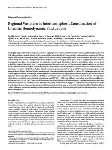
Skeletonized tracts displayed on group Fractional anisotropy in a region in the posterior callosum was found to differ between mean FA. Cluster shows results for a groups based on the RTE, with lesser FA in those showing a RTE. contrast Group1>Group2 (TFCE) thresholded at p