M. Vasantha et. al. / International Journal of Engineering Science and Technology Vol. 2(6), 2010, 2071-2076
Medical Image Feature, Extraction, Selection And Classification 1
M.VASANTHA*, 2DR.V.SUBBIAH BHARATHI, 3R.DHAMODHARAN 1. Research Scholar, Mother Teresa Women’s University, KodaiKanal, and
Asst.Professor, Department of Computer applications,St.Peters University , Chennai
[email protected], Phone No. 9884759409 2. Dean ,DMI College Of Engineering, Chennai 3. St. Petre’ s University, Chennai Abstract - Breast cancer is the most common type of cancer found in women. It is the most frequent form of cancer and one in 22 women in India is likely to suffer from breast cancer. This paper proposes a image classifier to classify the mammogram images. Mammogram image is classified into normal image, benign image and malignant image. Totally 26 features including histogram intensity features and GLCM features are extracted from mammogram image. A hybrid approach of feature selection is proposed in this paper which reduces 75% of the features. Decision tree algorithms are applied to mammography classification by using these reduced features. Experimental results have been obtained for a data set of 113 images taken from MIAS of different types. This technique of classification has not been attempted before and it reveals the potential of Data mining in medical treatment. Keywords- Breast cancer, Mammogram, Decision tree, Data mining, classification. 1. INTRODUCTION Breast cancer in India is in rise and rapidly becoming the leading cancer in females (MedIndia 2006) and death toll is increasing at fast rate (Gajalakshmi et al., 2009) and no effective way to treat this disease yet. So early detection becomes a critical factor to cure the disease and improving the surviving rate. Generally the X-ray mammography is a valuable and most reliable method in early detection. Data mining of medical images is used to collect effective models, relations, rules, abnormalities and patterns from large volume of data. This procedure can accelerate the diagnosis process and decision-making. Different methods of data mining have been used to detect and classify anomalies in mammogram images such as wavelets [2, 6], statistical methods and most of them used feature extracted using image processing techniques [5].Some other methods are based on fuzzy theory [1] and neural networks [3].Most of the Computer Aided Methods proved to be the powerful tool that assists the radiologist to speed up the treatment process. In this paper we have used classification method called Decision tree classifier for image classification. Classification process typically involves two phases: training phase and testing phase. In training phase the properties of typical image features are isolated and based on this training class is created .In the subsequent testing phase , these feature space partitions are used to classify the image. We have used supervised decision tree method by extracting low level image features for classification. The merits of this method are effective feature extraction, selection and efficient classification. The rest of the paper is organized as follows. Section 2 presents the preprocessing and section 3 presents the feature extraction phase. Section 4 discusses the proposed method of Feature selection and classification. In section5 the results are discussed and conclusion is presented in section 6. 2.PRE-PROCESSING The mammogram image for this study is taken from Mammography Image Analysis Society (MIAS), which is an UK research group organization related to the Breast cancer investigation. As mammograms are difficult to interpret, preprocessing is necessary to improve the quality of image and make the feature extraction phase as an easier and reliable one. The calcification cluster/tumor is surrounded by breast tissue that masks the calcifications
ISSN: 0975-5462
2071
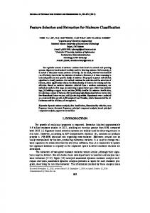
M. Vasantha et. al. / International Journal of Engineering Science and Technology Vol. 2(6), 2010, 2071-2076 preventing accurate detection and shown in Figures 2.1 .A pre-processing; usually noise-reducing step is applied to improve image and calcification contrast. In this work an efficient filter referred to as the low pass filter, was applied to the image that maintained calcifications while suppressing unimportant image features. Figures 2 shows representative output image of the filter for a image cluster in figure 1. By comparing the two images, we observe background mammography structures are removed while calcifications are preserved. This simplifies the further tumor detection step.
Fig. 1 ROI of a Benign
Fig. 2. ROI after Pre-processing Operation
2.1 Histogram Equalization Histogram equalization is a method in image processing of contrast adjustment using the image's histogram. Through this adjustment, the intensities can be better distributed on the histogram. This allows for areas of lower local contrast to get better contrast. Histogram equalization accomplishes this by efficiently spreading out the most frequent intensity values. The method is useful in images with backgrounds and foregrounds that are both bright or both dark. In particular, the method can lead to better views of bone structure in x-ray images, and to better detail in photographs that are over or under-exposed. In mammogram images Histogram equalization is used to make contrast adjustment so that the image abnormalities will be better visible. 3. FEATURE EXTRACTION Features, characteristics of the objects of interest, if selected carefully are representative of the maximum relevant information that the image has to offer for a complete characterization a lesion. Feature extraction methodologies analyze objects and images to extract the most prominent features that are representative of the various classes of objects. Features are used as inputs to classifiers that assign them to the class that they represent. In this Work intensity histogram features and Gray Level Co-Occurrence Matrix(GLCM) features are Extracted. 3.1 Intensity Histogram Features Intensity Histogram analysis has been extensively researched in the initial stages of development of this algorithm. Prior studies have yielded the intensity histogram features like mean, variance, entropy etc. These are summarized in Table 3.1(a) Mean values characterize individual calcifications; Standard Deviations (SD) characterize the cluster. Table 3.1(b) summarizes the values for those features.
ISSN: 0975-5462
2072
M. Vasantha et. al. / International Journal of Engineering Science and Technology Vol. 2(6), 2010, 2071-2076 Table 3.1(a) Intensity histogram features Feature Number assigned 1.
Feature Mean
2.
Variance
3.
Skewness
4.
Kurtosis
5.
Entropy
6.
Energy
In this paper , the value obtained from our work for different type of image is given as follows:
Table 3.1.(b) Intensity histogram features and their values Image Type
Features Mean
normal
7.2534
Varian ce 1.6909
malignan benign
6.8175 5.6279
4.0981 3.1830
Skewn ess -1.4745
Kurtosis
Entropy
Energy
7.8097
0.2504
1.5152
-1.3672 -1.4769
4.7321 4.9638
0.1904 0.2682
1.5555 1.5690
3.2 GLCM Features It is a statistical method that considers the spatial relationship of pixels is the gray-level co-occurrence matrix (GLCM), also known as the gray-level spatial dependence matrix. By default, the spatial relationship is defined as the pixel of interest and the pixel to its immediate right (horizontally adjacent), but you can specify other spatial relationships between the two pixels. Each element (I, J) in the resultant GLCM is simply the sum of the number of times that the pixel with value I occurred in the specified spatial relationship to a pixel with value J in the input image. The Following GLCM features were extracted in our research work: Autocorrelation, Contrast, Correlation, Cluster Prominence, ClusterShade, Dissimilarity Energy, Entropy, Homogeneity, Maximum probability , Sum of squares, Sum average, Sum variance, Sum entropy, Difference variance, Difference entropy, Information measure of correlation, information measure of correlation, Inverse difference normalized. The value obtained for the above features from our work for a typical image is given in the following table 3.2
ISSN: 0975-5462
2073
M. Vasantha et. al. / International Journal of Engineering Science and Technology Vol. 2(6), 2010, 2071-2076 Table 3.2 : GLCM Features and values Extracted from Mammogram Image Feature No 1) 2) 3) 4) 5) 6) 7) 8) 9) 10) 11) 12) 13) 14) 15) 16) 17) 18) 19) 20)
Feature Name
FeatureValues
autocd contrd corrpd cpromd cshad1 dissid energd entrod homopd maxprd sosvhd savghd svarhd senthd dvarhd denthd inf1hd inf2hd indncd idmncd
44.1530 1.8927 0.1592 37.6933 4.2662 0.8877 0.1033 2.6098 0.6645 0.6411 0.1973, 44.9329 13.2626 133.5676 1.8188 1.8927 1.2145 -0.0322 0.2863 0.9107
4. FEATURE SELECTION Feature selection helps to reduce the feature space which improves the prediction accuracy and minimizes the computation time. This is achieved by removing irrelevant, redundant and noisy features .i.e., it selects the subset of features that can achieve the best performance in terms of accuracy and computation time. It performs the Dimensionality reduction. Features are generally selected by search procedures. A number of search procedures have been proposed .Popularly used feature selection algorithms are Sequential forward Selection, Sequential Backward selection, Genetic Algorithm and Particle Swarm Optimization. In this work a combined approach of Greedy stepwise method and Genetic Algorithm is proposed to select the optimal features. The selected optimal features are considered for classification. 4.1 Proposed Hybrid Approach Algorithm:
1. Extract N number of features A1, A2, A3..AN from ROI Of the preprocessed Image 2. Apply Genetic algorithm to select the optimal set containing n1 number of features where n1