Acta Neurobiol. Exp. 2003, 63: 31-38
NEU OBIOLOGI E EXPE IMENT LIS
Memory for object and object-location after lesions to the ventromedial prefrontal cortex in humans Iwona Szatkowska1, Anna Grabowska1 and Olga Szymañska2 1
Nencki Institute of Experimental Biology, Department of Neurophysiology, 3 Pasteur St., 02-093 Warsaw, 2Warsaw University of Medicine, Department of Neurosurgery, 1a Banach St., 02-097 Warsaw, Poland
Abstract. The aim of the present study was to investigate the effect of small unilateral lesions to the ventromedial portion of the prefrontal cortex on two memory functions: memory for objects and memory for object locations. Patients, who had undergone surgery of the anterior communicating artery aneurysm, and normal control subjects, participated in the study. The patients were subdivided into two groups: with and without unilateral resection of the gyrus rectus. Subjects were presented with two memory tests, that required remembering either simultaneously presented visual stimuli (object memory test; OMT) or locations of the stimuli (location memory test; LMT). In the OMT, patients with resection of the gyrus rectus were impaired in comparison to patients without resection and normal control subjects. In the LMT, the three groups did not differ from each other. Our results suggest that the ventromedial prefrontal cortex is specifically involved in memory for objects.
The correspondence should be addressed to I. Szatkowska, Email:
[email protected]
Key words: ACoA aneurysm, gyrus rectus, orbitofrontal cortex, damage, patient
32
I. Szatkowska et al.
INTRODUCTION In recent years, substantial evidence has accumulated for the critical role of the prefrontal cortex in memory functions. Studies in nonhuman primates have shown that, within the prefrontal cortex, two separate systems could be identified for visual objects memory and visual location memory that are functionally and anatomically distinct. They are associated with the inferior (or more ventral) and with the superior (or more dorsal) lateral areas, respectively (Goldman-Rakic 1995, Wilson et al. 1987). An important question is whether functional segregation applies to the human prefrontal cortex as well. Positron emission tomography (PET) studies (Courtney et al. 1996, 1998) suggest that the human lateral prefrontal cortex shows a high similarity to the monkey’s lateral prefrontal cortex in that object memory occupies the more ventral area, whereas location memory the more dorsal part (but see Owen 1997, and D`Esposito et al. 1998, for different opinions). This conclusion was derived from finding that retention of information concerning the identity of face stimuli activated the right inferior and mid-prefrontal cortex whereas retention of information about the location of the same face stimuli activated an area of the superior frontal sulcus. It is worth noting that studies revealing the functional differentiation of the prefrontal cortex have mainly dealt with its lateral parts: dorsal or ventral. Little is known of what specific memory functions are subserved by the areas located in the ventromedial rather than in the ventrolateral region. Bachevalier and Mishkin (1986) found that, in monkeys, lesion to the ventromedial portion of the prefrontal cortex (including the orbital cortex, gyrus rectus, subcallosal cortex and anterior cingulate gyrus) impaired object recognition memory measured by delayed non-matching-to-sample tasks whereas dorso-lateral lesions affected spatial delayed responses. This data suggests that the ventromedial cortex in monkeys shows a high similarity to the ventrolateral part in that they are both engaged in retention of object rather than spatial information. It remains unknown, however, whether this also holds true for the human brain because most of the clinical research that has dealt with the role of the ventromedial prefrontal cortex in memory were performed on subjects with relatively large lesions. The effects of damage to the ventral portion of the prefrontal cortex were often investigated in patients who underwent rupture and repair of the an-
eurysm of the anterior communicating artery (ACoA). In most cases, the lesions were rather large due to infarction in the territory of the ACoA and they extended posteriorly, involving the basal forebrain region (Alexander and Freeman 1984, Drumm et al. 1993, £uria 1976, Petrides 2000). Patients with such lesions are impaired in remembering both object features and the spatial context in which objects occurred (Talland et al. 1967, Volpe and Hirst 1983) but it is difficult to attribute specific memory deficits to particular structures. In the present study, we could be much more precise because we had the cooperation of patients in whom brain damage was limited to the ventromedial prefrontal cortex (posterior part of the gyrus rectus) either in the left or right hemisphere. The aim of the present study was to investigate whether the ventromedial portion of the prefrontal cortex plays a role in memory functions. Additionally, we were interested whether this role would be limited exclusively to object memory, or it would be related both to memory for objects and memory for object locations.
METHODS Subjects PATIENTS
Twenty-one patients after surgery of ACoA aneurysm participated in the study. They were operated with ligation of the aneurysmal neck. During surgery for the ACoA aneurysm, partial resection of the gyrus rectus is frequently performed to facilitate exposure of the aneurysm. In other cases, the operation does not involve removal of neural tissue. Our patients, thus, constituted two groups: one with resection of the posterior 1/4 quarter of the gyrus rectus (GR+) and one without this resection (GR-). All patients were very similar in both patterns of their disease and the medical treatment they received. The comparison of the two groups offered the opportunity to test the effects of such small lesions on brain functions. As the resection was performed either on the left or right side, the GR+ group was further subdivided into two subgroups: left (LGR+) and right (RGR+). The sizes of the left- and right-sided lesions were very similar (1 cm diameter) because of the standard procedure used in such operations. Patients were selected from a large sample of brain-damaged subjects who had undergone an opera-
Object and object-location-memory after ventromedial prefrontal lesions 33 Table I Patients’ characteristics Sex
RGR+ LGR+ GR-
Age (years)
M
F
Mean
Range
Education (years) Mean Range
5 5 5
1 1 4
40.2 45.6 38.7
27-49 41-50 24-53
11.0 11.3 14.0
Time after operation (years) Mean Range
8-13 8-16 10-17
4.6 5.0 5.0
3-7 4-8 2-7
RGR+, patients with partial resection of the right gyrus rectus; LGR+, patients with partial resection of the left gyrus rectus; GR- , patients without resection of the gyrus rectus.
tion of the ACoA aneurysm at the Department of Neurosurgery of the Warsaw University of Medicine. High attention was devoted to careful selection of patients. Only patients who fulfilled the following criteria were included: no CT evidence of cerebral damage due to factors other than the operation; no evidence for clinical or angiographic post-hemorrhagic vasospasm; no gross memory loss as revealed by performance on memory subtests of WAIS-R (Wechsler Adult Intelligence Scale, Revised) (Brzeziñski et al. 1996) and by neuropsychological examination; no plegias or other neurological impairments; no aphasia symptoms; no other diagnoses likely to affect cognition (e.g., psychiatric disease, alcoholism or drug abuse); not older than 55 years. The patients were characterized by normal intellectual functions as measured with WAIS-R. All patients were right handed. All of them consented to enter the study. Table I presents data about the sociodemographic and clinical characteristics of patients.
CONTROL SUBJECTS
Fourteen right-handed individuals (9 men and 5 women) served as control subjects. They were matched as much as possible to the patients with respect of their age, education and intelligence (as measured by WAIS-R). None had any history of trauma affecting the central nervous system. Procedure Subjects were presented with two recognition memory tests that required remembering either visual stimuli per se (OMT - object memory test), or the locations of simultaneously presented stimuli (LMT - location memory test). Object and location memory tests used equivalent (similar) stimuli. They differed only in terms of the information that subjects were instructed to retain, namely the identity or the location of the stimuli.
OBJECT MEMORY TEST
LOCATION MEMORY TEST
(OMT)
(LMT)
/
/
\
\
VERBAL SESSION
NONVERBAL SESSION
VERBAL SESSION
NONVERBAL SESSION
(max. 9 trials)
(max. 9 trials)
(max. 9 trials)
(max. 9 trials)
Fig. 1. Experimental paradigm.
34
I. Szatkowska et al.
Fig. 2. The sequence of events in a single trial of the object memory test (A) and location memory test (B).
The test material consisted of verbal stimuli (3-letter Polish words) and nonverbal stimuli (Kanji characters). Both the object- and location-memory tests consisted of two experimental sessions – verbal and nonverbal (Fig. 1). The order of the tests and sessions was counterbalanced within each group. For both sessions, the procedure was the same. In the single trial, stimuli (verbal or nonverbal) were presented on a test card for 60 s and,
after a delay of about 5 s, a response card was exposed (Fig. 2). The task of the subject was to recognize either the previously presented stimuli or their spatial arrangement. Trials were repeated until the subject reached the criterion of two consecutive 100% correct performances. The intertrial interval was approximately 90 s. Participant was not informed about the number of errors committed. The session was interrupted if the subject
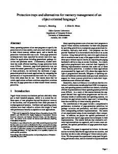
Object and object-location-memory after ventromedial prefrontal lesions 35
Fig. 3. Performance of four groups of subjects (controls, patients GR-, patients RGR+, patients LGR+) on the verbal and nonverbal sessions in the object memory test (A) and location memory test (B). The bars represent S.D.
had not reached the criterion up to the 9th trial. In the case of interrupted session, we arbitrary assumed that subject reached the criterion in the 10th trial. To match the sessions for the level of difficulty, the number of verbal and nonverbal stimuli (11 and 9 items, respectively) was determined in a pilot study. In that study, the test cards contained various numbers (8-12) of stimuli. Subjects obtained similar results in the verbal
and nonverbal session (i.e. they reached the criterion in the 4-5 trial) when the test cards contained 11 verbal and 9 nonverbal stimuli. In the OMT, all test cards contained the same vertically arranged stimuli, but the order of stimuli varied from trial to trial. Recognition cards contained both the stimuli previously exposed on the test card (1/3 of total number) and novel ones. The stimuli were arranged in
36
I. Szatkowska et al.
six rows. The content of recognition cards was fixed but the position of the stimuli varied, so that, as far as possible, across the trials, each stimulus would appear in every possible position. The subject’s task was to indicate which stimuli were previously presented on the test card. In the LMT, the test cards were always the same (they contained the same stimuli arranged in the same order). After a delay, subjects were simultaneously shown 16 recognition cards that contained the same stimuli as the test card, but arranged in a different order. Only one of the 16 recognition cards perfectly matched the test card. To avoid recognitions that would relay solely on the first or last item, the stimuli that began and finished each list (card) were always the same. The subject’s task was to indicate which recognition card matched the order of stimuli on the test card.
RESULTS The number of trials to criterion was analyzed. A three-way repeated measures analysis of variance MANOVA with group of subjects (Controls, GR-, LGR+, RGR+), test (OMT, LMT), and stimulus material (verbal, nonverbal) showed a significant main effect of group (F3,31=14.64, P