The Journal of Immunology
1,25-Dihyroxyvitamin D3 Promotes FOXP3 Expression via Binding to Vitamin D Response Elements in Its Conserved Noncoding Sequence Region Seong Wook Kang,*,†,1 Sang Hyun Kim,*,‡,1 Naeun Lee,*,1 Won-Woo Lee,*,x,1 Kyung-A Hwang,*,{ Min Sun Shin,* Seung-Hyun Lee,*,‖ Wan-Uk Kim,# and Insoo Kang* FOXP3-positive regulatory T (Treg) cells are a unique subset of T cells with immune regulatory properties. Treg cells can be induced from non-Treg CD4+ T cells (induced Treg [iTreg] cells) by TCR triggering, IL-2, and TGF-b or retinoic acid. 1,25-Dihyroxyvitamin D3 [1,25(OH)2VD3] affects the functions of immune cells including T cells. 1,25(OH)2VD3 binds the nuclear VD receptor (VDR) that binds target DNA sequences known as the VD response element (VDRE). Although 1,25(OH)2VD3 can promote FOXP3 expression in CD4+ T cells with TCR triggering and IL-2, it is unknown whether this effect of 1,25(OH)2VD3 is mediated through direct binding of VDR to the FOXP3 gene without involving other molecules. Also, it is unclear whether FOXP3 expression in 1,25(OH)2VD3-induced Treg (VD-iTreg) cells is critical for the inhibitory function of these cells. In this study, we demonstrated the presence of VDREs in the intronic conserved noncoding sequence region +1714 to +2554 of the human FOXP3 gene and the enhancement of the FOXP3 promoter activity by such VDREs in response to 1,25(OH)2VD3. Additionally, VD-iTreg cells suppressed the proliferation of target CD4+ T cells and this activity was dependent on FOXP3 expression. These findings suggest that 1,25(OH)2VD3 can affect human immune responses by regulating FOXP3 expression in CD4+ T cells through direct VDR binding to the FOXP3 gene, which is essential for inhibitory function of VD-iTreg cells. The Journal of Immunology, 2012, 188: 5276–5282. t is known that CD4+ T cells, which express the transcriptional factor FOXP3, are regulatory T (Treg) cells with the capacity to regulate immune responses (1). In addition to FOXP3, Treg cells express high levels of CD25 that serves as a cell surface marker for the identification of this cell subset (2, 3). FOXP3 is essential for the immune regulatory function of Treg cells. Transfection of FOXP3 into CD252CD4+ T cells, which do
I
*Department of Internal Medicine, Yale University School of Medicine, New Haven, CT 06520; †Department of Internal Medicine, College of Medicine, Chungnam National University, Daejeon 301-131, Republic of Korea; ‡Department of Microbiology, College of Medicine, Kangwon National University, Chuncheon 200-701, Republic of Korea; xDepartment of Microbiology and Immunology, College of Medicine, Seoul National University, Seoul 110-799, Republic of Korea; {Department of Agrofood Resources, National Academy of Agricultural Science, Suwon 441-853, Republic of Korea; ‖Department of Microbiology, Konkuk University School of Medicine, Chugju, Chungchungbuk-Do 380-701, Republic of Korea; and #Department of Internal Medicine, The Catholic University of Korea, Seoul 137-701, Republic of Korea 1
S.W.K., S.H.K., N.L., and W.-W.L. contributed equally to this work.
Received for publication April 26, 2011. Accepted for publication March 27, 2012. This work was supported in part by National Institutes of Health Grants AT005241, AG028069, AI075157, and U19AI082713 (all to I.K.). I.K. and W.-U.K. are participants of the World Class University Program of the Republic of Korea. Address correspondence and reprint requests to Dr. Insoo Kang, Section of Rheumatology, Department of Internal Medicine, Yale School of Medicine, 300 Cedar Street, New Haven, CT 06520. E-mail address:
[email protected] The online version of this article contains supplemental material. Abbreviations used in this article: ChIP, chromatin immunoprecipitation; CNS, conserved noncoding sequence; DC, dendritic cell; DR, direct repeat; ER, everted repeat; iTreg, induced regulatory T; MS, multiple sclerosis; nTreg, naturally occurring regulatory T; 1, 25(OH)2VD3; 1, 25-dihyroxyvitamin D3; RA, rheumatoid arthritis; RXR, retinoid X receptor; siRNA, small interfering RNA; SLE, systemic lupus erythematosus; Treg, regulatory T; VD, vitamin D; VDR, vitamin D receptor; VDRE, vitamin D response element. Copyright Ó 2012 by The American Association of Immunologists, Inc. 0022-1767/12/$16.00 www.jimmunol.org/cgi/doi/10.4049/jimmunol.1101211
not normally have regulatory function, conferred the immune regulatory property (4–6). Furthermore, mutations in the foxp3 gene have been found in scurfy mice with X-linked lymphoproliferative disease as well as in humans with immune dysregulation, polyendocrinopathy, enteropathy, and X-linked syndrome (7, 8). Whereas FOXP3+ Treg cells are normally generated in the thymus (naturally occurring Treg [nTreg] cells), FOXP3+ Treg cells can also be induced from non-Treg CD4+ T cells (induced Treg [iTreg] cells) in the presence of anti-CD3/CD28 Abs, IL-2, and TGF-b or retinoic acid (9–18). However, it has been controversial whether iTreg cells have inhibitory function in humans (9–14). 1,25-Dihyroxyvitamin D3 [1,25(OH)2VD3], the most physiologically active VD3 metabolite, exerts an inhibitory effect on immune cells including T cells (15). Of interest, low levels of circulating 25(OH)VD3 [precursor of 1,25(OH)2VD3] are found in patients with autoimmune diseases including rheumatoid arthritis (RA) and systemic lupus erythematosus (SLE) (16–20), suggesting a potential role for this vitamin in autoimmunity. In fact, increased generation of FOXP3+CD4+ T cells was reported in mice treated with oral or topical 1,25(OH)2VD3 (21–23) although the exact mechanism(s) for this finding is yet to be determined. In humans, an increased percentage of FOXP3+CD4+ T cells was noticed in PBMCs treated with 1,25(OH)2VD3 in vitro (24). Also, 1,25(OH)2VD3 enhanced FOXP3 expression in purified human CD252CD4+ T cells in the presence of anti-CD3/CD28 Abs (Abs) and IL-2 (25). 1,25(OH)2VD3 activates the vitamin D receptor (VDR) that heterodimerizes with retinoid X receptor (RXR) (15). The heterodimer then binds to its cognate DNA sequence known as vitamin D response elements (VDREs), leading to the regulation of gene expression. VDREs have two copies of a hexameric DNA sequence, referred to as core binding motif, with the consensus se-
The Journal of Immunology quence RGKTSA (R = A or G, K = G or T, and S = C or G) (26). VDREs can be formed by a direct repeat (DR) of two hexameric core binding motifs with three (DR3 type, e.g., 59-AGGTCA– NNN–AGGTCA-39) or four (DR4) intervening nucleotides (26). Also, VDR can bind to everted repeat (ER or inverted palindrome)-type responding elements with six, eight, or nine spacing nucleotides [e.g., ER9 AGGTCA–(N)9–TGACCT] (26). Despite the enhanced FOXP3 expression in CD4+ T cells by 1,25(OH)2VD3 and the capacity of this vitamin to regulate gene expression via direct binding to the genes, it is unknown whether 1,25(OH)2VD3 can directly induce FOXP3 gene expression without involving other molecules. Additionally, it is unclear whether FOXP3 expressed in 1,25(OH)2VD3-treated CD252CD4+ T cells is critical for the suppression of target T cells in that this vitamin can affect various functions of T cells such as cytokine production (15). In this study, we demonstrated the presence of VDREs in a conserved noncoding sequence (CNS) region that is analogous to the enhancer 1 of the mouse foxp3 gene (27). Such VDREs enhanced the FOXP3 promoter activity in the presence of 1,25(OH)2VD3. Additionally, FOXP3+CD4+ T cells generated by a combination of 1,25(OH)2VD3, anti-CD3/CD28 Abs, and IL-2 suppressed the proliferation of target CD4+ T cells dependent on FOXP3 and cell contact. These findings suggest that 1,25(OH)2VD3 can affect human immune responses by regulating FOXP3 expression in CD4+ T cells through direct VDR binding to the FOXP3 gene, which is essential for inhibitory function of 1,25(OH)2VD3induced FOXP3+ Treg (VD-iTreg) cells.
Materials and Methods Cell preparation, culture, and FOXP3 knockdown This work was approved by the Institutional Review Committee of Yale University. Human peripheral blood was obtained from the New York Blood Center or from healthy adult donors with informed consent. CD25–CD4+ T cells (.95% purity) were isolated from PBMCs using a cell purification kit (Miltenyi Biotec, Auburn, CA) or a FACSAria (BD Bioscience, San Jose, CA). For induction of FOXP3 and T cell suppression assay, purified CD252CD4+ T cells were suspended in RPMI 1640 medium with 10% FCS and stimulated for 5 d with 1,25(OH)2VD3 (10 nM or indicated; Sigma-Aldrich, St. Louis, MO), IL-2 (25 ng/ml; R&D Systems, Minneapolis, MN), anti-CD3 (4 mg/ml, OKT-3; National Cell Culture Center, Minneapolis, MN), and anti-CD28 Abs (3 mg/ml; BD Pharmingen, San Diego, CA). Cells (VD-iTreg cells) were then rested for 2 d and incubated for 5 additional days with autologous CD252CD4+ T cells (target cells) that were stained with CFSE (Invitrogen, Carlsbad, CA) in a transwell culture system separating the two cell populations or a regular culture plate in the presence of anti-CD3/CD28 Abs. To knock down FOXP3 expression, CD252CD4+ T cells that were stimulated for 3 d with 1,25(OH)2VD3, antiCD3/CD28 Abs, and IL-2 were transfected with control or FOXP3-specific small interfering RNA (siRNA) using the Amaxa transfection system (Lonza, Walkersville, MD). Transfected cells were cocultured for 5 d with CFSE-stained autologous CD252CD4+ T cells in the presence of anti-CD3/ CD28 Abs. Some transfected cells were stained with anti-human FOXP3 Abs (BioLegend, San Diego, CA) and analyzed on a flow cytometer. Target T cell proliferation was analyzed by flow cytometry.
Pull-down assay and immunobloting Biotinylated DNA fragments containing putative VDRE1 or VDRE2/3 in the intronic CNS region +1714 to +2554 of the FOXP3 gene were amplified by PCR using sense (59-biotin CTCCATATGTGGGTCCATGT-39 for VDRE1; 59-biotin TTTGCTGATTGTTGCTTTGC-39 for VDRE2/3) and anti-sense primers (59-biotin TAAAGAAGGGCAAGGTGCCA-39 for VRDE1; 59-biotin CATGTGTGCGAGAGGAGG-39 for VDRE2/3). The sequences in VDRE1, VDRE2, and VDRE3 were mutated using the GeneArt site-directed mutagenesis system (Invitrogen). Nuclear extracts from human CD252CD4+ T cells that were treated for 5 d with 1,25 (OH)2VD3 were obtained using a commercially available kit (Thermo Scientific, Loughborough, U.K.) and mixed with the biotinylated DNA fragments (27). Beads conjugated with streptoavidin were mixed with the DNA-nuclear extract reaction. VDR and RXR were detected by immu-
5277 noblotting using anti-VDR and anti-RXR rabbit polyclonal Abs (Santa Cruz Biotechnology, Santa Cruz, CA) as previously described (27).
Chromatin immunoprecipitation assay As previously described (27), chromatin immunoprecipitation (ChIP) assay was done using a commercially available kit (Upstate Biotechnology, Lake Placid, NY). Briefly, human CD252CD4+ T cells were treated for 5 d with or without 1,25(OH)2VD3.in the presence of anti-CD3/CD28 Abs and IL-2. Cells were then treated for 10 min with 1% formaldehyde at 37˚C and washed twice in ice-cold PBS containing protease inhibitors. Cell pellets were lysed in SDS lysis buffer and sonicated under conditions that reduced DNA length to between 200 and 1000 bp. The lysates were incubated overnight at 4˚C with anti-VDR rabbit polyclonal Abs and normal rabbit IgG Ab (both from Santa Cruz Biotechnology). Immune complexes were precipitated with protein G-agarose (Upstate Biotechnology), and DNA was purified using spin columns. PCR was performed to detect VDRE1 and VDRE2/3 using the following primers: VDRE1, sense (59-CTCCATATGTGGGTCCATGTCCAA-39) and anti-sense (59-TAAAGAAGGGCAAGGTGCCAGGGA-39); and VDRE2/3, sense (59-TTTGCTGATTGTTGCTTTGCAATA-39) and anti-sense (59- TGGACACATGCATGGAGAGCCAGA-39). A 150-bp DNA fragment in the FOXP3 promoter region (2145 to +5) that did not contain a VDRE was amplified by PCR as a negative control using the following primers: sense (59-ACCCCGTGATTATCAGCGCACACA-39) and antisense (59-CCTGGCTTGTGGGAAACTGTCACG-39).
Reporter gene assay The promoter (2511 to +176) and a highly CNS region (+1714 to +2554) of the FOXP3 gene were cloned into pGL3 basic vector (Promega, Madison, WI) followed by transfection to primary human CD252CD4+ T cells, which were stimulated for 5 d with anti-CD3/CD28 Abs and IL-2 in the presence or absence of 1,25(OH)2VD3 using the Amaxa transfection system (Lonza) (27, 28). In some experiments, VDRE1, 2, and/or 3 sequences in the CNS region were mutated using the GeneArt site-directed mutagenesis system (Invitrogen) and inserted into the vector for FOXP3 gene reporter assay. Transfected cells were analyzed for luciferase activity and normalized against Renilla activity (Promega).
Statistical analysis A paired t test was used to analyze data with a significance level of p , 0.05.
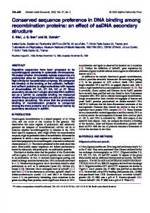
Results Identification of putative VDREs in the human FOXP3 CNS region +1714 to +2554 that increase the FOXP3 promoter activity Previous studies reported enhanced induction of FOXP3 in FOXP3-negative CD252CD4+ T cells by 1,25(OH)2VD3 in the presence of anti-CD3/CD28 Abs and IL-2 (25). However, it is unknown whether VDR binds directly to the FOXP3 gene, leading to such an upregulation. We thus searched for potential binding sites of the VDR in the human FOXP3 gene in silico using NUBIScan (http://www.nubiscan.unibas.ch), an algorithm predicting nuclear receptor binding elements (29). We focused our search on the previously reported promoter (2511 to +176) and an intronic CNS region of the human FOXP3 gene (+1714 to +2554) (27, 28). This CNS region had been identified as an enhancer in the mouse foxp3 gene with the binding sites for Smad3 and NFAT that promoted the foxp3 gene expression (27). Three potential response elements for VDR were identified in the CNS region of the human FOXP3 gene. These were ER6 types and designated as VDRE1, VDRE2, and VDRE3 (located at +2380 to +2397, +2504 to +2521, and +2527 to +2544 in the human FOXP3 gene, respectively) (Fig. 1A). We constructed a vector containing the FOXP3 promoter and the CNS region and then measured the promoter activity in human primary CD252CD4+ T cells in the presence or absence of 1,25(OH)2VD3 (Fig. 1B). 1,25(OH)2VD3 increased the promoter activity in transfected CD252CD4+ T cells in response to anti-CD3/CD28 Abs and IL-2. This suggests possible VDR binding to the putative VDREs in the CNS region.
5278
VDRE AND FOXP3
FIGURE 1. Identification of putative VDREs in the intronic CNS region +1714 to +2554 of the human FOXP3 gene that increases the FOXP3 promoter activity in response to 1,25(OH)2VD3. (A) The locations and sequences of three putative VDREs in the intronic CNS region +1714 to +2554 of the human FOXP3 gene. The sequences of the putative VDREs are in boldface type. (B) The CNS region +1714 to +2554 increases the human FOXP3 promoter (FP, 2511 to +176) activity in the presence of 1,25(OH)2VD3, anti-CD3/CD28 Abs, and IL-2 as measured by luciferase reporter assay. Human primary CD252 CD4+ T cells were stimulated for 5 d with anti-CD3/CD28 Abs and IL-2 (25 ng/ml) in the presence or absence of 1,25(OH)2VD3 (Vit D, 10 nM) and transfected with pGL3 basic vector (PGL3), vector with an insert containing the FOXP3 promoter (FP), or vector containing the FOXP3 promoter and the CNS region +1714 to +2554 (FP+CNS). Cells were additionally incubated for 2 d in the presence of the same stimulation, with or without 1,25(OH)2VD3. Luciferase activity was measured and normalized against Renilla activity. Data represent the means 6 SEM from six independent experiments.
VDR directly binds to VDRE1, 2, and 3 in the CNS region +1714 to +2554 of the FOXP3 gene We next determined whether VDR and RXR could bind VDRE1, VDRE2, and VDRE3 using a pull-down assay. We constructed biotinylated DNA fragments that contained VDRE1, VDRE2/3, or mutated VDRE1, VDRE2, or VDRE3 (Fig. 2A). The biotinylated fragment for VDRE2/3 had both VDRE2 and VDRE3 since these VDREs are separated by only five nucleotides. The constructed fragments were incubated with nuclear extracts of human CD252 CD4+ T cells treated with 1,25(OH)2VD3 and anti-CD3/CD28 Abs
and pulled down with streptavidin. Western blot analysis showed that the pull down with the fragment for VDRE1 or VDRE2/3 had VDR and RXR proteins whereas the pull down with the fragments containing mutated VDRE1, VDRE2, or VDRE3 had substantially decreased protein binding (Fig. 2B). These findings suggest VDR binding to VDRE1 and VDRE2/3 in the CNS +1714 to +2554 of the human FOXP3 gene. To further demonstrate VDR binding to VDRE1, 2, and 3, a ChIP assay was done using lysates of 1,25(OH)2VD3-treated CD252 CD4+ T cells. In the sample immunoprecipitated with anti-VDR
FIGURE 2. Pull-down assay shows the binding of the VDR to DNA fragments containing VDREs in the CNS region +1714 to +2554 of the human FOXP3 gene. (A) Biotinylated DNA fragments that contained putative VDRE1 (+2380 to +2397) or VDRE2/3 (+2504 to +2521 and +2527 to +2544, respectively) in the intronic CNS region +1714 to +2554 of the FOXP3 gene were amplified by PCR. The sequences in VDRE1, VDRE2, and VDRE3 were mutated (VDRE1m, VDRE2m, VDRE3m) using the GeneArt site-directed mutagenesis system (Invitrogen). (B) Human CD252CD4+ T cells were treated for 5 d with 1,25(OH)2VD3 (VD, 10 nM) in the presence of anti-CD3/CD28 Abs and IL-2. Nuclear extracts of the cells were mixed with biotinylated DNA fragments from (A). Beads conjugated with streptoavidin (Dynabeads M-280 Streptavidin Dynal Biotech) were mixed with the DNA nuclear extracts. VDR and RXR were detected by immunoblotting. Representative data are from two independent experiments.
The Journal of Immunology
5279
Abs, we detected VDRE1 and VDRE2/3 by PCR (VDRE1 and VDRE2/3 fragments, respectively; Fig. 3). However, we did not detect a DNA sequence without a VDRE that exists outside the CNS (non-VDRE fragment, 2145 to +5, negative control) in the same sample. These results of the ChIP assay further support VDR binding to VDRE1 and VDRE2/VDRE3 in the CNS +1714 to +2554 of the human FOXP3 gene. VDRE1, 2, and 3 in the human FOXP3 CNS region +1714 to +2554 enhance the FOXP3 promoter activity in the presence of 1,25(OH)2VD3 To determine the role for VDRE1, 2, and 3 in the CNS region in promoting the FOXP3 promoter activity, we constructed a vector containing the FOXP3 promoter and the CNS region with mutations in VDRE1, VDRE2, and/or VDRE3. The construct with mutated VDRE1, VDRE2, and/or VDRE3 had decreased FOXP3 promoter activity compared with the construct with the intact CNS region (Fig. 4). These findings indicate that VDRE1, VDRE2, and VDRE3 in the FOXP3 CNS region +1714 to +2554 are essentially involved in promoting the FOXP3 promoter activity in the presence of 1,25(OH)2VD3. Suppression of CD4+ T cell proliferation by CD252CD4+ T cells treated with 1,25(OH)2VD3, TCR triggering, and IL-2 is dependent on FOXP3 and cell contact Although a previous study reported the inhibitory function of FOXP3+ iTreg cells induced by the combination of 1,25 (OH)2VD3, anti-CD3/CD28 Abs, and IL-2 (25), it is still unknown whether FOXP3 is essential for this phenomenon. Thus, we induced FOXP3 in purified human CD252CD4+ T cells by treating cells with anti-CD3/CD28 Abs, IL-2, and 1,25(OH)2VD3. As previously reported (25), FOXP3 expression was higher in the presence of 1,25(OH)2VD3 compared with the absence of this molecule (Fig. 5A). A dose-dependent effect of 1,25(OH)2VD3 on FOXP3 expression was observed (Fig. 5B). Also, CD252CD4+ T cells stimulated with anti-CD3/CD28 Abs, IL-2, and 1,25(OH)2VD3 had increased expression of FOXP3 and VDR over time (Supplemental Fig. 1). We then cocultured iTreg cells induced by the combination of anti-CD3/CD28 Abs, IL-2, and 1,25(OH)2VD3 (VD-iTreg cells) with target CD252CD4+ T cells. The former cells suppressed proliferation of the target cells (Fig. 5C, left panel). Such suppression was largely dependent on cell contact since separating the two cell populations during cell culture reduced the inhibitory effect of VD-iTreg cells on the target cells (Fig. 5C, right panel). To determine the direct role for FOXP3 in inhibiting the target cell proliferation, we next knocked down the FOXP3 gene in VD-iTreg cells using siRNA technique (Fig. 5D). VD-iTreg cells treated with FOXP-specific siRNA had
FIGURE 3. Detection of VDR binding to VDRE1 and VDRE2/3 in the CNS region +1714 to +2554 of the human FOXP3 gene using a ChIP assay. Human CD252CD4+ T cells were treated for 5 d with or without 1,25(OH)2VD3 (Vit D, 10 nM) in the presence of anti-CD3/CD28 Abs and IL-2. Cell lysates were immunoprecipitated with anti-VDR Abs or control IgG. Immune complexes were collected, and PCR analysis was performed to determine the presence of VDRE1, VDRE2/3, and non-VDRE (2145 to +5, control) regions against input DNA. Representative data are from three independent experiments.
FIGURE 4. VDRE1, 2, and 3 in the CNS region +1714 to +2554 of the FOXP3 gene are required for enhancing the FOXP3 promoter activity in the presence of 1,25(OH)2VD3. Human primary CD252CD4+ T cells were incubated for 5 d with anti-CD3/CD28 Abs and IL-2 (25 ng/ml) in the presence of 1,25(OH)2VD3 (Vit D, 10 nM) and transfected with a pGL3 basic vector (PGL3), a vector with an insert containing the FOXP3 promoter (FP), or a vector containing the FOXP3 promoter and the CNS region +1714 to +2554 (FP+CNS) with intact or mutated VDRE1, 2, and/or 3 (VDRE1m, 2m, 3m). Cells were incubated for an additional 2 d in the presence of the same stimulation. Luciferase activity was measured and normalized against Renilla activity. Data represent the means 6 SEM from seven independent experiments.
a decreased inhibitory effect on the target cells compared with the same cells treated with control siRNA (Fig. 5E). These findings indicate that human VD-iTreg cells suppress proliferation of target CD4+ T cells and that this suppression is dependent on cell contact and FOXP3 expression.
Discussion 1,25(OH)2VD3 can affect the functions of immune cells including T cells (15). 1,25(OH)2VD3 binds the nuclear VDR that binds target DNA sequences known as VDRE. Although previous studies reported that 1,25(OH)2VD3 promoted FOXP3 expression in CD4+ T cells in the presence of anti-CD3/CD28 Abs and IL-2 (21–25), it is unknown whether this effect of 1,25(OH)2VD3 is mediated through direct binding of VDR to the FOXP3 gene without involving other molecules. Also, it is unclear whether FOXP3 expression in VD-iTreg cells is critical for the inhibitory function of these cells. In this study, we demonstrated the presence of VDREs in the intronic human FOXP3 CNS region +1714 to +2554 and the enhancement of the FOXP3 promoter activity by such VDREs in response to 1,25(OH)2VD3. Furthermore, VDiTreg cells suppressed the proliferation of target CD4+ T cells, and this effect was dependent on FOXP3 expression and cell contact. These findings suggest that 1,25(OH)2VD3 can affect human immune responses by directly binding the FOXP3 gene and regulating its expression in CD4+ T cells and that FOXP3 is essential for the inhibitory function of VD-iTreg cells. 1,25(OH)2VD3 is a relatively small lipophilic molecule that can easily penetrate the cell membrane by simple diffusion and complex with VDR (15). The VDR then heterodimerizes with RXR and binds to VDREs in genes. In fact, 1,25(OH)2VD3 suppressed the expression of IL-2 and IFN-g mRNA and protein in T cells by binding of VDR to the VDRE in the promoters of the IL2 and IFNG genes (30, 31). Previous studies reported increased FOXP3 expression in human and mouse T cells by treating with 1,25(OH)2VD3 (21–25). This phenomenon appeared to be secondary to the effect of this vitamin on T cells and dendritic cells (DCs). 1,25(OH)2VD3-treated DCs induced FOXP3+ iTreg cells with suppressive activity (22), although the exact mechanism was
5280
VDRE AND FOXP3
FIGURE 5. 1,25(OH)2VD3 promotes FOXP3 expression in human CD252CD4+ T cells in the presence of anti-CD3/CD28 Abs and IL-2, and these vitamin D-treated cells (VD-iTreg cells) suppress target T cell proliferation dependently of cell contact and FOXP3. Human CD252CD4+ T cells were purified from peripheral blood of healthy individuals. (A and B) Cells were treated for 5 d with 1,25(OH)2VD3 (10 nM or indicated), anti-CD3/CD28 Abs, and IL-2 to generate 1,25(OH)2VD3-induced Treg (VD-iTreg) cells. Intracellular FOXP3 expression was determined by flow cytometry. (C) VD-iTreg cells were generated as in (A), rested for 2 d, and then cocultured for 5 d with CFSE-stained autologous CD252CD4+ T cells (target) at different ratios in a transwell culture system separating the two cell populations or a regular culture plate in the presence of anti-CD3/CD28 Abs. (D and E) Cells were stimulated for 3 d with 1,25(OH)2VD3 (10 nM), anti-CD3/CD28 Abs, and IL-2. Cells were then transfected with control or FOXP3-specific siRNA (Stealth Select RN, HSS 121458; Invitrogen,) using electroporation (Amaxa Biosystems). (D) Cells were fixed, permeabilized, and stained with Abs to FOXP3 or isotype controls and analyzed on a flow cytometer. (E) CD252CD4+ T cells that were transfected with control or FOXP3-specific siRNA and treated with 1,25(OH)2VD3, anti-CD3/CD28 Abs, and IL-2 were incubated for 5 d with CFSE-stained CD252CD4+ T cells (target cells) in the presence of anti-CD3/ CD28 Abs. Cells were analyzed on a flow cytometer. Representative data are from seven (A) or 3 (B–E) independent experiments (one experiment per donor).
not elucidated. In humans, increased expression of FOXP3 in CD4+ T cells was found in PBMCs treated with 1,25(OH)2VD3 (24). This effect was dependent on the production of IDO from DCs that could suppress immune responses and induce FOXP3+ Treg cells (32, 33). Similar to our finding, a combination of 1,25(OH)2VD3, IL-2, and anti-CD3/CD28 Abs synergistically upregulated FOXP3 expression in human CD252CD4+ T cells in the absence of other immune cells including DCs (25), demonstrating the direct effect of this vitamin on regulating FOXP3 expression in CD4+ T cells. Such a direct effect of 1,25(OH)2VD3 on CD4+ T cells could be secondary to altered expression of molecules that can regulate the FOXP3 promoter activity in CD4+ T cells. Alternatively, but not mutually exclusively, 1,25(OH)2VD3 could directly bind VDREs in the FOXP3 gene, leading to increased FOXP3 expression. Indeed, the results of our studies demonstrate that the intronic CNS region +1714 to +2554 of the FOXP3 gene has VDREs and that 1,25(OH)2VD3 promotes the FOXP3 gene promoter activity via directly binding to these VDREs. In our study, we initially identified three putative VDREs in the CNS region +1714 to +2554 of the FOXP3 gene. These are VDRE1, 2, and 3 that are located at +2380 to +2397, +2504 to +2521, and +2527 to +2544, respectively. The results of our ChIP assay and pull-down assay using DNA fragments containing these VDREs showed binding of VDR to VDRE1. Furthermore, mutating the sequence of VDRE1 reduced FOXP3 promoter activity
in the presence of 1,25(OH)2VD3. These findings suggest the role for VDRE3 in promoting FOXP3 expression in human CD4+ T cells in response to 1,25(OH)2VD3. We also showed the binding of VDR to the region covering both VDRE2 and VDRE3. The results of the FOXP3 promoter assay indicate that both VDRE2 and VDRE3 are involved in enhancing FOXP3 promoter activity in the presence of 1,25(OH)2VD3 because mutating individual sequences reduced the promoter activity. These findings suggest that VDRE1, VDRE2, and VDRE3 are essential for the 1,25(OH)2VD3-mediated promotion of FOXP3 in human CD4+ T cells. In our study, CD252CD4+ T cells that were treated with 1,25(OH)2VD3 in the presence of anti-CD3/CD28 Abs and IL-2 suppressed proliferation of CD4+ T cells in a cell number-dependent manner. Although a large number of these vitamin-treated CD4+ T cells expressed FOXP3, this molecule might not be necessary for the anti-proliferative effect. Of interest, cell-to-cell contact was required for suppressing the proliferation of target T cells by VD-iTreg cells, suggesting that soluble factors are not critically involved in this phenomenon. To determine the specific role for FOXP3 in suppressing the proliferation of target T cells, we knocked down FOXP3 gene expression in CD252CD4+ T cells treated with 1,25(OH)2VD3, anti-CD3/CD28 Abs, and IL-2. Indeed, the FOXP3 knock down decreased the anti-proliferative function of such 1,25(OH)2VD3-treated cells. These findings in-
The Journal of Immunology dicate the indispensable role for FOXP3 in executing the suppressive function of VD-iTreg cells in humans. In addition to inducing FOXP expression in non-Treg cells, 1,25(OH)2VD3 appears to have an effect on nTreg cells that express high levels of CD25 and FOXP3. In fact, 1,25(OH)2VD3 decreased the proliferation of human nTreg cells in the presence of anti-CD3/CD28 Abs and IL-2 (34). In the same study, nTreg cells treated with this vitamin had increased production of IL-10 without any effect on their activation status and inhibitory capacity. It is disputable that 1,25(OH)2VD3 could affect a small number of non-CD252CD4+ T cells such as nTreg cells in our study since the purity of CD252CD4+ T cells may not be 100%. However, we think this possibility has no significant effect on our findings in that 1,25(OH)2VD3 suppressed the expansion of nTreg cells (34). A recent mouse study that conditionally targeted VDR in T cells showed no change in the frequency of Foxp3+CD4+ T cells although intact VDR function in hematopoietic cells was necessary for 1,25(OH)2VD3 to inhibit experimental allergic encephalitis, a mouse model of multiple sclerosis (MS) (35). These findings suggest that 1,25(OH)2VD3 may act directly on pathogenic CD4+ T cells to suppress experimental allergic encephalitis. Decreased blood levels of vitamin D were reported in patients with autoimmune diseases including type I diabetes mellitus, SLE, RA, and MS (15, 20). Given the effects of 1,25(OH)2VD3 on immune cells, it is conceivable that vitamin D deficiency may have a role in the pathogenesis of autoimmunity. In fact, 1,25 (OH)2VD3 or its analogs have been tried as treatments for autoimmune diseases. For instance, in a murine model of lupus, administration of 1,25(OH)2VD3 decreased proteinuria and prolonged life span (36, 37). Also, 1,25(OH)2VD3 prevented disease or reduced disease severity in animal models of MS and type I diabetes mellitus (38–40). Although this therapeutic effect of 1,25(OH)2VD3 appears to be mediated by several mechanisms, it could be associated with enhanced numbers and/or functions of Treg cells in that topical 1,25(OH)2VD3 or its analog enhanced suppressive activity of Treg cells and induced the expansion of this cell subset (21–23). Of interest, decreased numbers and/or function of FOXP3+ Treg cells were reported in patients with MS, RA, and SLE (41–46), raising a possible pathogenic association of vitamin D deficiency with quantitative and qualitative defects of FOXP3+ Treg cells in these patients. However, in humans, the trial of VD3 supplement for autoimmune diseases including MS and RA showed conflicting results (20). This controversial issue on the potential therapeutic effect of vitamin D administration in autoimmunity could be clarified with additional clinical studies. Taken together, the results of our studies demonstrate the presence of VDREs in the intronic CNS region +1714 to +2554 of the human FOXP3 gene and the enhancement of the FOXP3 promoter activity by such VDREs in response to 1,25(OH)2VD3. Additionally, VD-iTreg cells suppressed the proliferation of target CD4+ T cells, and this effect was dependent on FOXP3 expression and cell contact. These findings suggest that 1,25(OH)2VD3 can affect human immune responses by regulating FOXP3 expression in CD4+ T cells through direct VDR binding to the FOXP3 gene, which is essential for the inhibitory function of VD-iTreg cells.
Acknowledgments We thank Dr. Alexia Belperron for critical review of this manuscript.
Disclosures The authors have no financial conflicts of interest.
5281
References 1. Sakaguchi, S., M. Miyara, C. M. Costantino, and D. A. Hafler. 2010. FOXP3+ regulatory T cells in the human immune system. Nat. Rev. Immunol. 10: 490– 500. 2. Zhou, L., M. M. Chong, and D. R. Littman. 2009. Plasticity of CD4+ T cell lineage differentiation. Immunity 30: 646–655. 3. Valencia, X., and P. E. Lipsky. 2007. CD4+CD25+FoxP3+ regulatory T cells in autoimmune diseases. Nat. Clin. Pract. Rheumatol. 3: 619–626. 4. Fontenot, J. D., M. A. Gavin, and A. Y. Rudensky. 2003. Foxp3 programs the development and function of CD4+CD25+ regulatory T cells. Nat. Immunol. 4: 330–336. 5. Hori, S., T. Nomura, and S. Sakaguchi. 2003. Control of regulatory T cell development by the transcription factor Foxp3. Science 299: 1057–1061. 6. Khattri, R., T. Cox, S. A. Yasayko, and F. Ramsdell. 2003. An essential role for Scurfin in CD4+CD25+ T regulatory cells. Nat. Immunol. 4: 337–342. 7. Bennett, C. L., J. Christie, F. Ramsdell, M. E. Brunkow, P. J. Ferguson, L. Whitesell, T. E. Kelly, F. T. Saulsbury, P. F. Chance, and H. D. Ochs. 2001. The immune dysregulation, polyendocrinopathy, enteropathy, X-linked syndrome (IPEX) is caused by mutations of FOXP3. Nat. Genet. 27: 20–21. 8. Brunkow, M. E., E. W. Jeffery, K. A. Hjerrild, B. Paeper, L. B. Clark, S. A. Yasayko, J. E. Wilkinson, D. Galas, S. F. Ziegler, and F. Ramsdell. 2001. Disruption of a new forkhead/winged-helix protein, scurfin, results in the fatal lymphoproliferative disorder of the scurfy mouse. Nat. Genet. 27: 68–73. 9. Pillai, V., S. B. Ortega, C. K. Wang, and N. J. Karandikar. 2007. Transient regulatory T-cells: a state attained by all activated human T-cells. Clin. Immunol. 123: 18–29. 10. Tran, D. Q., H. Ramsey, and E. M. Shevach. 2007. Induction of FOXP3 expression in naive human CD4+FOXP3 T cells by T-cell receptor stimulation is transforming growth factor-b dependent but does not confer a regulatory phenotype. Blood 110: 2983–2990. 11. Wang, J., A. Ioan-Facsinay, E. I. van der Voort, T. W. Huizinga, and R. E. Toes. 2007. Transient expression of FOXP3 in human activated nonregulatory CD4+ T cells. Eur. J. Immunol. 37: 129–138. 12. Kang, S. G., H. W. Lim, O. M. Andrisani, H. E. Broxmeyer, and C. H. Kim. 2007. Vitamin A metabolites induce gut-homing FoxP3+ regulatory T cells. J. Immunol. 179: 3724–3733. 13. Mucida, D., Y. Park, G. Kim, O. Turovskaya, I. Scott, M. Kronenberg, and H. Cheroutre. 2007. Reciprocal TH17 and regulatory T cell differentiation mediated by retinoic acid. Science 317: 256–260. 14. Schambach, F., M. Schupp, M. A. Lazar, and S. L. Reiner. 2007. Activation of retinoic acid receptor-a favours regulatory T cell induction at the expense of IL-17-secreting T helper cell differentiation. Eur. J. Immunol. 37: 2396–2399. 15. Mora, J. R., M. Iwata, and U. H. von Andrian. 2008. Vitamin effects on the immune system: vitamins A and D take centre stage. Nat. Rev. Immunol. 8: 685– 698. 16. Huisman, A. M., K. P. White, A. Algra, M. Harth, R. Vieth, J. W. Jacobs, J. W. Bijlsma, and D. A. Bell. 2001. Vitamin D levels in women with systemic lupus erythematosus and fibromyalgia. J. Rheumatol. 28: 2535–2539. 17. Mu¨ller, K., N. J. Kriegbaum, B. Baslund, O. H. Sørensen, M. Thymann, and K. Bentzen. 1995. Vitamin D3 metabolism in patients with rheumatic diseases: low serum levels of 25-hydroxyvitamin D3 in patients with systemic lupus erythematosus. Clin. Rheumatol. 14: 397–400. 18. Kerr, G. S., I. Sabahi, J. S. Richards, L. Caplan, G. W. Cannon, A. Reimold, G. M. Thiele, D. Johnson, and T. R. Mikuls. 2011. Prevalence of vitamin D insufficiency/deficiency in rheumatoid arthritis and associations with disease severity and activity. J. Rheumatol. 38: 53–59. 19. Haque, U. J., and S. J. Bartlett. 2010. Relationships among vitamin D, disease activity, pain and disability in rheumatoid arthritis. Clin. Exp. Rheumatol. 28: 745–747. 20. Arnson, Y., H. Amital, and Y. Shoenfeld. 2007. Vitamin D and autoimmunity: new aetiological and therapeutic considerations. Ann. Rheum. Dis. 66: 1137– 1142. 21. Daniel, C., N. A. Sartory, N. Zahn, H. H. Radeke, and J. M. Stein. 2008. Immune modulatory treatment of trinitrobenzene sulfonic acid colitis with calcitriol is associated with a change of a T helper (Th) 1/Th17 to a Th2 and regulatory T cell profile. J. Pharmacol. Exp. Ther. 324: 23–33. 22. Penna, G., A. Roncari, S. Amuchastegui, K. C. Daniel, E. Berti, M. Colonna, and L. Adorini. 2005. Expression of the inhibitory receptor ILT3 on dendritic cells is dispensable for induction of CD4+Foxp3+ regulatory T cells by 1,25-dihydroxyvitamin D3. Blood 106: 3490–3497. 23. Gorman, S., L. A. Kuritzky, M. A. Judge, K. M. Dixon, J. P. McGlade, R. S. Mason, J. J. Finlay-Jones, and P. H. Hart. 2007. Topically applied 1,25dihydroxyvitamin D3 enhances the suppressive activity of CD4+CD25+ cells in the draining lymph nodes. J. Immunol. 179: 6273–6283. 24. Correale, J., M. C. Ysrraelit, and M. I. Gaita´n. 2009. Immunomodulatory effects of vitamin D in multiple sclerosis. Brain 132: 1146–1160. 25. Jeffery, L. E., F. Burke, M. Mura, Y. Zheng, O. S. Qureshi, M. Hewison, L. S. Walker, D. A. Lammas, K. Raza, and D. M. Sansom. 2009. 1,25-Dihydroxyvitamin D3 and IL-2 combine to inhibit T cell production of inflammatory cytokines and promote development of regulatory T cells expressing CTLA-4 and FoxP3. J. Immunol. 183: 5458–5467. 26. Carlberg, C., and S. Seuter. 2007. The vitamin D receptor. Dermatol. Clin. 25: 515–523, viii (viii.). 27. Tone, Y., K. Furuuchi, Y. Kojima, M. L. Tykocinski, M. I. Greene, and M. Tone. 2008. Smad3 and NFAT cooperate to induce Foxp3 expression through its enhancer. Nat. Immunol. 9: 194–202.
5282 28. Mantel, P. Y., N. Ouaked, B. Ru¨ckert, C. Karagiannidis, R. Welz, K. Blaser, and C. B. Schmidt-Weber. 2006. Molecular mechanisms underlying FOXP3 induction in human T cells. J. Immunol. 176: 3593–3602. 29. Turunen, M. M., T. W. Dunlop, C. Carlberg, and S. Va¨isa¨nen. 2007. Selective use of multiple vitamin D response elements underlies the 1 a,25-dihydroxyvitamin D3-mediated negative regulation of the human CYP27B1 gene. Nucleic Acids Res. 35: 2734–2747. 30. Alroy, I., T. L. Towers, and L. P. Freedman. 1995. Transcriptional repression of the interleukin-2 gene by vitamin D3: direct inhibition of NFATp/AP-1 complex formation by a nuclear hormone receptor. Mol. Cell. Biol. 15: 5789–5799. 31. Cippitelli, M., and A. Santoni. 1998. Vitamin D3: a transcriptional modulator of the interferon-g gene. Eur. J. Immunol. 28: 3017–3030. 32. Baban, B., P. R. Chandler, M. D. Sharma, J. Pihkala, P. A. Koni, D. H. Munn, and A. L. Mellor. 2009. IDO activates regulatory T cells and blocks their conversion into Th17-like T cells. J. Immunol. 183: 2475–2483. 33. Chung, D. J., M. Rossi, E. Romano, J. Ghith, J. Yuan, D. H. Munn, and J. W. Young. 2009. Indoleamine 2,3-dioxygenase-expressing mature human monocyte-derived dendritic cells expand potent autologous regulatory T cells. Blood 114: 555–563. 34. Khoo, A. L., I. Joosten, M. Michels, R. Woestenenk, F. Preijers, X. H. He, M. G. Netea, A. J. van der Ven, and H. J. Koenen. 2011. 1,25-Dihydroxyvitamin D3 inhibits proliferation but not the suppressive function of regulatory T cells in the absence of antigen-presenting cells. Immunology 134: 459–468. 35. Mayne, C. G., J. A. Spanier, L. M. Relland, C. B. Williams, and C. E. Hayes. 2011. 1,25-Dihydroxyvitamin D3 acts directly on the T lymphocyte vitamin D receptor to inhibit experimental autoimmune encephalomyelitis. Eur. J. Immunol. 41: 822–832. 36. Abe, J., K. Nakamura, Y. Takita, T. Nakano, H. Irie, and Y. Nishii. 1990. Prevention of immunological disorders in MRL/l mice by a new synthetic analogue of vitamin D3: 22-oxa-1 a,25-dihydroxyvitamin D3. J. Nutr. Sci. Vitaminol. (Tokyo) 36: 21–31.
VDRE AND FOXP3 37. Lemire, J. M., A. Ince, and M. Takashima. 1992. 1,25-Dihydroxyvitamin D3 attenuates the expression of experimental murine lupus of MRL/l mice. Autoimmunity 12: 143–148. 38. Lemire, J. M., and D. C. Archer. 1991. 1,25-Dihydroxyvitamin D3 prevents the in vivo induction of murine experimental autoimmune encephalomyelitis. J. Clin. Invest. 87: 1103–1107. 39. Spach, K. M., F. E. Nashold, B. N. Dittel, and C. E. Hayes. 2006. IL-10 signaling is essential for 1,25-dihydroxyvitamin D3-mediated inhibition of experimental autoimmune encephalomyelitis. J. Immunol. 177: 6030–6037. 40. Mathieu, C., C. Gysemans, A. Giulietti, and R. Bouillon. 2005. Vitamin D and diabetes. Diabetologia 48: 1247–1257. 41. Valencia, X., G. Stephens, R. Goldbach-Mansky, M. Wilson, E. M. Shevach, and P. E. Lipsky. 2006. TNF downmodulates the function of human CD4+CD25hi T-regulatory cells. Blood 108: 253–261. 42. Venken, K., N. Hellings, M. Thewissen, V. Somers, K. Hensen, J. L. Rummens, R. Medaer, R. Hupperts, and P. Stinissen. 2008. Compromised CD4+ CD25high regulatory T-cell function in patients with relapsing-remitting multiple sclerosis is correlated with a reduced frequency of FOXP3-positive cells and reduced FOXP3 expression at the single-cell level. Immunology 123: 79–89. 43. Huan, J., N. Culbertson, L. Spencer, R. Bartholomew, G. G. Burrows, Y. K. Chou, D. Bourdette, S. F. Ziegler, H. Offner, and A. A. Vandenbark. 2005. Decreased FOXP3 levels in multiple sclerosis patients. J. Neurosci. Res. 81: 45–52. 44. Lyssuk, E. Y., A. V. Torgashina, S. K. Soloviev, E. L. Nassonov, and S. N. Bykovskaia. 2007. Reduced number and function of CD4+CD25highFoxP3+ regulatory T cells in patients with systemic lupus erythematosus. Adv. Exp. Med. Biol. 601: 113–119. 45. Zhang, B., X. Zhang, F. Tang, L. Zhu, and Y. Liu. 2008. Reduction of forkhead box P3 levels in CD4+CD25high T cells in patients with new-onset systemic lupus erythematosus. Clin. Exp. Immunol. 153: 182–187. 46. Valencia, X., C. Yarboro, G. Illei, and P. E. Lipsky. 2007. Deficient CD4+ CD25high T regulatory cell function in patients with active systemic lupus erythematosus. J. Immunol. 178: 2579–2588.