Clinical Immunology (2015) 156, 85-97
available at www.sciencedirect.com
Clinical Immunology www.elsevier.com/locate/yclim
Pulmonary tuberculosis patients with a vitamin D deficiency demonstrate low local expression of the antimicrobial peptide LL-37 but enhanced FoxP3 + regulatory T cells and IgG-secreting cells Sayma Rahman a , Anders Rehn a , Jubayer Rahman a , Jan Andersson a,b , Mattias Svensson a , Susanna Brighenti a,⁎ a
Center for Infectious Medicine, Department of Medicine, Karolinska Institutet, Karolinska University Hospital Huddinge, Stockholm, Sweden b Division of Infectious Diseases, Department of Medicine, Karolinska Institutet, Karolinska University Hospital Huddinge, Stockholm, Sweden Received 31 August 2014; accepted with revision 3 December 2014 Available online 12 December 2014 KEYWORDS Tuberculosis; Human; Lung; Antimicrobial peptide; T cell; B cell
Abstract Control of human tuberculosis (TB) requires induction and maintenance of both macrophage and T cell effector functions. We demonstrate that pulmonary TB patients with a vitamin D deficiency had significantly reduced local levels of the vitamin D-inducible antimicrobial peptide LL-37 in granulomatous lesions compared to distal parenchyma from the infected lung. Instead, TB lesions were abundant in CD3+ T cells and FoxP3+ regulatory T cells as well as IgG-secreting CD20+ B cells, particularly in sputum-smear positive patients with cavitary TB. Mycobacteria-specific serum IgG titers were also elevated in patients with active TB. An up-regulation of the B cell stimulatory cytokine IL-21 correlated with mRNA expression of CD20, total IgG and also IL-10 in the TB lesions. Altogether, vitamin D-deficient TB patients expressed a weak antimicrobial response but an IL-21 associated expansion of IgG-secreting B cells combined with a rise in FoxP3+ regulatory T cells at the local site of infection. © 2014 The Authors. Published by Elsevier Inc. This is an open access article under the CC BY-NC-ND license (http://creativecommons.org/licenses/by-nc-nd/3.0/).
Abbreviations: TB, tuberculosis; Mtb, Mycobacterium tuberculosis; NO, nitric oxide; CTL, cytolytic T cell; Th, T helper; Treg, regulatory T cell; Breg, regulatory B cell; IL, interleukin; IFN, interferon; TNF, tumor necrosis factor; TGF, transforming growth factor; Ig, immunoglobulin; HIV, human immunodeficiency virus; iNOS, inducible nitric oxide synthase; Ct, cycle threshold; ACIA, acquired computerized image analysis; BCG, Bacillus Calmette Guerin; PBS, phosphate buffered saline; FCS, fetal calf serum; OD, optical density; IQR, interquartile range; LA, lymphoid aggregates. ⁎ Corresponding author at: Center for Infectious Medicine (CIM), F59, Department of Medicine, Karolinska Institutet, Karolinska University Hospital Huddinge, 141 86 Stockholm, Sweden. E-mail address:
[email protected] (S. Brighenti). http://dx.doi.org/10.1016/j.clim.2014.12.003 1521-6616/© 2014 The Authors. Published by Elsevier Inc. This is an open access article under the CC BY-NC-ND license (http://creativecommons.org/licenses/by-nc-nd/3.0/).
86
1. Introduction Tuberculosis (TB) remains a major global health problem and thus an improved understanding of the pathogenic mechanisms involved in the progression of infection and disease is required to develop new therapeutic strategies. Protective host immunity in Mycobacterium tuberculosis (Mtb) infection is dependent on both innate [1] and adaptive [2] immune responses. This includes important antimicrobial responses of both activated macrophages and T cells to produce potent bactericidal compounds such as reactive oxygen and nitrogen intermediates, antimicrobial peptides and granule-associated cytolytic effector molecules, respectively [3,4]. Macrophages kill intracellular Mtb bacilli primarily through production of nitric oxide (NO) [5] as well as the cationic antimicrobial peptide human cathelicidin, LL-37 [6]. The importance of LL-37 in human TB has been revealed partly through studies on the immunomodulatory effects of vitamin D, which is known to promote LL-37 expression and intracellular killing of Mtb [7,8]. Importantly, low levels of vitamin D have been shown to be associated with an enhanced susceptibility to develop active TB [7]. LL-37 disrupts bacterial membrane integrity and also induces autophagy, which is a physiological process known to enhance intracellular degradation of mycobacteria [9]. Cytolytic T cells (CTLs) are also important in TB immune control and execute killing of Mtb-infected cells by the coordinated release of the poreforming protein perforin and the antimicrobial peptide granulysin [10,11]. We have previously demonstrated defective expression of perforin and granulysin in CD8+ CTLs at the site of Mtb infection in lung [12] and lymph nodes [13] from patients with progressive TB. Impaired CTL responses may be the consequence of excess regulatory T cell (Treg) responses including local expansion and infiltration of FoxP3+ Treg cells [13,14] that have been reported to suppress immune responses in TB [14,15]. Although Treg cells could reduce local immunopathology in chronic infections such as TB, a high abundance of Treg cells could also prevent protective Th1 responses and result in a failure to control the infection [16]. Whereas protective immunity in human TB requires proper infiltration and activation of CD4+ Th1 cells and CD8+ CTLs [3,4], the need for B cells and humoral immune responses remains controversial [17]. Since Mtb is an intracellular pathogen, antibody-mediated immune responses may not confer efficient protection. Instead, Mtb-specific antibody responses may be useful for diagnostic purposes or as biomarkers of active disease and/or progression of disease [18,19]. However, B cells can also act as antigen-presenting cells that may have a role to enhance activation of local T cell responses in Mtb-infected tissues [17]. Recently, it has become evident that a subset of regulatory B cells (Breg cells) control inflammation and autoimmunity in both mice [20] and humans [21]. There may also be a functional link between certain rare subsets of Breg cell and Treg cells that may contribute to the immunological balance in different human diseases [22]. A variety of inflammatory and immunoregulatory cytokines are produced during active TB infection that could influence the outcome of disease at the local site of infection. Typically, Th1 (IFN-γ, TNF-α) and Th17 (IL-17) cytokines promote bactericidal functions in macrophages and T cells and also regulate granuloma formation in TB, while Th2 cytokines (IL-4, IL-13) and anti-inflammatory
S. Rahman et al. cytokines (IL-10, TGF-β) counteract Th1 responses and promote humoral responses [4,23]. While IL-17 has been shown to regulate the production of antimicrobial peptides [24] as well as the activation of IFN-γ expressing T cells at mucosal sites [25], the potential role of IL-21 in mycobacterial infections has been less investigated. Interestingly, IL-21 has been found to be highly potent to promote activation and differentiation of human B cells [26] including antibody-secretion and Ig-class switch [27]. In this study, we aimed to discover the nature of the unfavorable immune responses present at the local site of infection in patients with chronic pulmonary TB. For the first time we demonstrate that active TB patients with low serum levels of vitamin D had significantly reduced expression of LL-37 in pulmonary lesions where mycobacterial antigens were accumulated. Instead, lymphoid aggregates consisting of both CD3+ T and FoxP3+ T cells as well as CD20+ B cells and IgG-secreting cells were significantly increased in the TB lesions compared to distal lung parenchyma of the Mtb-infected lungs. We also detected elevated mycobacteria-specific IgG titers in serum samples from active TB patients compared to healthy controls. Interestingly, we found a significant up-regulation of IL-21 as well as IL-10 in the granulomatous TB lesions, which may promote humoral and regulatory immune responses at the local site of Mtb infection. The findings from this study may be used to describe novel immune response profiles that are associated with progression of active TB disease in humans.
2. Materials and methods 2.1. Patients Lung tissue biopsies were obtained from patients with active pulmonary TB (n = 19) who underwent surgical treatment for chronic TB at the Department of Thoracic Surgery at St Petersburg State City TB Hospital, Russia, as previously described [12]. Inclusion criteria: HIV-negative patients N 18 years with a TB diagnosis based on TB contact and clinical symptoms (severe weight loss, N 8 weeks of productive cough and hemoptysis), typical chest x-ray findings, positive sputum-smear microscopy (11/19), positive Mtb culture of lung tissue homogenate (6/19), histopathology consistent with TB (19/19) and/or a positive PCR (19/19). Despite extensive chemotherapy including ≥ 5 first- and second-line drugs for more than 6 months, patients with a confirmed TB diagnosis failed to respond to conventional treatment and thus surgical treatment was performed on all study subjects to limit mycobacterial load and disease progression. From the resected lung segments, tissue biopsies were collected from the granulomatous TB lesion (pathological site) and also from a distal, macroscopically normal area of the lung parenchyma (unaffected site). Thus, we compared paired observations obtained from different sites of the Mtb-infected lung of the same individual. The discrimination between pathological TB lesion and macroscopically normal lung parenchyma was made by the surgeon and a TB specialist based on visual examination of the resected lung segments. Tissue biopsies were immediately frozen and stored at − 85 °C for immunological analysis. Blood samples were also collected from the patients at the
Impaired local immunity in vitamin D-deficient TB patients time of surgery, from which serum was obtained and stored at − 85 °C for molecular analysis. Serum samples were also obtained from n = 10 Swedish healthy controls recruited at the Karolinska University Hospital Huddinge. Patients were recruited into the study after informed consent and ethical approval in both Russia and Sweden.
2.2. Quantitative real-time PCR of lung tissue sections Cryopreserved lung tissues were embedded in OCT-compound (Tissue-TEK, Sakura, Alphen aan den rijn, the Netherlands) and sectioned in 2×50 μm thick sections that were used for RNA extraction (Ambion RiboPure extraction kit, Invitrogen, Carlsbad, CA). Reverse transcription of RNA was performed using a high capacity cDNA reverse transcription kit (Applied Biosystems, Foster City, CA). Amplification of 18S, CD68, CD3, CD20, FoxP3, iNOS, LL-37, granzyme A, perforin, granulysin, total IgG, TNF-α, IFN-γ, IL-17A, IL-21, IL-4, IL-13, IL-10 and TGF-β cDNA was performed using the ABI PRISM 7700 sequence detection system and commercial 6-carboxyfluorescein dye-labeled TaqMan MGB probes and primers (Applied Biosystems). Cycle threshold (Ct) values for the target genes were normalized to the Ct value for the housekeeping gene 18S. The relative expression of the target genes was calculated by comparing the Ct value for the TB lesion to that for the distal lung tissue from the same patient [28]. Data are presented as fold change of mRNA in the TB lesions compared to the mRNA expression in the distal lung parenchyma.
2.3. Immunohistochemistry and in situ computerized image analysis of lung tissue sections Immunohistology and in situ computerized image analysis were used to study tissue morphology and cellular phenotypes and to quantify the functional expression of effector molecules in TB infected tissues. OCT-embedded lung tissues were sectioned in 8 μm thick sections and mounted on HTC microscope slides (Histolabs, Gothenburg, Sweden) and fixed in 4% formaldehyde (Sigma, Stockholm, Sweden) for 15 minutes. Immunohistochemistry was performed according to the ABC-method [12]. Primary antibodies were CD68, MAC387, CD3 (BD, San Diego, CA), neutrophil elastase, polyclonal Mycobacterium bovis bacille Calmette-Guérin (pAb-BCG) (Dako, Glostrup, Denmark), CD20 and FoxP3 (Abcam, Cambridge, UK), IgG and IL-21 (Jackson ImmunoResearch Laboratories, West grove, PA), IL-10 (Nordic Biosite, Stockholm, Sweden) as well as iNOS (BD/Transduction Laboratories, San Jose, CA). LL-37 was kindly provided by Dr. Andreas Cederlund at the Department of Medical Biochemistry and Biophysics (MBB) at Karolinska Institutet. Biotinylated secondary antibodies included goat anti-mouse IgG, rabbit anti-goat IgG and swine anti-rabbit F(ab´)2 from Dako. Tissue sections stained with secondary antibodies only were used as negative controls. Positive immunostaining (brown) was developed using a diaminobenzidine substrate (Vector Laboratories, Burlingame, CA) and hematoxylin was used for nuclear counterstaining (blue). Immunohistochemical stainings were analysed by acquired computerized image analysis (ACIA) using a DMR-X microscope and a digital computerized Quantimet 5501 W
87 image analyser (Leica Microsystems, Wetzlar, Germany) [29]. Single-cell protein expression was assessed in 20 to 50 high-power fields using a Qwin 550 software program (Leica Imaging Systems, Germany). Protein expression was determined in the total relevant cell area (fibrotic and necrotic tissue areas were excluded) where the total cell area was defined as the nucleated and cytoplasmic area within the tissue biopsy. Data are presented as ACIA values, calculated as the percentage of the positively stained area in the cell area multiplied by the total mean intensity of positive staining. Tissues included in the software analysis had a mean size of 5.0×106 μm2. Two-color staining was performed using indirect immunofluorescence and analysis performed using a Nikon A1R spectral detector confocal microscope (Nikon Instruments, Amstrelveen, Netherlands). For dual staining, tissues were stained with mouse anti-human CD138 and CD20 (Abcam) or rabbit anti-human IgG (Jackson ImmunoResearch Laboratories) followed by the appropriate Alexa Fluor-conjugated secondary antibodies (Molecular Probes, Eugene, Oregon).
2.4. BCG-specific IgG ELISA The release of mycobacteria-specific IgG antibodies in serum samples obtained from the study subjects was quantified using a Bacillus Calmette Guerin (BCG)-specific ELISA [18]. Briefly, a BCG vaccine (Japan BCG Laboratories, Tokyo, Japan) was used to coat Maxisorb plates (Nunc, Roskilde, Denmark) overnight at 4 °C. The plates were washed with PBS–0.05%Tween-20 (Sigma) and blocked with PBS + 10%FCS. Serum samples from patients and controls were added (100 μl/well, diluted 1:1800) and incubated for 2 h at 37 °C before washing and addition of a rabbit anti-human IgG horseradish peroxidase conjugate (Jackson ImmunoResearch Laboratories) for 2 h at room temperature. Dilution buffer (PBS + 10%FCS) was used as negative control. The enzyme-substrate reaction was developed after 20 minutes using O-phenylenediamine (OPD) (Sigma) substrate solution. BCG-specific IgG titers were expressed as optical density (OD) measured at 492 nm multiplied by the dilution factor.
2.5. Vitamin D levels in serum samples Concentrations of 25-hydroxyvitamin D2 and D3 in serum samples obtained from the study subjects were measured using the DiaSorin assay performed at the Chemical Laboratory, Karolinska University Hospital Solna. Serum samples were collected from TB patients and uninfected controls during the first six calendar months of the year (January-July).
2.6. Statistical analysis Data that passed a normal distribution test (D´Agostino and Pearson omnibus normality test) were analyzed using a parametric test. Statistical significance of differences in serum vitamin D levels in sputum-smear positive (n = 11) and sputum-smear negative (n = 8) TB patients compared to controls (n = 10) was determined by a non-parametric Kruskal-Wallis test. Fold changes of mRNA determined as the ratio of mRNA expression in the TB lesions compared to distal lung parenchyma (n = 19) were analyzed using a
88 Table 1
S. Rahman et al. Demographics of pulmonary TB patients a.
Chest X-ray findings Cavitary TB Non-cavitary TB
b
Number 10 9
Gender (M/F) c
Age (yr) (mean)
Sputum positive
6/4 8/1
31 46
8 3
a
Culture positive 5 1
a
PCR positive 10 9
a
Histology positive a 10 9
a All patients had clinical symptoms of active pulmonary TB at the time of surgery despite prior treatment with ≥ 5 first- and second-line anti-TB drugs. TB diagnosis was based on clinical history, sputum-smear microscopy, Mtb culture of tissue homogenate, PCR, histopathology of tissue sections and chest X-ray data. b The clinical forms of pulmonary TB were diagnosed using chest X-ray as either cavitary TB (formed cavern N 3 cm, with a fibrotic capsule) or non-cavitary TB (pulmonary infiltrates or multiple nodular densities with various forms and sizes). Cavitary TB was considered to include extensive tissue destruction with formation of a fibrotic cavern whereas non-cavitary TB included limited fibrotic processes. c M, male; F, female.
paired t test and the data are presented as a box and whisker plot (median and range). ACIA values in the TB lesions compared to distal lung parenchyma (n = 19) were analyzed using a paired t test and the data are presented in bar graphs (mean +/− SE). Values from two individual experiments are shown. ACIA values determined in the TB lesions from sputum-smear positive (n = 11) compared to sputumsmear negative (n = 8) TB patients was analyzed using a non-parametric Mann–Whitney test and the data are presented in bar graphs (median +/− IQR). Spearman’s correlation test was used for the correlation analyses. Differences between groups were considered to be statistically significant at p b 0.05. The statistical analyses were performed in GraphPad Prism-5.
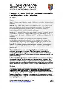
3. Results 3.1. Decreased serum levels of vitamin D is associated with low expression of LL-37 in TB lesions that have a high content of mycobacterial antigens In this study, lung tissue biopsies and serum samples were collected from 19 patients with cavitary or non-cavitary forms of chronic pulmonary TB (Table 1) [12]. First, we assessed serum levels of 25-hydroxyvitamin D in samples obtained from the TB patients and healthy controls (n = 10). Vitamin D is an immunomodulatory molecule that regulates both innate and adaptive immune responses and low levels of vitamin D have been shown to be associated with an enhanced
susceptibility to develop active TB [7]. Accordingly, we found that all TB patients were vitamin D deficient (b 25 nmol/l), and both sputum-positive and sputum-negative patients expressed vitamin D levels that were significantly lower compared to the control cohort (p b 0.001 and p b 0.01, respectively) (Fig. 1A). Next, we assessed local immune responses including distribution and functional expression of myeloid cells and lymphocyte subsets in cryopreserved tissue sections from the Mtb-infected lungs using quantitative mRNA and in situ computerized image analysis. Microscopic examination of gross TB lesions at a lower magnification revealed large necrotic granulomas, with CD3+ T cells in the mantel area and neutrophils in the core region (Fig. 1B). Immunostaining using a polyclonal M. bovis BCG antibody with documented cross-reactivity to Mtbantigens [30], revealed a significantly (p b 0.01) higher antigen expression in pulmonary TB lesions compared to the distal lung parenchyma (Fig. 1C), which suggest persistence of mycobacteria primarily at the local site of infection. Contrary, significantly (p b 0.0001) lower levels of the antimicrobial peptide LL-37 were detected in the TB lesions compared to the distal sites (Fig. 1B), which may suggest a specific downregulation of LL-37 at the site of mycobacterial infection. Quantitative real-time PCR demonstrated that mRNA levels of innate immune cells and effector molecules including markers of macrophages (CD68), inducible nitric oxide synthase (iNOS/ NOS2) and LL-37, were all expressed at similar levels in the TB lesions compared to distal lung parenchyma (Fig. 1D). Despite comparable mRNA levels of CD68, in situ image analysis revealed that protein expression of CD68 was significantly (p = 0.003) lower in the TB lesions compared to the distal
Figure 1 Pulmonary TB patients with a vitamin D deficiency express low local levels of the vitamin D-inducible antimicrobial peptide LL-37 in granulomatous lesions from the Mtb-infected lung. (A) S-vitamin D levels (median and range) in sputum-smear positive (sputum-pos; n = 11) and sputum-smear negative (sputum-neg; n = 8) TB patients compared to healthy Swedish controls (n = 10). The dotted lines mark the threshold for vitamin D deficiency (b 25 nmol/l) and insufficiency (25–75 nmol/l), respectively. Representative immunohistochemical images demonstrate expression of (B) CD3+ T cells and neutrophil elastase in granulomatous TB lesions (magnification × 25) and (C) BCG antigens and LL-37-expressing cells in the Mtb-infected lung (magnification × 125). In situ computerized image analysis was used to determine the expression (mean ± SE) of BCG antigens and LL-37 in the TB lesions compared to distal lung (n = 19). (D) Relative mRNA expression (median and range) of CD68, iNOS and LL-37 in the TB lesions compared to distal lung parenchyma (n = 19) was quantified using real-time PCR. The dotted line represents a relative difference of 1. (E) Immunohistochemical images illustrates CD68+ macrophages and iNOS-expressing cells in the Mtb-infected lung (magnification × 125) while in situ imaging was used to determine the expression (mean ± SE) of CD68 and iNOS in the TB lesions compared to distal lung (n = 19). Arrows in the images indicate positive cells (brown) whereas negative cells (blue) were counterstained with hematoxylin. p b 0.05 *, p b 0.01 **, p b 0.001 ***.
Impaired local immunity in vitamin D-deficient TB patients sites (Fig. 1E). In addition, MAC387, which is a protein found on monocytes, reactive macrophages, neutrophils and mucosal epithelia, were also significantly (p = 0.01) lower in the TB lesions (data not shown); while iNOS expression was similar in the TB lesions and distal lung parenchyma (Fig. 1E).
A.
89
3.2. Elevated levels of FoxP3+ Treg cells and IgG-secreting cells in pulmonary TB lesions We have previously demonstrated an impaired expression of perforin and granulysin in pulmonary TB lesions despite
Vitamin D status
25-hydroxyvitam in D (nmol/l)
*** ** 100 75 vitD insufficiency (25-75)
50 vitD deficiency (