The Application of the Genetic Algorithm Tool Box at Target Enhancement Processing in Breast Cancer Microwave Imaging Zhongling Han 1, Zhifu Tao1, Meng Yao1*, Yizhou Yao2, Blair Fleet3, Erik D. Goodman3, and John R. Deller4 1 Institute of information science and technology East China Normal University, Shanghai, China 2 Weiyu high school, Shanghai, China 3 BEACON Michigan State University, East Lansing, MI 4 ECE Michigan State University, East Lansing, MI *Corresponding Author, e-mail:
[email protected] Abstract - Genetic algorithms, as kind of fast, simply and highly fault-tolerant algorithms for near-field microwave detection of breast cancer; are useful processing method. For the image has not been dealt with high demands, which can quickly detect the interested area, and by bypassing the nonline Inversion of the problem brought about difficulties. The article mechanism to the imaging reflecting from analyzing a microwave starts off, analyses principle and process applying the Genetic algorithm detecting targets. Keywords: Microwave reflection; genetic algorithm; Image inversion
1
2
Breast Cancer Detection Microwave Imaging
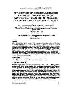
Assumed microwave reflectivity function of the cross section of the imaged object is f(x, y). The coordinate is fixed on the imaged object, and the non-directional transceiver antenna is located in the point (x0, y0). The antenna can only be rattling around the imaged object in a circle of radius R, whose moving can change the azimuth angle φ between the microwave beam and the imaged object. The reflect signal wave p(ρ, φ) received by the transceiver antenna represents line integral of f(r, θ) along a concentric circle arc s which the center is (x0, y0) (Shown in Figure 1).
Introduction
Microwave imaging sounding is an emerging breast cancer detection research method. This method might present a safe, convenient and cost-effective supplement to traditional BC imaging diagnoses methods. Different tissues can be imaged, which is based on a very high dielectric contrast between normal tissue and malignant tissue. It is known that the dielectric properties of tissues with high (tumor) and low (normal tissue) water content are significantly different. Reflection and refraction will occur and consequently the electromagnetic wave propagation path will be change, when the electromagnetic wave encounters all sorts of medium layer in process of propagation, such as skin, fat, breast lobules and tissue etc. Based on this property, we can detect and locate the breast tumor by detecting the microwaves reflect signal energy in different dielectric contrast mediums interface. The reflect signal energy also can convert into the transmitting waves propagation distance. In this paper, we start from analyzing the mechanism of microwave reflection imaging, and then using the genetic algorithm [1, 2] to optimize the inversion image. It has been found that this method can increase the detectable rate of breast cancer.
Figure1. It gives the geometric scheme of microwave imaging. The microwave reflectivity function is f(x, y). The transceiver is located in point (x0, y0), and it can only be rattling around the imaged object in a circle of radius R. the tumor is located in point (r, θ). The angle between the microwave beam and the imaged object is φ. The projection is expressed as follow. p( , ) f ( x, y )ds s
2
0
0
f (r , ) ( r 2 R 2 2rR cos )rdrd
r 2 R2 2rR cos ( x x0 )2 ( y y 0 )2
( )
(1)
, (2)
Where δ(d-ρ) is δ-function. We can get the equation (3) by the Fourier transform of projection S(ω,φ) and paraxial approximation.
S (, ) e Where,
jR
[F (u, v) jF (u, v) F (u, v) ...] (1)
S ( , )
F (u , v)
F (1) (u, v)
p ( , )e
j
( 2)
(3)
d
f ( x, y )e j ( ux vy ) dxdy
F ( 2) (u, v)
2
G ( x, y ) f ( x, y )e j (ux vy ) dxdy
G 2 ( x, y ) f ( x, y )e j (ux vy ) dxdy
F
(u , v)
n
G ( x, y ) f ( x, y ) e
G( x, y) ( x sin y cos ) u cos , v sin
j ( ux vy )
dxdy
2
2R
We can use Fourier transform to rebuild the profile distribution f(x, y) of the target by detecting the backscattering field (projection) p(ρ, φ). It can be ignored such as F(1)(u, v) and F(2)(u, v), when R is very big, and f(x, y) is limited band function. Then equation (1) can be seen as Fourier slice theory of CT imaging. The theory shows that the value can be given by the one-dimensional Fourier transform of the signal p(ρ, φ), which is the two-dimensional Fourier transform of the function f(x, y) in spatial frequency. u equals to ωcosφ, v equals to ωsinφ. We could get the function f(x, y) of detection space after inversing above process.
3
principle of breast cancer detection
The function f(x, y), which is the distribution function of reflection coefficient for each point in the detection space can be obtained from inversion. According to microwave propagation principle, f(x, y) can be calculated by equation (4).
f ( x, y )
1( x, y ) 2 ( x, y ) 1( x, y ) 2 ( x, y )
Genetic algorithm process and results
The goal of the microwave breast tumor detection is to separate cancerous target from inversion image f(x, y). So the function f(x, y) can be divided into two categories: one belongs to the cancerous target distribution C1 which the image intensity is greater than M; Another kind does not belong to the cancerous target distribution C2 which the image intensity is less than or equal to M. Considering actual inversion image grey levels of 256, the threshold value of the gray-level image coding for an 8 bits binary code string. Fitness function is shown in equation (5).
f (M ) w1 (M ) * w2 (M ) *[u1 (M ) u2 (M )]2
… (n)
4
(4)
Where ε1(x, y) and ε2(x, y) are the dielectric constant of two sides of the interface, respectively. Through calculation, the echo coefficient of cancerous tissue is greater than 0.49, the echo coefficient of lobules and gland tissue is between 0.2018 and 0.49 and the echo coefficient of blood vessels tissue is between 0.15 and 0.2018. Detecting breast cancer can be transformed into detecting some particular value distribution of f(x, y) in reflection space. So, cancerous tissue target is located in those points which echo coefficient is greater than 0.49.
(5)
Where w1 (M) is pixel number of C1, w2 (M) is pixel number of C2, u1 (M) is the average gray value in C1, u2 (M) is the average gray value in C2. Following is the genetic algorithm processing function [3,4] of MATLAB code, which used genetic algorithm tool box developed by the University of Sheffield. function C= Segmentation_GA (X, downth, upth); % input: X is waiting process image matrix, % downth and upth is the threshold value of detection target % output: C is the marked image NIND=40; % Individual number NVAR=1; % variable number MAXGEN=50; % Maximum number of generations PRECI=8; % bit number of variable GGAP=0.9; % generation gap FielD=[8*NVAR;downth;upth;1;0;1;1]; % Establishment of regional described device Chrom=crtbp(NIND,PRECI*NVAR); % Establish initial population gen=0; phen=bs2rv(Chrom,FielD); % The initial population decimal conversion ObjV=target(X,phen); % Calculate population fitting function while genM
X(i,j)=upth; % Using maximum mark end end end C=X; % Output marked image
5
Results
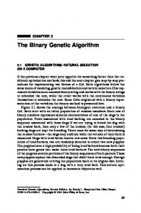
With the genetic algorithm method, we have succeeded in target enhancement. Figure 2 is simulation results of the data inversion when the microwave transceiver is located in circumference sampling uniformity of eight sampling test points. Form figure 2, we can see that the breast cancer is obviously enhanced after fifty iterations, and the background is smoother than ahead two imaging.
iterations by the genetic algorithm is shown in (a), (b) and (c), respectively.
6
Conclusion
In this study, we analyzed the microwave imaging theory and the microwave breast cancer detection theory. Then we use the simple genetic algorithm into microwave breast cancer detection to enhance tumor emerging area. As is a rapid, easy and fault-tolerance strong robust algorithm, the simple genetic algorithm can very good solve the nonlinear inversion problem in detection. A great deal of further research effort is needed to elucidate certain aspects of the genetic algorithm application to stability and target detection error.
7
Acknowledgments
This work has been performed while Prof. M. Yao was a visiting scholar in Michigan State University, thanks to a visiting research program from Prof. Erik D. Goodman. M. Yao would also like to acknowledge the support of Shanghai Science and Technology Development Foundation under the project grant numbers 03JC14026 and 08JC1409200, as well as the support of TI Co. Ltd through TI (China) Innovation Foundation.
8 (a)
(b)
(c) Figure2. It is inversion images of eight sampling test points. The distribution of the original detection space, the inversion detection space and the detection space processed after fifty
Reference
[1] Ming Zhou. Principle and application of the genetic algorithm Defense industry press 1999.6, pp. 36–37 [2] Rajarshi Das, Melanie Mitchell, and James P. Crutch_eld, Parallel Problem Solving from Nature---PPSN III. Berlin: Springer-Verlag. 1994 [3] Das, R. and Whitley, L.D. The Only Challenging Problems Are Deceptive: Global Search by Solving Order-1 Hyperplanes. Proceedings of ICGA 1991, pp.166-173. [4] Christopher R. Houck, Jeffery A.Joines, and Michael G. Kay A Genetic Algorithm for Function Optimization- A Matlab Implementation Technical Report NCSU-IE-TR-9509, North Carolina State University, Raleigh, NC (1995)