The attentional-relevance and temporal dynamics of visualtactile crossmodal interactions differentially influence early stages of somatosensory processing Christina Popovich & W. Richard Staines Department of Kinesiology, University of Waterloo, 200 University Ave. W, Waterloo, Ontario N2L 3G1, Canada
Keywords Attention, crossmodal, ERPs, event-related potentials, sensorimotor integration, somatosensory cortex, tactile, visual Correspondence W. Richard Staines, Department of Kinesiology , University of Waterloo, 200 University Ave W, BMH 3031, Waterloo, Ontario N2L 3G1, Canada. Tel: (519) 888-4567 (ext. 37756); Fax: (519) 885-0470; E-mail:
[email protected] Funding Information This research was supported by NSERC. Received: 23 August 2013; Revised: 6 December 2013; Accepted: 15 December 2013 Brain and Behavior 2014; 4(2): 247–260 doi: 10.1002/brb3.210
Abstract Background: Crossmodal interactions between relevant visual and tactile inputs can enhance attentional modulation at early stages in somatosensory cortices to achieve goal-oriented behaviors. However, the specific contribution of each sensory system during attentional processing remains unclear. We used EEG to investigate the effects of visual priming and attentional relevance in modulating somatosensory cortical responses. Methods: Healthy adults performed a sensory integration task that required scaled motor responses dependent on the amplitudes of tactile and visual stimuli. Participants completed an attentional paradigm comprised of 5 conditions that presented sequential or concurrent pairs of discrete stimuli with random amplitude variations: 1) tactile-tactile (TT), 2) visual-visual (VV), 3) visual-tactile simultaneous (SIM), 4) tactile-visual delay (TVd), and 5) visual-tactile delay (VTd), each with a 100 ms temporal delay between stimulus onsets. Attention was directed to crossmodal conditions and graded motor responses representing the summation of the 2 stimulus amplitudes were made. Results: Results of somatosensory ERPs showed that the modality-specific components (P50, P100) were sensitive to i) the temporal dynamics of crossmodal interactions, and ii) the relevance of these sensory signals for behaviour. Conclusion: Notably, the P50 amplitude was greatest in the VTd condition, suggesting that presentation of relevant visual information for upcoming movement modulates somatosensory processing in modality-specific cortical regions, as early as the primary somatosensory cortex (SI).
Introduction It is well-known that attention can modulate neurophysiological responses in modality-specific cortices including: visual (Motter 1993; Gazzaley et al. 2007; Andersen et al. 2008), auditory (Woldorff et al. 1993; Ja¨ncke et al. 1999; Petkov et al. 2004), and somatosensory cortices (Josiassen et al. 1990; Hsiao et al. 1993; Johansen-Berg et al. 2000; Staines et al. 2002). However, recent investigations have begun to examine whether attention influences neural responses across sensory modalities when sensory input from more than one modality is present. Behavioral studies have shown that crossmodal input can also improve performance as indexed by faster reaction times (Hershenson 1962; Gielen et al. 1983), improved detection of weak stimuli (Frens and Van Opstal 1995; Driver and Spence 1998; McDonald et al. 2000), and improved sensory-perception
of illusory effects such as the ventriloquist or McGurk illusions (Howard and Templeton 1966; McGurk and MacDonald 1976). Human and animal studies have shown that the mere presence of additional sensory input even when it is irrelevant for performance of a task can enhance neural excitability in the attended sensory modality (Calvert et al. 1997; Macaluso et al. 2000, 2002; Calvert 2001; Foxe et al. 2002; Kayser et al. 2005, 2007; Pekkola et al. 2006; Lehmann et al. 2006; Lakatos et al. 2007; Meehan and Staines 2009), suggesting that interactions between modality-specific cortical representations exist. By contrast, other studies have shown crossmodal enhancement in modality-specific sensory cortex only occurs when both stimuli events are relevant for behavior (Dionne et al. 2010, 2013). These findings suggest that crossmodal processing is likely governed by both bottom-up sensorysensory interactions and top-down attentional mechanisms
ª 2014 The Authors. Brain and Behavior published by Wiley Periodicals, Inc. This is an open access article under the terms of the Creative Commons Attribution License, which permits use, distribution and reproduction in any medium, provided the original work is properly cited.
247
Interaction of Vision, Touch, and Attention in Sensory Cortex
in order to allow for the selection, amplification, and integration of sensory input relevant for initiating goal-oriented responses. Bottom-up interactions can occur when salient stimuli from an unattended sensory modality influence neural excitability in the attended modality, while top-down processing occurs when attention is voluntarily directed toward relevant stimuli in the presence of environmental distracters. However, while both these attentional mechanisms can modulate neural responses in modality-specific sensory cortex, it remains unclear how these attentional mechanisms interact during sensory processing of crossmodal stimuli. Neurophysiological research in the primary auditory cortex of monkeys has provided evidence that sensory-tosensory interactions exist. Recent studies have shown that neural responses in regionally distinct areas of the primary auditory cortex are enhanced when visual and/or tactile stimuli are paired with auditory stimuli (Kayser et al. 2005, 2007). Lakatos et al. (2007) showed that presentation of somatosensory stimuli increased auditory neural responses when the two stimuli were simultaneously combined versus when the auditory stimulus was presented in isolation. Furthermore, Bizley et al. (2007) reported a 15% neuronal increase in the ferret primary auditory cortex following simultaneous presentation of visuo-auditory stimuli (Bizley et al. 2007). Neuroimaging studies in humans complement the sensory-to-sensory interactions reported in animal findings by showing that the presence of crossmodal input can modulate neural excitability in modality-specific sensory cortices. For example, several functional magnetic resonance imaging (fMRI) studies have reported increased blood oxygenation level-dependent (BOLD) responses in modality-specific cortices due to the mere presence of stimuli from another modality. These interactions have been found between: visual and auditory cortices (Calvert et al. 1997; Calvert 2001; Lehmann et al. 2006; Pekkola et al. 2006), auditory and somatosensory cortices (Foxe et al. 2002; Sch€ urmann et al. 2006), as well as visual and somatosensory cortices (Macaluso et al. 2000, 2002). However, a recent fMRI study investigated crossmodal effects on BOLD responses generated in the primary somatosensory cortex (SI) when both stimuli were relevant for guiding a motor response. Here, relevant unimodal (visual or tactile) and crossmodal stimuli (simultaneous visual + tactile) were presented and participants were required to summate both stimuli by squeezing a pressure-sensitive bulb. In order to ensure that stimulus associations were successfully learned prior to testing, participants completed a brief sensorimotor training session that required them to judge the amplitude of visual and vibrotactile stimuli and make a graded motor response representing the perceived amplitude of the stimuli. Results showed that the greatest BOLD
248
C. Popovich & W. R. Staines
responses were elicited in SI during crossmodal versus unimodal interactions suggesting that combining visual-tactile information relevant for behavior enhances modality-specific excitability in SI (Dionne et al. 2010). In a follow-up study, Dionne et al. (2013); used electroencephalography (EEG) and the same sensory-to-motor task to investigate the time course of crossmodal effects in SI. Results showed that crossmodal interactions between vibrotactile and visual stimuli enhanced the amplitude of the somatosensory P50 component, generated in SI, at contralateral parietal electrode sites only when both stimuli were task-relevant. By contrast, the amplitude of the P100, likely generated in SII, increased bilaterally at parietal electrode sites during presentation of crossmodal stimuli but was not sensitive to the task-relevance of the stimuli. These findings suggest that crossmodal modulation occurs at very early stages in the somatosensory processing stream if both stimuli are relevant for behavior (Dionne et al. 2013). Several other EEG studies support the finding that crossmodal stimuli can modulate neural excitability at very early stages of sensory processing. For example, Giard and Peronnet (1999); found that visual modulation for audio-visual stimuli, occurred as early as 40-msec post stimulus onset, while audio-tactile modulation has been found at 50 msec (Foxe et al. 2000; Molholm et al. 2002). Kennett et al. (2001); found modulation of visual eventrelated potentials (ERPs) by irrelevant but spatially aligned tactile stimuli at approximately 140-msec post visual onset, while McDonald et al. (2000); reported modulation of visual ERPs was possible with spatially aligned auditory stimuli. In summary, crossmodal interactions can improve behavioral performance and enhance neural excitability at early stages in modality-specific cortices to achieve goal-oriented behaviors (Dionne et al. 2010, 2013). However, the specific contribution of each sensory system during attentional processing in modalityspecific sensory cortices remains unclear. In this study, we manipulated the attentional relevance and temporal onsets of visual and tactile stimuli to examine whether both top-down and bottom-up mechanisms can modulate early stages of somatosensory processing. The specific aim of this study was to explore the relative contributions of visual priming (bottom-up sensory input) and task-relevance (top-down attention) on influencing early somatosensory cortical responses, namely the P50 somatosensory ERP generated in SI. We hypothesized that somatosensory activity would be modulated based on the temporal onset and stimulus order of task-relevant crossmodal (visual-tactile) events. To test whether bottom-up sensory-sensory interactions influence crossmodal modulation of the P50 component, we manipulated the temporal onsets of visual and tactile events in two cross-
ª 2014 The Authors. Brain and Behavior published by Wiley Periodicals, Inc.
C. Popovich & W. R. Staines
modal conditions. In one condition, visual stimuli preceded tactile stimuli by 100 msec to examine whether the presentation of relevant visual information prior to tactile information influenced crossmodal modulation of the P50 component. In the other condition, tactile stimuli preceded visual stimuli by 100 msec. This condition acted as a control to the previously described condition since the onset of the P50 component would have already occurred prior to the presentation of visual information, thus P50 modulation in this case would not be due to crossmodal influences. If bottom up and top-down mechanisms influence early somatosensory ERPs in contralateral SI, then the P50 amplitude should be greatest for relevant crossmodal interactions where visual information preceded tactile information and smallest for the irrelevant unimodal interactions.
Material and Methods Participants EEG was collected from 20 self-reported right-handed healthy participants (mean age = 26, 10 males). Five subjects were excluded due to either excessive artifacts found during inspection of the raw EEG collection, or the absence of clearly defined somatosensory ERPs of interest (i.e., P50 and/or P100 components). The final sample consisted of 15 healthy participants (mean age = 27.5, 7 men). Experimental procedures were approved by the University of Waterloo Office of Research Ethics. All subjects provided informed written consent.
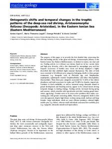
Behavioral paradigm The behavioral paradigm consisted of five conditions that presented pairs of discrete visual and/or tactile stimuli with random amplitude variations. Stimuli were always presented in pairs, either sequentially (unimodal conditions) or simultaneously (crossmodal conditions): (1) tactile-tactile (TT; 500 msec each, 30-msec interstimulus interval [ISI]), (2) visual-visual (VV; 500 msec each, 30msec ISI), (3) visual-tactile simultaneous (SIM; 1000 msec concurrent), (4) visual-tactile with a 100-msec temporal delay between stimulus onsets (visual-tactile delay, [VTd]; 500 msec each, visual presented first), and (5) tactilevisual with a 100-msec temporal delay between stimuli (tactile-visual delay, [TVd], tactile presented first) (refer to Fig. 1A–D). Participants were instructed to only attend to the crossmodal stimuli (i.e., TT/VV conditions were ignored), judge the amplitude of the two stimuli, and then make a graded motor response representing the sum of these amplitudes by squeezing a pressure-sensitive bulb with their right hand (Fig. 1E). Prior to the EEG collec-
ª 2014 The Authors. Brain and Behavior published by Wiley Periodicals, Inc.
Interaction of Vision, Touch, and Attention in Sensory Cortex
tion, participants underwent a 5-min training session with visual feedback in a sound attenuated booth to learn the relationship between the amplitudes of the stimuli and the corresponding force required to apply to the bulb. During training, a horizontal target bar appeared on the computer monitor and subjects were instructed to squeeze the pressure-sensitive bulb with enough force to raise another visual horizontal bar to the same level as the target bar. At the same time, as subjects applied force to the bulb with their right hand the vibrotactile device vibrated against the volar surface of their left index finger with corresponding changes in amplitude. In other words, as they squeezed harder on the bulb the amplitude of the vibration increased proportionately. Subjects were instructed to pay attention to these changes in amplitude as they related to the force they were applying to the bulb. This training allowed subjects to become familiar with the relationship between the vibrotactile stimulus amplitude and the corresponding force applied to the bulb. To control for force related trial to trial differences, stimulus amplitudes were scaled such that no single stimulus required a squeeze of more than 25% of an individual’s maximum force, thus the response for adding two stimuli was never more than 50% of an individual’s maximum force. Stimuli were always presented in pairs, either unimodally (two visual or two tactile) presented sequentially, or crossmodally (one visual and one tactile), presented simultaneously or with a 100-msec temporal offset between each stimuli.
Experimental paradigm During the experiment, participants sat comfortably in a sound attenuated booth and were instructed to visually fixate on the computer monitor, rest the volar surface of their left index finger gently on the vibrotactile device, and hold the pressure-sensitive response bulb in their right hand (Fig. 1F). Participants were instructed to attend only to crossmodal interactions, judge the amplitude of both the visually presented horizontal bars and the vibrotactile stimuli, and produce force graded motor responses using the pressure-sensitive bulb that represented the summation of both stimulus amplitudes. Stimuli were presented for 1 sec after which participants were required to make their motor response immediately following presentation of the crossmodal stimuli during a 2.5 sec window prior to the start of the next trial, for a total of 3.5 sec per trial. Each condition was randomized and performed in six blocks of 120 trials with each block lasting approximately 5 min. The order of the conditions was counterbalanced across each block and all subjects performed the same six blocks in sequential order.
249
Interaction of Vision, Touch, and Attention in Sensory Cortex
(A)
C. Popovich & W. R. Staines
(E)
(B)
(C)
(D)
(F)
Figure 1. Experimental paradigm. (A) shows the unimodal conditions (VV, TT), (B) shows the crossmodal condition with simultaneously presented visual-tactile stimuli, (C) shows the crossmodal condition where tactile stimuli are presented 100 msec before visual stimuli (TVd), (D) shows the crossmodal condition where visual stimuli are presented 100 msec before tactile stimuli (VTd) between visual-tactile condition (VT). Participants were required to ignore all unimodal conditions and only respond to the crossmodal conditions. To depict the behavioral task, the columns are intended to represent examples of the temporal onset and amplitudes of stimulus events while the dotted trace is a schematic of the corresponding force applied to the squeeze-bulb when making the motor response to those stimuli. (E) shows an example of a bimodal simultaneous condition (SIM) and a unimodal tactile-tactile condition (TT). Subjects were to attend only to bimodal conditions and make a graded motor response with a pressure bulb representing the summation of each stimuli. (ITI; intertrial interval, ISI; interstimulus interval). (F) The experimental setup is depicted for the positioning of participants to receive the tactile and visual stimulation.
Stimuli Visual stimuli consisted of a centrally presented horizontal bar (6 cm wide), which raised to varying heights on a computer monitor positioned 50 cm in front of the subject and represented different visual amplitudes. Vibrotactile stimuli consisted of discrete vibrations delivered by a custom made vibrotactile device applied to the volar surface of the left index finger. Vibrotactile stimulation was controlled by converting digitally generated waveforms to an analog signal (DAQCard 6024E; National Instruments, Austin, TX) and then amplifying the signal (Bryston
250
2BLP, Peterborough, Ontario, Canada) using a custom program written in LabVIEW (version 8.5; National Instruments). Varying the amplitude of the driving voltage to the vibrotactile device produced proportional changes in vibration of the device on the finger. The amplitude of each discrete vibration was constant within a trial and varied randomly between trials. The average stimulus amplitude across all trials including a tactile stimulus did not differ between the experimental conditions. The frequency of the vibration was held constant at 25 Hz. Participants received 70 db whitenoise (Stim2; Neuroscan, Compumedics USA, Charlotte, NC)
ª 2014 The Authors. Brain and Behavior published by Wiley Periodicals, Inc.
C. Popovich & W. R. Staines
Interaction of Vision, Touch, and Attention in Sensory Cortex
throughout the training session and the experiment to prevent auditory perception of the vibrotactile stimulus.
Data acquisition and recording parameters EEG data were recorded from 64 electrode sites (64-channel Quick-Cap, Neuroscan, Compumedics USA) in accordance with the international 10–20 system for electrode placement, and referenced to the linked mastoids (impedance