Neuroscience 163 (2009) 977–984
“THE DEVELOPMENTAL AND FUNCTIONAL LOGIC OF NEURONAL CIRCUITS”: COMMENTARY ON THE KAVLI PRIZE IN NEUROSCIENCE J. C. GLOVER*
adhesion molecules, sensorimotor reflex circuits, sonic hedgehog, shh, motor control, muscle spindles, stretch reflex, reticulospinal, vestibulospinal, mammalian, cat, fish, lamprey, supraspinal centers, swimming, locomotion, anatomy, electrophysiology, fictive locomotion, bistable membrane, excitatory aminoacid, NMDA receptors, rhythmicity, computer simulation, laser scanning confocal microscopy, spinal cord injury, striatum, goal-directed behavior, informatics, neuroinformatics, neuromechanical model.
Department of Physiology, Institute of Basic Medical Sciences, University of Oslo, PB 1103, Blindern, 0317 Oslo, Norway
Abstract—The first Kavli Prize in Neuroscience recognizes a confluence of career achievements that together provide a fundamental understanding of how brain and spinal cord circuits are assembled during development and function in the adult. The members of the Kavli Neuroscience Prize Committee have decided to reward three scientists (Sten Grillner, Thomas Jessell, and Pasko Rakic) jointly “for discoveries on the developmental and functional logic of neuronal circuits”. Pasko Rakic performed groundbreaking studies of the developing cerebral cortex, including the discovery of how radial glia guide the neuronal migration that establishes cortical layers and for the radial unit hypothesis and its implications for cortical connectivity and evolution. Thomas Jessell discovered molecular principles governing the specification and patterning of different neuron types and the development of their synaptic interconnection into sensorimotor circuits. Sten Grillner elucidated principles of network organization in the vertebrate locomotor central pattern generator, along with its command systems and sensory and higher order control. The discoveries of Rakic, Jessell and Grillner provide a framework for how neurons obtain their identities and ultimate locations, establish appropriate connections with each other, and how the resultant neuronal networks operate. Their work has significantly advanced our understanding of brain development and function and created new opportunities for the treatment of neurological disorders. Each has pioneered an important area of neuroscience research and left a legacy of exceptional scientific achievement, insight, communication, mentoring and leadership. © 2009 IBRO. Published by Elsevier Ltd. Open access under CC BY-NC-ND license.
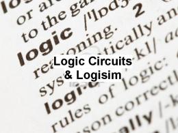
Pasko Rakic received his MD in 1960 and his PhD in 1969 from the University of Belgrade. During this period he held posts as Clinical and Research Fellow at Harvard Medical School and as Assistant Professor at the University of Belgrade. From the beginning of his research career he was drawn to human brain anatomy and development. Early work while at Harvard focused on the histogenesis of cortical structures in both humans and mice, using among other techniques the labeling of newly synthesized DNA with tritiated thymidine to determine the birthdates of neurons. Rakic continued to use this technique to study the timing of neuron production, particularly in the monkey, publishing a long list of papers that has provided a spatiotemporal map of neurogenesis throughout much of the primate brain. Many of these seminal studies remain to this day principle reference works for researchers studying the development of specific brain, and especially cortical, areas. Noting that neurons occupying different cortical layers are generated during sequential epochs of proliferation, Rakic proposed that a neuron’s eventual physical destination and phenotype are specified at the time of its last mitotic division (Rakic, 1974), thereby launching efforts to understand the role of intrinsic determinants of neuronal identity which continue to this day. In 1971, now an Assistant Professor at Harvard, Rakic published a seminal paper describing an intimate anatomical relationship between migrating neurons and radial glia in the macaque monkey (Rakic, 1971a,b). Prior to this, the radial glia were enigmatic, with no known function despite their peculiar morphology, stretching from the inner surface to the outer surface of the developing brain. Rakic’s work clearly implicated the radial glia as substrates for the radial migration of newly generated neurons (Fig. 1). The mechanism and its functional implications would preoccupy Rakic for many years to come, and attract a substantial number of other researchers to the question of how neurons migrate to their ultimate destinations in the cortex and elsewhere in the brain. Rakic was the first to discover a specific gene whose mutation disrupts neuronal migration (Rakic and Sidman, 1973), and has continued to make seminal contributions in this area, extending to the role of
Key words: Grillner S, Jessell TM, Rakic P, cerebral cortex, cortical layers, radial glia, radial unit hypothesis, neuronal migration, connectivity, evolution, spinal cord, sensorimotor circuits, molecular specification of neurons, locomotor, central pattern generator, network organization, brain anatomy, human, monkey, primate, histogenesis, tritiated thymidine, neurogenesis, spatiotemporal map, last mitotic division, neuronal identity, “migration disorders”, ocular dominance columns, cytoarchitectonic, ependyma, proliferative unit, prot-map, neuronal progenitors, substance P, sensory transmitters, sensory neurons, pain, opiates, capsaicin, acetylcholine receptors, recognition molecules, embryonic development, hybridoma, monoclonal antibodies, epitopes, surface molecules, molecular markers, commissural interneurons, chemotropic, floor plate, axonal pathfinding, nerve fiber trajectories, notochord, signaling gradient, nuclear transcription factors, dorsoventral patterning, motoneuron pools, anteroposterior patterning, retionoic acid, hox genes, motoneuron diseases, amyotrophic lateral sclerosis, *Corresponding author. Tel: ⫹47- 22851230; fax: ⫹47-22851249. E-mail address:
[email protected] (J. C. Glover).
0306-4522/09 © 2009 IBRO. Published by Elsevier Ltd. Open access under CC BY-NC-ND license. doi:10.1016/j.neuroscience.2009.07.047
977
978
J. C. Glover / Neuroscience 163 (2009) 977–984
Fig. 1. The radial unit hypothesis as depicted by Rakic. Panel A shows the radial migratory pathways that map the inner array of neuronal progenitors (LV) to the mature cortical layers (CP). Panel B shows the intimate relationship between a migrating neuron (N) and a radial glial fiber (RF). From Rakic (2003) Developmental and evolutionary adaptations of cortical radial glia. Cerebral Cortex 13:541–549.
intrinsic and extrinsic molecular factors that influence, regulate, and disrupt the migratory process. His work has in large part established the foundation for current efforts at understanding the etiology of various human brain malformations and disorders that result from disturbances of neuronal migration, now known collectively as “migration disorders”. While working on neurogenesis in the monkey visual cortex, Rakic used his skill at intrauterine surgery to test whether connections in the visual system that subserve the formation of ocular dominance columns form prenatally in primates. Ocular dominance had been studied previously by eventual Nobel Prize winners David Hubel and Torstein Weisel, who had shown that the relevant connections form postnatally in kittens, when visual experience can exercise a pivotal activity-dependent influence. Rakic’s finding that the ocular dominance columns in the primate visual cortex form prenatally (Rakic, 1981) argued for an innate contribution to the ocular dominance pattern, a proposal that has since been supported by experiment in other species including the cat, and also set the stage for later work addressing the fundamental role of spontaneous activity in shaping brain circuits, before sensory experience becomes a major factor.
In 1978, Rakic became Professor at Yale University, where he has continued to work since. There he continued to pioneer the study of cortical development, not only regarding neuronal migration per se, but also on how this contributes to the partitioning of cortex into functional areas (Rakic, 1988). His insight into the four-dimensional process of cortical development and its regulation by sensory input led eventually to the formulation of a conceptual framework for corticogenesis, the “radial unit hypothesis” (Fig. 1). In his own words: “According to this hypothesis, the ependymal layer of the embryonic cerebral ventricle consists of proliferative units that provide a proto-map of prospective cytoarchitectonic areas. The output of the proliferative units is translated via glial guides to the expanding cortex in the form of ontogenetic columns, whose final number for each area can be modified through interaction with afferent input”. In his seminal review article in 1988 (Rakic, 1988) he integrated findings from numerous studies and methodological approaches to show how these supported the hypothesis. He expanded on this theme to propose that radial units could form the basis for cortical microcircuits, as well as the coinage of the evolutionary expansion in cortical area that has led to ever more complex and sophisticated vertebrate brains (Rakic, 1995;
J. C. Glover / Neuroscience 163 (2009) 977–984
2003). The radial unit hypothesis has guided a generation of researchers in their efforts to piece together the intricacies of cortical function, and continues to serve as a viable platform for posing new questions and applying new technologies to answer them. With arguably the broadest perspective on primate neurogenesis of anyone alive, Rakic naturally was drawn to more recent discussions regarding the possibility of adult neurogenesis in humans, which followed the discovery of neural stem cells in the adult human brain. He has publicly insisted on the utmost methodological rigor in documenting the presence of newly generated neurons in the adult human brain and, armed with solid quantitative information, tempered speculations on the extent to which adult neurogenesis can contribute to brain plasticity in humans (Rakic, 2002). His authoritative role in this arena has provided a sober reminder that scientific theories are only as good as the experimental data on which they are founded and by which they are eventually substantiated. By establishing conceptual frameworks for the interplay of neurogenesis, migration, activity and the formation of functional circuits in cortical development, Pasko Rakic has made a fundamental contribution to our understanding of how the cortex, seat of our consciousness, takes form during development and has been shaped by evolution. He continues to make important contributions to this understanding (Letinic et al., 2009; Rakic, 2009; Rakic et al., 2009; Torii et al., 2009). Thomas M. Jessell received his PhD in 1977 from Cambridge University, where he studied the distribution and release of neurotransmitters in the CNS while working in the laboratory of Leslie Iversen. His graduate career at Cambridge was extremely fruitful, generating within just a few years an impressive list of publications that contributed importantly to the understanding of neurotransmitter actions in the mammalian brain. One of the transmitters that received special attention was substance P, which had been implicated as a neurotransmitter in sensory fibers carrying pain signals. Together with Iversen, A. Claudio Cuello, Masanori Otsuka and others, Jessell demonstrated that substance P-containing sensory fibers were profoundly affected by opiates, which inhibit the release of sensory transmitters, and by capsaicin, the substance that puts the zing in chili peppers, which depleted substance P and caused a prolonged analgesia (Jessell et al., 1978). Important enough as a seminal contribution to pain research, these experiments marked a turning point in Jessell’s career, drawing his attention from the brain to the spinal cord, which has remained the principal focus of his work up to the present. Following a stint in Gerald Fishbach’s laboratory at Harvard University, where he made another seminal contribution, this time regarding nerve-derived proteins that influence the clustering of acetylcholine receptors on muscle (Jessell et al., 1979), Jessell took an Assistant Professorship at Harvard where he started to devote his energies to the study of sensory fiber function and diversity. Combining physiological and molecular techniques, he and his collaborators began dissecting the population of sensory
979
neurons present in spinal ganglia into subclasses based on neurotransmitter phenotypes and biochemical properties. It was already known that sensory fibers of different types had characteristic termination patterns within the spinal cord, a feature upon which Jessell’s studies elaborated. Realizing that these patterns implied a capacity to selectively recognize the appropriate central target neurons during embryonic development, Jessell set out to identify candidate recognition molecules. Using the recently developed hybridoma approach for generating monoclonal antibodies, a panel of markers was obtained that defined molecularly distinct subclasses of the sensory neurons (Dodd et al., 1984). Many of the epitopes involved were found to be surface molecules that were also expressed in the central target regions innervated by the sensory neurons. A molecular lock-and-key mechanism that might explain how sensory fibers make specific connections with central neurons seemed to be close at hand. Jessell and his coworkers, now at Columbia University, also discovered other molecular markers expressed on central neurons and used these to elucidate how the growth of central axons, notably those of commissural interneurons whose axons cross the midline, is directed (Dodd et al., 1988). In a series of elegant papers, the existence of chemotropic factors that coax commissural axons to the midline was demonstrated (Tessier-Lavigne et al., 1988). These factors were shown to derive from the floor plate, an embryonic structure that gives rise to the midline seam that runs along the length of the ventral surface of the spinal cord. The floor plate immediately became the object of intense investigation in many laboratories. In discovering this simple mechanism for selectively orienting the growth of a specific class of nerve fibers, Jessell and his coworkers not only created a fertile field for the investigation of axonal pathfinding in general, but also contributed profound insight into how the intricate network of axon tracts within the vertebrate brain could be built up from a series of pathfinding decisions, among the first of which was the decision to cross or not cross the midline. With a new focus on floor plate-derived molecules as driving force, Jessell began addressing how not only nerve fiber trajectories but also neuron identities are patterned within the spinal cord. A seminal contribution was the finding that the floor plate and another ventrally situated structure, the notochord, were the source of diffusible proteins that, entering at the ventral surface and diffusing dorsally, created a signaling gradient that instructed naïve neuronal precursor cells within the spinal cord to generate specific types of neurons (Yamada et al., 1991). In a remarkable series of papers, Jessell and his coworkers identified an opposing signaling gradient derived from the dorsal midline (Basler et al., 1993; Liem et al., 1997), discovered a panel of nuclear transcription factors that define a series of neuronal progenitors, and established a working model for how their activation leads to the specification of different types of motoneurons and interneurons. This model, in which a bipolar signaling gradient induces or represses the expression of specific transcription factors at characteristic locations along the dorsoven-
980
J. C. Glover / Neuroscience 163 (2009) 977–984
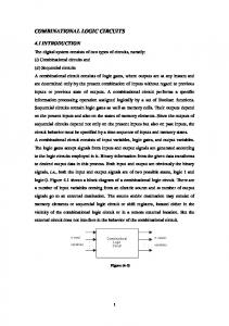
Fig. 2. The molecular logic of neuronal specification and sensorimotor circuit formation. The top panels show how a ventral-to-dorsal gradient of a signal molecule (sonic hedgehog, or shh) secreted by the notochord (N) and floor plate (FP) is translated into a sequence of sharply demarcated neuron progenitor domains each of which expresses a specific transcription factor (colors) and gives rise to a specific subset of neurons (MN, motoneurons; V1, V2, V3, specific types of ventral interneurons). From Briscoe and Ericson (1999) The specification of neuronal identity by graded sonic hedgehog signaling. Seminars in Cell and Developmental Biology 10:353–362. The lower panels show how the neurons derived from specific progenitor domains become specified to make synaptic connections with each other and muscle to create functional sensorimotor circuits. From Goulding et al. (2002) The formation of sensorimotor circuits. Current Opinion in Neurobiology 12:508 –515.
tral axis, was then elaborated with the finding that the transcription factors in question exert mutually repressive actions on each other (Briscoe et al., 2000; Jessell, 2000). Thus, the gradient is converted into a series of stripes, each containing progenitors destined to give rise to a molecularly distinct subset of spinal neurons (Fig. 2). It has since become clear that the brain is patterned in the same way, making this a universal mechanism for dorsoventral patterning in the CNS. Jessell had at the same time an eye to the other main axis of the spinal cord: the anteroposterior axis. The motoneurons of the spinal cord are arranged in columns with different anteroposterior domains, and these columns are further subdivided into coherent motoneuron pools that each innervate a specific muscle. Jessell and his coworkers had already shown that the columns were molecularly distinct on the basis of a combinatorial expression of transcription factors, and soon found that a molecular distinction could be extended to the level of individual motoneuron pools. Taking advantage of earlier work that had implicated retinoic acid and other signaling molecules in the anteroposterior patterning of the brain and spinal cord, and
knowing that Hox genes provide different addresses along the longitudinal axis of the nervous system, Jessell and his coworkers showed how the sequential action and interaction of these molecular systems partitions the motoneurons of the spinal cord into columns and pools (Liu et al., 2001; Dasen et al., 2005). The insight gained from these studies was also used to induce embryonic stem cells to generate motoneuron precursors that differentiate into functional motoneurons in vivo (Wichterle et al., 2002), yielding a promising avenue towards the treatment of motoneuron diseases such as amyotropic lateral sclerosis. Never losing sight of his early interest in how neurons make synaptic connections with the proper specificity, Jessell and his coworkers went on to show that the combinatorial code of transcription factors that defines motoneuron subtypes drives the expression of cell– cell adhesion molecules that leads to their clustering into pools, regulates the expression of receptors that govern the growth of their axons to the appropriate target muscles, and is matched by the expression of genes in muscle sensory neurons that instruct these to innervate the appropriate motoneurons (Price et al., 2002; Kania and Jessell, 2003; Arber et al.,
J. C. Glover / Neuroscience 163 (2009) 977–984
2000). Although many details remain to be discovered, the molecular logic that sets up the specific connectivity of sensorimotor reflex circuits is fast emerging. And, in a return to an earlier theme, the molecular basis for selective targeting of sensory projections involved in pain transmission is also yielding to Jessell’s current efforts (Chen et al., 2006). In addressing these relatively simple circuits, Thomas Jessell’s astounding synthesis of molecular patterning mechanisms is paving the way to understanding how complex circuits throughout the brain are assembled (Agalliu et al., 2009; Dasen et al., 2009; Friese et al., 2009; Pecho-Vrieseling et al., 2009). Sten Grillner received his PhD and MD in 1969 from the University of Göteborg, a major center for the study of motor control in mammals. His earliest studies were focused on the descending and reflex control of activity in motoneurons, including the gamma-motoneurons that set the tone of muscle spindles, important for the regulation of muscle stretch reflex activity. Still at the University of Göteborg, within a few years he had published a comprehensive list of articles characterizing the anatomical origins and synaptic effects of reticulospinal and vestibulospinal pathways, and the functional interactions between these and with the sensory fibers from muscle spindles in the cat (Grillner et al., 1968; 1970; Grillner, 1969). In 1971 he was a visiting scientist at the Academy of Science in Moscow, during which he shifted his attention to the specific problem of locomotion and its neural control. Together with coworkers in Moscow and Göteborg, he showed that locomotion in the cat could be triggered from anatomically distinct supraspinal centers (Grillner and Shik, 1973), and that the mammalian spinal cord could generate locomotor movements in the absence of descending influences (Grillner, 1975). The latter finding was consistent with the presence of a “central pattern generator” for locomotion, that is, a circuit contained within the spinal cord that was sufficient to generate the patterned activity in motoneurons responsible for locomotory movements. Although he would continue for several years to investigate the descending and reflex control of motoneurons, particularly during locomotion, his interest became more and more focused on the nature of the central pattern generator for locomotion. While still working on the cat, Grillner also performed experiments in the dogfish, showing again that the spinal cord could generate locomotor activity in the absence of descending influences from supraspinal centers, as well as without sensory feedback through the sensory dorsal roots (Grillner, 1974). Fish clearly provided a more convenient preparation for such studies, as well as a simpler locomotor pattern, since locomotion involved side-to-side body contractions and not the complex activation of limbs. Now a young professor at the Karolinska Institute in Stockholm, Grillner quickly realized the advantages of studying a simpler vertebrate system, and in a brilliant move chose the lamprey, a jawless fish thought to resemble a primitive fish ancestor, as the preparation for his further studies of vertebrate locomotion (Grillner and Wallén, 1980). The lamprey has an easily characterized swimming movement and a thin and nearly transparent
981
spinal cord that is perfect for electrophysiological recording from individual neurons. Instead of recording blindly from spinal neurons in the mammal, it was now possible to aim electrodes at specific types of neurons, characterize their patterns of activity and synaptic connections, and then inject them with a dye to reveal their anatomy. A new chapter had been opened. Grillner continued to perform experiments on mammalian locomotion for several years, but by the mid-1980s the lamprey had monopolized his research activity. What ensued is nothing short of a tour de force. The existence of a central pattern generator (CPG) was demonstrated definitively by showing that the isolated spinal cord could alone produce the motoneuron firing patterns characteristic of locomotor activity—so-called “fictive locomotion” since the motor nerves no longer contact muscles and no movements are actually generated. Intracellular recordings of motoneuron and interneuron activity led to the identification of candidate CPG neurons, and systematic pair-wise recordings began to reveal the synaptic connections. Already by 1983, Grillner and coworkers could provide a preliminary report of the connectivity pattern (“The neural generation of locomotion in the lamprey—an incomplete account”, Grillner et al., 1983). Pharmacological perturbations showed the importance of excitatory amino acid neurotransmission in the circuit, and led to the discovery that activation of NMDA receptors creates bistable membrane properties in key neurons, an effect that contributes to the rhythmicity of the CPG (Sigvardt et al., 1985). As the CPG components were identified, membrane properties and neurotransmitter phenotypes were ascertained for each neuron type. Parallel studies were made of descending inputs and sensory inputs involved in initiating and modulating CPG activity. By the mid-1980s the CPG for lamprey swimming was established as one of the best understood motor circuits in the literature (Grillner and Wallén, 1985) (Fig. 3), an impressive achievement given that most efforts in that direction had been made in simpler invertebrate systems. Armed with an appreciable amount of detailed information on the ion channels, neurotransmitter receptors, firing properties and synaptic connectivity of the CPG neurons, Grillner and his coworkers then pushed on to develop a computer simulation of the circuit (Grillner et al., 1988; 1990). The ambitious aim to bridge from single neuron models to a network model helped prompt the development of a platform for laser scanning confocal microscopy for generating three-dimensional neuron reconstructions—a platform that became one of the first commercially available LSCM and helped propagate the use of confocal microscopy among neuroscientists (Wallén et al., 1988). The simulation, which sprung from close collaborations with computer scientists, allowed Grillner and his coworkers to predict and validate the properties of the proposed segmental CPG, and provided insight into intersegmental coordination and the effects of sensory feedback and descending inputs. As new experimental data surfaced, they were added to improve and refine the simulation. Grillner’s lucid and elegant use of
982
J. C. Glover / Neuroscience 163 (2009) 977–984
tai l
head
spiking
inhibited
depolarized
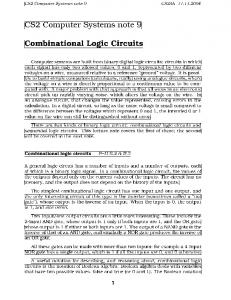
Fig. 3. Deciphering and modeling the CPG for locomotion in the lamprey. The top panels show (left) a schematic of the CPG network, with descending inputs mediated by basal ganglia, locomotor centers (MLR, DLR) and reticulospinal neurons (RS) impinging onto the spinal network, and (right) the output firing pattern of the computer simulation of that network. The lower panels show (left) two visual representations of the simulated firing patterns of all CPG neurons in a set of successive segments of the lamprey spinal cord, with color-coded excitability states, and (right) a bird’s-eye view of a swimming digital lamprey created by the neuromechanical simulation. From Grillner et al. (2007) Modeling a vertebrate motor system: pattern generation, steering and control of body orientation. Progress in Brain Research 165:221–234, and S. Grillner (personal communication).
the model to explain the basic operating principles of the CPG was a highlight of many a symposium proceedings on motor control. The realization that the human spinal cord must also contain a CPG for locomotion has prompted the development of training programs for spinal cord injury patients aimed at re-activating this network in ways that can help them walk again. With his strong background in motor control in mammals, Grillner sought to place his work on the lamprey in a richer functional context. The lamprey locomotor CPG now became a gold mine for elucidating the higher order control of movement. Grillner and coworkers focused increasingly on the descending systems involved in the initiation and modulation of the CPG, describing their transmitters and synaptic connections and their functional roles in postural control and locomotor steering (Brodin et al., 1988; Deliagina et al., 1992; Ullén et al., 1995). In
more recent years, Grillner has also begun investigating the coordination of locomotion with other motor programs such as eye movements (Saitoh et al., 2007). Moving ever higher in the motor hierarchy, Grillner eventually moved to the striatum—to attack the question of how motor programs such as locomotion are selected from the motor palette to generate goal-directed behavior (Grillner et al., 2005a). Grillner’s studies of motor control in the lamprey provide a guiding light that influences researchers studying the same questions in mammals including man (Gardella et al., 2008; Dietz et al., 2009). Ever pushing the envelope and making the fullest possible use of informatics, Grillner initiated another impressive project, to create a computer simulation of the musculoskeletal system of the lamprey that could be integrated with the neural network simulation. The re-
J. C. Glover / Neuroscience 163 (2009) 977–984
sulting “neuromechanical model” truly bridged the gap from ion channels to networks to behavior, generating cinematic representations of digital swimming lampreys that could be used to test the behavioral effects of sensory inputs and descending modulation (Grillner et al., 2007). As a natural extension of his pioneering use of informatics to model the neural basis of behavior, Grillner now holds a key position in formulating international strategy and policy in the field of neuroinformatics, and has been a prime mover in the development of an International Neuroinformatics Coordinating Facility based at the Karolinska Institute (Bjaalie et al., 2008). Throughout this remarkable march towards a detailed description of motor control in the lamprey, Grillner has constantly sought to extend the principles gleaned from the jawless fish to the context of motor control in mammals. His background makes him the preeminent expert in this endeavour. Recent advances include the production of digital models of walking in limbed vertebrates and important treatises on the organization and function of microcircuits in the CNS, from the lamprey spinal cord to the mammalian cerebral cortex (Grillner et al., 2005b). As a coordinator both of ideas and of colleagues, Grillner embodies one of his own published tenets, that of defining “unifying concepts, organizing principles and emerging tools” to advance the understanding of neuroscience (Silver et al., 2007). Also see interview with the first winners of the Kavli Prize in Neuroscience (Rakic et al., 2008).
REFERENCES Agalliu D, Takada S, Agalliu I, McMahon AP, Jessell TM (2009) Motor neurons with axial muscle projections specified by Wnt4/5 signaling. Neuron 61(5):708 –720. Arber S, Ladle DR, Lin JH, Frank E, Jessell TM (2000) ETS gene Er81 controls the formation of functional connections between group Ia sensory afferents and motor neurons. Cell 101(5):485– 498. Basler K, Edlund T, Jessell TM, Yamada T (1993) Control of cell pattern in the neural tube: regulation of cell differentiation by dorsalin-1, a novel TGF beta family member. Cell 73(4):687–702. Bjaalie JG, Grillner S, Usui S (2008) Neuroinformatics: databases, tools, and computational modeling for studying the nervous system. Neural Netw 21(8):1045–1046. Briscoe J, Ericson J (1999) The specification of neuronal identity by graded sonic hedgehog signaling. Sem Cell Dev Biol 10:353–362. Briscoe J, Pierani A, Jessell TM, Ericson J (2000) A homeodomain protein code specifies progenitor cell identity and neuronal fate in the ventral neural tube. Cell 101(4):435– 445. Brodin L, Grillner S, Dubuc R, Ohta Y, Kasicki S, Hökfelt T (1988) Reticulospinal neurons in lamprey: transmitters, synaptic interactions and their role during locomotion. Arch Ital Biol 126(4): 317–345. Chen CL, Broom DC, Liu Y, de Nooij JC, Li Z, Cen C, Samad OA, Jessell TM, Woolf CJ, Ma Q (2006) Runx1 determines nociceptive sensory neuron phenotype and is required for thermal and neuropathic pain. Neuron 49(3):365–377. Dasen JS, Jessell TM (2009) Hox networks and the origins of motor neuron diversity. Curr Top Dev Biol 88:169 –200. Dasen JS, Tice BC, Brenner-Morton S, Jessell TM (2005) A Hox regulatory network establishes motor neuron pool identity and target-muscle connectivity. Cell 123(3):477– 491. Deliagina TG, Orlovsky GN, Grillner S, Wallén P (1992) Vestibular control of swimming in lamprey. III. Activity of vestibular afferents:
983
convergence of vestibular inputs on reticulospinal neurons. Exp Brain Res 90(3):499 –507. Dietz V, Grillner S, Trepp A, Hubli M, Bolliger M (2009) Changes in spinal reflex and locomotor activity after a complete spinal cord injury: a common mechanism? Brain 132(Pt 8):2196 –2205. Dodd J, Morton SB, Karagogeos D, Yamamoto M, Jessell TM (1988) Spatial regulation of axonal glycoprotein expression on subsets of embryonic spinal neurons. Neuron 2:105–116. Dodd J, Solter D, Jessell TM (1984) Monoclonal antibodies against carbohydrate differentiation antigens identify subsets of primary sensory neurones. Nature 311(5985):469 – 472. Friese A, Kaltschmidt JA, Ladle DR, Sigrist M, Jessell TM, Arber S (2009) Gamma and alpha motor neurons distinguished by expression of transcription factor Err3. Proc Natl Acad Sci U S A 106(32):13588 –13593. Gardella E, Rubboli G, Francione S, Tassi L, Lo Russo G, Grillner S, Tassinari CA (2008) Seizure-related automatic locomotion triggered by intracerebral electrical stimulation. Epileptic Disord 10(4):247–252. Goulding M, Lanuza G, Sapir T, Narayan S (2002) The formation of sensorimotor circuits. Curr Opin Neurobiol 12:508 –515. Grillner S (1969) Supraspinal and segmental control of static and dynamic gamma-motoneurones in the cat. Acta Physiol Scand Suppl 327:1–34. Grillner S (1975) Locomotion in vertebrates: central mechanisms and reflex interaction. Physiol Rev 55(2):247–304. Grillner S, Buchanan JT, Lansner A (1988) Simulation of the segmental burst generating network for locomotion in lamprey. Neurosci Lett 89(1):31–35. Grillner S, Hellgren J, Ménard A, Saitoh K, Wikström MA (2005a) Mechanisms for selection of basic motor programs—roles for the striatum and pallidum. Trends Neurosci 28(7):364 –370. Grillner S, Hongo T, Lund S (1968) Reciprocal effects between two descending bulbospinal systems with monosynaptic connections to spinal motoneurones. Brain Res 10(3):477– 480. Grillner S, Hongo T, Lund S (1970) The vestibulospinal tract. Effects on alpha-motoneurones in the lumbosacral spinal cord in the cat. Exp Brain Res 10(1):94 –120. Grillner S, Kozlov A, Dario P, Stefanini C, Menciassi A, Lansner A, Hellgren Kotaleski J (2007) Modeling a vertebrate motor system: pattern generation, steering and control of body orientation. Prog Brain Res 165:221–234. Grillner S, Markram H, De Schutter E, Silberberg G, LeBeau FE (2005b) Microcircuits in action—from CPGs to neocortex. Trends Neurosci 28(10):525–533. Grillner S, Shik ML (1973) On the descending control of the lumbosacral spinal cord from the “mesencephalic locomotor region”. Acta Physiol Scand 87(3):320 –333. Grillner S, Wallén P (1980) Does the central pattern generation for locomotion in lamprey depend on glycine inhibition? Acta Physiol Scand 110(1):103–105. Grillner S, Wallén P (1985) Central pattern generators for locomotion, with special reference to vertebrates. Annu Rev Neurosci 8: 233–261. Grillner S, Wallén P, McClellan A, Sigvardt K, Williams T, Feldman J (1983) The neural generation of locomotion in the lamprey: an incomplete account. Symp Soc Exp Biol 37:285–303. Grillner S, Wallén P, Viana di Prisco G (1990) Cellular network underlying locomotion as revealed in a lower vertebrate model: transmitters, membrane properties, circuitry, and simulation. Cold Spring Harb Symp Quant Biol 55:779 –789. Jessell TM (2000) Neuronal specification in the spinal cord: inductive signals and transcriptional codes. Nat Rev Genet 1:20 –29. Jessell TM, Iversen LL, Cuello AC (1978) Capsaicin-induced depletion of substance P from primary sensory neurones. Brain Res 152(1):183–188. Jessell TM, Siegel RE, Fischbach GD (1979) Induction of acetylcholine receptors on cultured skeletal muscle by a factor extracted
984
J. C. Glover / Neuroscience 163 (2009) 977–984
from brain and spinal cord. Proc Natl Acad Sci U S A 76(10): 5397–5401. Kania A, Jessell TM (2003) Topographic motor projections in the limb imposed by LIM homeodomain protein regulation of ephrin-A: EphA interactions. Neuron 38(4):581–596. Letinic K, Sebastian R, Toomre D, Rakic P (2009) Exocyst is involved in polarized cell migration and cerebral cortical development. Proc Natl Acad Sci U S A 106(27):11342–11347. Liem KF Jr, Tremml G, Jessell TM (1997) A role for the roof plate and its resident TGFbeta-related proteins in neuronal patterning in the dorsal spinal cord. Cell 91(1):127–138. Liu JP, Laufer E, Jessell TM (2001) Assigning the positional identity of spinal motor neurons: rostrocaudal patterning of Hox-c expression by FGFs, Gdf11, and retinoids. Neuron 32(6):997–1012. Pecho-Vrieseling E, Sigrist M, Yoshida Y, Jessell TM, Arber S (2009) Specificity of sensory-motor connections encoded by Sema3ePlxnd1 recognition. Nature 459(7248):842– 846. Price SR, De Marco Garcia NV, Ranscht B, Jessell TM (2002) Regulation of motor neuron pool sorting by differential expression of type II cadherins. Cell 109(2):205–216. Rakic P (1971a) Guidance of neurons migrating to the fetal monkey neocortex. Brain Res 33(2):471– 476. Rakic P (1971b) Neuron-glia relationship during granule cell migration in developing cerebellar cortex. A Golgi and electronmicroscopic study in Macacus Rhesus. J Comp Neurol 141(3):283–312. Rakic P (1974) Neurons in rhesus monkey visual cortex: systematic relation between time of origin and eventual disposition. Science 183(123):425– 427. Rakic P (1981) Development of visual centers in the primate brain depends on binocular competition before birth. Science 214(4523):928–931. Rakic P (1988) Specification of cerebral cortical areas. Science 241(4862):170 –176. Rakic P (1995) A small step for the cell, a giant leap for mankind: a hypothesis of neocortical expansion during evolution. Trends Neurosci 18(9):383–388. Rakic P (2002) Neurogenesis in adult primate neocortex: an evaluation of the evidence. Nat Rev Neurosci 3(1):65–71. Rakic P (2003) Developmental and evolutionary adaptations of cortical radial glia. Cereb Cortex 13(6):541–549.
Rakic P (2009) Evolution of the neocortex: a perspective from developmental biology. Nat Rev Neurosci 10:724 –735. Rakic P, Ayoub AE, Breunig JJ, Dominguez MH (2009) Decision by division: making cortical maps. Trends Neurosci 32(5):291–301. Rakic P, Grillner S, Jessell T (2008) The Kavli prize winners. Nat Rev Neurosci 9(12):893– 897. Rakic P, Sidman RL (1973) Weaver mutant mouse cerebellum: defective neuronal migration secondary to abnormality of Bergmann glia. Proc Natl Acad Sci U S A 70(1):240 –244. Saitoh K, Ménard A, Grillner S (2007) Tectal control of locomotion, steering, and eye movements in lamprey. J Neurophysiol 97(4): 3093–3108. Sigvardt KA, Grillner S, Wallén P, Van Dongen PA (1985) Activation of NMDA receptors elicits fictive locomotion and bistable membrane properties in the lamprey spinal cord. Brain Res 336(2):390 – 395. Silver R, Boahen K, Grillner S, Kopell N, Olsen KL (2007) Neurotech for neuroscience: unifying concepts, organizing principles, and emerging tools. J Neurosci 27(44):11807–11819. Tessier-Lavigne M, Placzek M, Lumsden AG, Dodd J, Jessell TM (1988) Chemotropic guidance of developing axons in the mammalian central nervous system. Nature 336(6201):775–778. Torii M, Hashimoto-Torii K, Levitt P, Rakic P (2009) Integration of neuronal clones in the radial cortical columns by EphA and ephrin-A signalling. Nature 2009, Sep 16; [Epub ahead of print]. Ullén F, Deliagina T, Orlovsky G, Grillner S (1995) Spatial orientation in the lamprey. II. Visual influence on orientation during locomotion and in the attached state. J Exp Biol 198 (Pt 3):675– 681. Wallén P, Carlsson K, Liljeborg A, Grillner S (1988) Three-dimensional reconstruction of neurons in the lamprey spinal cord in wholemount, using a confocal laser scanning microscope. J Neurosci Methods 24(2):91–100. Wichterle H, Lieberam I, Porter JA, Jessell TM (2002) Directed differentiation of embryonic stem cells into motor neurons. Cell 110(3): 385–397. Yamada T, Placzek M, Tanaka H, Dodd J, Jessell TM (1991) Control of cell pattern in the developing nervous system: polarizing activity of the floor plate and notochord. Cell 64(3):635– 647.
(Accepted 24 July 2009)