Journal of General Virology (2003), 84, 1351–1366
Review
DOI 10.1099/vir.0.18922-0
Triple gene block: modular design of a multifunctional machine for plant virus movement Sergey Yu. Morozov and Andrey G. Solovyev
Correspondence Sergey Yu. Morozov
A. N. Belozersky Institute of Physico-Chemical Biology, Moscow State University, Moscow 119899, Russia
[email protected]
Many plant virus genera encode a ‘triple gene block’ (TGB), a specialized evolutionarily conserved gene module involved in the cell-to-cell and long-distance movement of viruses. The TGB-based transport system exploits the co-ordinated action of three polypeptides to deliver viral genomes to plasmodesmata and to accomplish virus entry into neighbouring cells. Although data obtained on both the TGB and well-studied single protein transport systems clearly demonstrate that plant viruses employ host cell pathways for intra- and intercellular trafficking of genomic nucleic acids and proteins, there is no integral picture of the details of molecular events during TGB-mediated virus movement. Undoubtedly, understanding the molecular basis of the concerted action of TGB-encoded proteins in transporting viral genomes from cell to cell should provide new insights into the general principles of movement protein function. This review describes the structure, phylogeny and expression of TGB proteins, their roles in virus cell-to-cell movement and potential influence on host antiviral defences.
INTRODUCTION Plant viruses require virus-encoded proteins to move from cell to cell via plasmodesmata (PD). The non-virion 30 kDa protein of Tobacco mosaic virus (TMV) was the first specific viral protein identified that could support intercellular plant virus spread (Leonard & Zaitlin, 1982; Ohno et al., 1983; Deom et al., 1987) and, therefore, was defined as a ‘transport protein’, or, currently, ‘movement protein’ (MP) (Hull, 1989; Atabekov & Taliansky, 1990). Functions that have been definitively or tentatively assigned to the 30 kDalike MPs (Melcher, 2000) include targeting of viral RNA to PD and increase in the effective PD pore size (SEL, size exclusion limit) to allow trafficking of the RNA or an RNA– MP complex (ribonucleoprotein complex, RNP) through the pore (Carrington et al., 1996; Lazarowitz & Beachy, 1999; Leisner, 1999; Lucas, 1999; Tzfira et al., 2000; Blackman & Overall, 2001; Haywood et al., 2002; Heinlein, 2002a). A number of positive-stranded RNA viruses have been found to lack gene products with similarity to the TMV MP (Mushegian & Koonin, 1993). Comparisons of genomic sequences in some such viruses revealed a strikingly similar element of three partially overlapping ORFs called the ‘triple gene block’ (TGB) (Bouzoubaa et al., 1986; Morozov et al., 1987, 1989; Forster et al., 1988; Huisman et al., 1988; Skryabin et al., 1988; Rupasov et al., 1989). TGB-encoded proteins are referred to as TGBp1, TGBp2 and TGBp3, according to the positions of their genes (Solovyev et al., Published ahead of print on 18 March 2003 as DOI 10.1099/ vir.0.18922-0
0001-8922 G 2003 SGM
Printed in Great Britain
1996). Further accumulation of plant virus genome sequence data revealed TGBs in the genera Potexvirus, Carlavirus, Allexivirus, Foveavirus, Hordeivirus, Benyvirus, Pomovirus and Pecluvirus (Fig. 1). While arrangement of the TGB cistrons relative to each other is well conserved, TGB positions in the genomes of viruses of different genera can vary considerably (Fig. 1) (Morozov et al., 1989; Morozov & Solovyev, 1999). Mutational analyses of infectious cDNA clones of virus genomes demonstrate that all three TGB proteins are essential for the virus movement process (Petty & Jackson, 1990; Petty et al., 1990; Beck et al., 1991; Gilmer et al., 1992; Herzog et al., 1998). Thus, movement functions carried on the single TMV MP are likely to be distributed over three proteins in TGB-containing viruses, a feature that makes such viruses an attractive model to investigate the movement process. Phylogeny and sequence comparisons of TGB proteins The TGB is found in only some viruses of the ‘alpha-like’ or ‘Sindbis-like’ supergroup (Fig. 1) (Koonin & Dolja, 1993; Mushegian & Koonin, 1993; Morozov & Solovyev, 1999), a feature that might reflect emergence of the TGB in virus(es) of this phylogenetic branch followed by co-adaptation between replication and movement genes. TGBp1 contains a NTPase/helicase sequence domain that is closely related to the replicative helicases of alpha-like viruses and belongs to helicases of superfamily I (SF-I) (Fig. 2) (Gorbalenya et al., 1989; Gorbalenya & Koonin, 1993; Koonin & Dolja, 1993). Of seven typical motifs in this 1351
S. Yu. Morozov and A. G. Solovyev
PVX (Potexvirus)
12K 25K
165K
8K 8K
ASPV (Foveavirus)
13K 25K
247K
7K
PVM (Carlavirus)
11K 25K
223K
13K
7K
ShVX (Allexivirus)
BNYVV (Benyvirus)
26K 11K
195K
RNA1 42K
13K
RNA2
14K
130K RNAa
14K
RNAb
17K
206K
142K RNA1
54K
RNA2
13K
RNA3 131K
RNA1
39K
17K
87K RNAc RNA
91K
PCV (Pecluvirus)
RNA4
RNA3
15K
58K
PMTV (Pomovirus)
15K
237K
75K
BSMV (Hordeivirus)
42K
8K
21K
cys
15K
191K 14K
51K
RNA2
17K
domain, motif I, with a characteristic GKS/T tripeptide, and motif II are responsible for binding ATP and Mg2+ and correspond to the ‘Walker A’ and ‘Walker B’ sites found in numerous ATP-binding proteins (Figs 2 and 3) (Gorbalenya & Koonin, 1993; Kadare & Haenni, 1997). Phylogenetic analysis of the NTPase/helicase sequences allows clustering of TGBp1 into two major groups, corresponding to filamentous viruses (genera Potexvirus, Carlavirus, Foveavirus and Allexivirus) and rod-shaped viruses (genera Hordeivirus, Benyvirus, Pomovirus and Pecluvirus). Furthermore, the molecular masses of TGBp1 in filamentous viruses range from 24 to 26 kDa and the NTPase/helicase domain
Potexvirus, Carlavirus, Allexivirus, Foveavirus 24 _ 26 kDa
I
IA
II
III IV
V
VI
Hordeivirus PSLV 63 kDa
I
IA
II
III
IV
V
VI
Pecluvirus PCV 51 kDa
I
IA
II
III
IV
V
VI
IV
V
VI
Benyvirus BNYVV 42 kDa
I
IA
II
III
Fig. 2. Molecular organization of TGBp1. The dark grey region indicates the position of the helicase sequence domain with the seven conserved motifs I–VI. Black boxes labelled ‘+’ indicate positively charged stretches in the N-terminal protein regions. PSLV, Poa semilatent virus; PCV, Peanut clump virus; BNYVV, Beet necrotic yellow vein virus. 1352
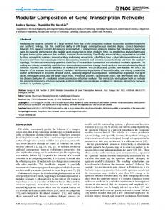
Fig. 1. TGB in viruses of different genera. Genes are shown as boxes and molecular masses of encoded proteins are indicated. TGB is shown in green. Genes of replicative proteins are shown in yellow and the locations of conserved protein sequence domains of methyltransferase (MT), protease (PRO), helicase (HEL) and polymerase (POL) are indicated. CP, coat protein gene; cys, genes of cysteine-rich proteins. Arrows depict weak terminator codons that can undergo readthrough. Readthrough domains of CPs are shown in dark blue. The third TGB gene of Shallot virus X (ShVX) lacks the initiator AUG codon and is indicated by an open box. PVX, Potato virus X (X05198); ASPV, Apple stem pitting virus (D21829); PVM, Potato virus M (X53062); ShVX, Shallot virus X (M97264); BNYVV, Beet necrotic yellow vein virus (X05147, X04197, M36894 and M36897); BSMV, Barley stripe mosaic virus (U35768, U35772 and U13918); PMTV, Potato mop-top virus (AJ238607, NC_003724 and AJ277556); PCV, Peanut clump virus (X78602 and L07269).
comprises the entire sequence, whereas TGBp1s of rodshaped viruses are substantially larger – from 39 to 63 kDa – and contain additional long N-terminal domains (Figs 1 and 2) (Solovyev et al., 1996; Wong et al., 1998; Erhardt et al., 1999b). Peculiar features of these extensions are (i) the presence of arginine/lysine-rich clusters, possibly involved in binding of nucleic acids, and (ii) a region of sequence similarity just upstream of the helicase domain (Figs 2 and 3) (Bleykasten et al., 1996; Solovyev et al., 1996). TGBp2 and TGBp3 contain hydrophobic sequences predicted to be involved in interaction of protein with membranes (Morozov et al., 1987, 1989). All TGBp2s contain two hydrophobic segments, with a conserved central region between them (Fig. 4a), which exhibit the highest degree of sequence conservation among the TGB proteins (Morozov et al., 1987; Skryabin et al., 1988; Solovyev et al., 1996). Unlike TGBp2, which shows almost uniform molecular organization in viruses of different genera, sequences of TGBp3 form two main groups. In filamentous viruses of the genera Potexvirus, Carlavirus, Foveavirus and Allexivirus, the 6–13 kDa TGBp3 contains one hydrophobic sequence at the N terminus followed by a conserved region with the characteristic signature CX5GX8C (Fig. 4a) (Morozov et al., 1991a). Another type of TGBp3 characteristic of rod-shaped viruses of the genera Hordeivirus, Pomovirus and Pecluvirus consists of 18–24 kDa proteins with two transmembrane segments, a conserved sequence in the N-terminal region containing invariant cysteine and histidine residues and the central conserved region with a typical tetrapeptide QDLN (Fig. 4a) (Solovyev et al., 1996; Koenig et al., 1998). Note that the conserved sequences in the TGBp3 hydrophilic regions of these two groups are not similar to one another. A third type of TGBp3 molecular organization is found in the genus Benyvirus, with two transmembrane segments but no significant sequence similarity with TGBp3 of the other rod-shaped viruses (Fig. 4a). Hence, a polyphyletic origin of TGBp3 can be proposed, whereas TGBp2, similarly to Journal of General Virology 84
Review: Triple gene block
Fig. 3. Amino acid alignments of the sequences located upstream of conserved motif I of the TGBp1 helicase domain in hordei-like TGB (a) and potex-like TGB (b). Asterisks show the position of the conserved GKS tripeptide. The conserved arginine residue involved in RNA binding (see text) is indicated by ‘#’. Numbers in parentheses indicate distances from the N terminus. BSMV, Barley stripe mosaic virus; PSLV, Poa semilatent virus; LRSV, Lychnis ringspot virus; PMTV, Potato mop-top virus; BSBV, Beet soil-borne virus; BBNV, Broad bean necrosis virus; BVQ, Beet virus Q; PCV, Peanut clump virus; BNYVV, Beet necrotic yellow vein virus; NVMV, Nicotiana velutina mosaic virus; PVX, Potato virus X; PlAMV, Plantago asiatica mosaic virus; WClMV, White clover mosaic virus; CYMV, Clover yellow mosaic virus; PMV, Papaya mosaic virus; NMV, Narcissus mosaic virus; PAMV, Potato aucuba mosaic virus; BMV, Bamboo mosaic virus; FMV, Foxtail mosaic virus; PVM, Potato virus M; PVS, Potato virus S; CVB, Chrysanthemum virus B; LSV, Lily symptomless virus; ShVX, Shallot virus X; GVA, Garlic virus A; GVB, Garlic virus B; ASPV, Apple stem pitting virus; CNRMV, Cherry necrotic rusty mottle virus.
TGBp1 (Koonin & Dolja, 1993), most probably originates from a common ancestor (Solovyev et al., 1996). Based upon rules for topology of integral membrane proteins (Sipos & Von Heijne, 1993; Gafvelin et al., 1997), hordeivirus TGBp2 and TGBp3 molecules are proposed to be integrated into the lipid bilayer in a U-like conformation with their N terminus exposed to the cytoplasm and endoplasmic reticulum (ER) lumen/extracellular space, respectively (Solovyev et al., 1996). Another protein topology can be predicted for the potexvirus TGBp3s (Fig. 4b). Thus, primary structure comparisons of the three TGB proteins allow us to distinguish two classes of TGBs: http://vir.sgmjournals.org
‘hordei-like’ (class 1) and ‘potex-like’ (class 2) (Figs 1, 2 and 4) (Solovyev et al., 1996; Erhardt et al., 1999b). The biological relevance of this classification is confirmed by the different role played by viral coat protein (CP) in cell-to-cell movement of the viruses with different types of TGB (Callaway et al., 2001). The CP is dispensable for cell-to-cell movement mediated by hordei-like TGBs (Petty & Jackson, 1990; Schmitt et al., 1992; Herzog et al., 1998; McGeachy & Barker, 2000). In contrast, the CP of potexviruses and, presumably, of other viruses with potex-like TGBs, is necessary for intercellular transport of the viral genome (Chapman et al., 1992; Forster et al., 1992; Sit & Abouhaidar, 1993) (see below). 1353
S. Yu. Morozov and A. G. Solovyev
(a)
TGBp2
12_14 kDa
TGBp3
18_24 kDa Hordeivirus, Pomovirus, Pecluvirus
15 kDa Benyvirus
6_13 kDa Potexvirus, Carlavirus, Allexivirus, Foveavirus
(b)
appropriate sizes have been found in infected plants, whereas TGBp3-specific sgRNA is not commonly detected (Guilford & Forster, 1986; Dolja et al., 1987; Gilmer et al., 1992; Zhou & Jackson, 1996). Detailed studies of TGB expression demonstrated that two sgRNAs are sufficient for translation of the three TGB proteins: the longer sgRNA serves as the template for translation of TGBp1, while the shorter sgRNA is the messenger for both TGBp2 and TGBp3 (Morozov et al., 1991b; Zhou & Jackson, 1996; Verchot et al., 1998; Agranovsky & Morozov, 1999). Expression of TGBp3 by leaky ribosome scanning through the TGBp2 gene was proposed (Skryabin et al., 1988; Morozov et al., 1989). This translation strategy maintains a low level of TGBp3 expression. For example, in vitro translation of a sgRNA transcript yields TGBp2 and TGBp3 in the ratio 10 : 1 (Zhou & Jackson, 1996). Furthermore, the expression level of TGBp3 in infected plants may be even lower, as suggested by the inability to detect TGBp3, while TGBp2 is detected easily (Niesbach-Klo¨sgen et al., 1990; Donald at al., 1993; Gorshkova et al., 2003). Functions of TGBp1 Biochemical activities of TGBp1 in vitro
cytoplasm
TGBp2 hordei-like
potex-like
TGBp3 hordei-like
potex-like
Fig. 4. Molecular organization of TGBp2 and TGBp3. (a) Conserved regions of the proteins. Molecular masses of proteins in different genera are shown. Dark grey boxes indicate hydrophobic transmembrane sequence segments. Characteristic signature sequences are shown. ‘???’ indicates lack of information on sequence conservation in TGBp3 of benyviruses. (b) Predicted topology of TGBp2 and TGBp3 molecules in the cell membrane. Transmembrane helixes are depicted as cylinders; ‘+’ and ‘2’ show the net charge of hydrophilic regions of the proteins. The short N-terminal hydrophilic segment of hordeilike TGBp2 in rod-shaped viruses has a positive net charge of 2 to 3, the corresponding region of potex-like TGBp2 in filamentous viruses is neutral or has a positive net charge of 1. Conversely, the C-terminal hydrophilic segment in filamentous viruses possesses a net charge of 2 to 5.
Expression of TGB proteins in virus-infected plants TGB proteins are expressed simultaneously at the early stages of infection (Niesbach-Klo¨sgen et al., 1990; Donald at al., 1993), as are MPs of other viruses, such as TMV (Lehto et al., 1990). Similarly to other Sindbis-like viruses, expression of 59-distal genes in TGB-containing viruses occurs via subgenomic RNAs (sgRNAs) that are 39 coterminal with genomic RNAs (Buck, 1996; Agranovsky & Morozov, 1999). For TGBp1 and TGBp2, sgRNAs of the 1354
Cell-to-cell movement of plant viruses was postulated to involve specific non-virion transport RNPs (Atabekov & Dorokhov, 1984; Citovsky & Zambryski, 1993), and further experiments have demonstrated that all tested MPs of the ‘30K superfamily’ are nucleic acid-binding proteins (Carrington et al., 1996; Ghoshroy et al., 1997; Tzfira et al., 2000). The TGBp1 proteins, similarly to 30K superfamily MPs, can bind ssRNA non-specifically in a co-operative manner and have affinity for ssDNA as well (Rouleau et al., 1994; Bleykasten et al., 1996; Kalinina et al., 2001; Donald et al., 1997). For the potex-like TGBp1s, stability of in vitro co-operative binding to RNA is lower than for the 30K superfamily MPs (Rouleau et al., 1994; Kalinina et al., 1996, 1998; Lough et al., 1998; Wung et al., 1999). The RNAbinding site of potexviral TGBp1 has been mapped to the N-terminal protein region containing positively charged residues essential for interaction with RNA. One of these residues is an arginine, conserved in all potex-like TGBp1, 17–19 aa upstream of the GKS/T tripeptide (Fig. 3) (Morozov et al., 1999; Wung et al., 1999). Multiple RNA-binding sites are found in hordeiviral TGBp1 (Donald et al., 1995, 1997). The isolated C-terminal helicase domain of a hordeiviral TGBp1 shows co-operative RNA binding similar to that of potex-like TGBp1, while the N-terminal extension domain demonstrates strong nonco-operative RNA binding, so that the whole protein exhibits both types of binding (Kalinina et al., 2001). Similarly, other hordei-like TGBp1s are capable of strong, salt-resistant RNA binding (Bleykasten et al., 1996; Donald et al., 1997; Cowan et al., 2002). In hordeiviral TGBp1s, the two short arginine/lysine-rich regions are essential for N-terminal extension domain-specific RNA binding (Solovyev et al., 1996; Kalinina et al., 2001). Likewise, RNA Journal of General Virology 84
Review: Triple gene block
binding of a benyvirus TGBp1 is specified by an arginine/ lysine-rich region positioned 6–18 residues from the N terminus (Fig. 2) (Bleykasten et al., 1996).
(a)
Both potex- and hordei-like TGBp1s have RNA helicase activity in vitro (Kalinina et al., 2002). Importantly, the hordeiviral TGBp1 helicase unwinds the duplex in both the 59R39 and the 39R59 directions, with respect to the chain used for entry, and is unable to unwind DNA duplexes (Kalinina et al., 2002). In contrast, superfamily I (SF-I) DNA helicases and RNA virus SF-II helicases operate in the 39R 59 direction only (Gorbalenya & Koonin, 1993; Kadare & Haenni, 1997). Generally, the duplex unwinding activity of helicases depends on the hydrolysis of NTPs, preferentially ATP (Gorbalenya et al., 1989; Gorbalenya & Koonin, 1993; Kadare & Haenni, 1997; Soultanas & Wigley, 2001; Caruthers & McKay, 2002). Accordingly, the RNA helicase activity of TGBp1 requires ATP and Mg2+ (Kalinina et al., 2002), and NTP-binding and Mg2+-dependent NTPase activities have been detected for TGBp1 in vitro (Rouleau et al., 1994; Bleykasten et al., 1996; Kalinina et al., 1996; Donald et al., 1997; Morozov et al., 1999; Solovyev et al., 1999; Liou et al., 2000). Finally, many helicases can form homodimers or oligomers (Gorbalenya & Koonin, 1993) and both hordei- and potex-like TGBp1 are capable of self-interactions (Cowan et al., 2002; our unpublished data).
PcrA
Four structural domains have been identified in SF-I DNA helicases. The N-terminal domain 1A includes helicase motifs I–III, while the C-terminal domain 2A carries motifs IV–VI. The sequences of domains 1A and 2A are interrupted by the inserted domains 1B and 2B (Fig. 5) (Soultanas & Wigley, 2001; Caruthers & McKay, 2002). Alignment with the sequence of PcrA, a bacterial SF-I DNA helicase with known three-dimensional structure, demonstrates that TGBp1 shows (i) conservation of the helicase motifs in domains 1A and 2A and (ii) the absence of domains 1B and 2B, which are precisely ‘deleted’ from the TGBp1 sequences (Fig. 5) (Kalinina et al., 2002). Importantly, deletion of the 2B domain introduced in a bacterial SF-I DNA helicase (Rep protein) had no effect on helicase activity in vitro and in vivo (Cheng et al., 2002). Hence, TGBp1 represents a naturally ‘simplified’ version of a SF-I helicase with just two structural domains. Interestingly, a similar structure has been described for the cellular eIF-4A helicase, the prototype member of the ‘DEAD’ SF-II family of RNA helicases, which shares with TGBp1 two other features exceptional among RNA helicases, namely the ability to discriminate between RNA and DNA and to operate in both directions (Gorbalenya & Koonin, 1993; Kadare & Haenni, 1997; Caruthers & McKay, 2002; Du et al., 2002; Kalinina et al., 2002). Activities of TGBp1 in vivo
RNA-binding activity of TGBp1 is thought to be responsible for formation of movement-competent genomic RNPs; such structures composed of viral RNA and TGBp1 were isolated from hordeivirus-infected plants (Brakke et al., http://vir.sgmjournals.org
V VI
II III IV
I IA
TGBp1 I IA II III IV
V VI
(b)
IV
I
III
II IA
1A
2A
VI V
Fig. 5. Subdomain structure of TGBp1 helicases. (a) Comparison of TGBp1 helicase and PcrA, a bacterial DNA helicase. Conserved sequence motifs I–VI are indicated. Domains 1A and 2A are shown in red and yellow, respectively. Domains 1B and 2B, absent from TGBp1, are shown in grey. (b) Putative secondary structure of the helicase domain of TGBp1. In the modified model of the secondary structure of PcrA (Caruthers & McKay, 2002), sequence elements represented by parallel a–b structures and conserved in domains 1A and 2A of TGBp1 are shown in colour. Arrows point to the positions of conserved sequence motifs I–VI. Domains 1B and 2B, absent from TGBp1, are shown by grey circles.
1988). Although the nature of movement-related RNPs in potexviruses remains obscure, they also contain TGBp1, which has been suggested to either interact with non-virion complexes that also contain CP (Lough et al., 1998, 2000) or to bind to and modify virions in a manner that allows them to transport to and through PD (Fig. 6) (Santa Cruz et al., 1998; Atabekov et al., 2000). The role of the other TGBp1 activities (NTP binding, NTPase and RNA helicase) in cell-to-cell movement remains unclear. According to recent views, the cell-tocell movement of viral genomes is an energy-dependent process (Carrington et al., 1996; Ghoshroy et al., 1997). There are at least two steps of MP-mediated translocation of nucleic acids where such ATP/NTP-dependent events may be involved: (i) intracellular transport of MP and virusspecific RNP to PD or to a region in the vicinity of PD and (ii) trafficking of proteins and RNP through PD involving both protein/RNA unfolding and microchannel dilation (Fig. 6) (Ghoshroy et al., 1997; Lazarowitz & Beachy, 1999; Lucas, 1999; Kragler et al., 1998; Tzfira et al., 2000; Haywood et al., 2002; Heinlein, 2002b; Roberts & Oparka, 2003). No ability to modify PD and move cell to cell has been 1355
S. Yu. Morozov and A. G. Solovyev
translation on free polysomes
binding to genomic RNA
binding to genomic RNA
translation on ER-bound polysomes
ER to Golgi cycling of TGBp2
budding of TGB-specific containers RNP binding to membranous movement-related compartments
travel along cytoskeleton
increase of PD permeability
Fig. 6. General scheme of TGB-mediated cell-to-cell movement and formation of transport RNP complexes. Processes specific for potex-like and hordei-like TGBs are shown in pink and green, respectively. Transport steps common for both potex- and hordei-like TGBs are shown in blue. Processes that are not involved directly in virus cell-to-cell movement are shown in grey. Cell-to-cell movement initiates with the synthesis of virus genomic and subgenomic RNAs in the replication complexes located on membranes of the ER or other organelles (Plante et al., 2000; Dunoyer et al., 2002b; Schwartz et al., 2002). TGBp1 is synthesized on free polysomes. Most probably, the hydrophobic TGBp2 and TGBp3 MPs enter the ER cotranslationally, and the hydrophobic regions of TGBp2 and TGBp3 migrate into the ER membrane (Vitale & Denecke, 1999). TGBp2 and TGBp3 travel to their destinations in specific membrane containers (Jiang & Rogers, 1998; Mitsuhashi et al., 2001; Stephens & Pepperkok, 2001; Nebenfu¨hr, 2002; Zamyatnin et al., 2002). Trafficking to the cell periphery may exploit the cytoskeleton-based pathway (Ploubidou & Way, 2001; von Bargen et al., 2001; Heinlein, 2002a). TGBp2/p3-specific membrane containers may bind movement-competent virions (or RNPs) containing TGBp1, either through protein–protein interactions or via direct interaction of TGBp2 and RNA (Solovyev et al., 2000; Cowan et al., 2002; Zamyatnin et al., 2002). These complexes are delivered to the neck region of PD and fuse to cortical ER (Solovyev et al., 2000; Zamyatnin et al., 2002; Lee et al., 2003). TGBp1 reaches the orifice of a PD microchannel and binds to receptor(s) involved in SEL increase (Lough et al., 1998; Lucas, 1999; Lee et al., 2003; Roberts & Oparka, 2003). TGBp1 and viral RNA are unfolded and then translocated through the PD microchannels until reaching the neighbouring cell (Kragler et al., 1998; Lucas, 1999). For further details, see text.
reported for individually expressed hordei-like TGBp1, suggesting that this protein is incapable of intracellular trafficking to PD by itself and depends on TGBp2 and TGBp3 for this function (discussed below). Conversely, potexviral TGBp1 is capable of interacting with PD and increasing PD SEL (Angell et al., 1996; Lough et al., 1998, 2000; Malcuit et al., 1999; Morozov et al., 1999; Yang et al., 1356
2000). Mutations influencing the potexviral TGBp1 helicase motif I (disabling both helicase and NTPase activities of the protein) block the protein’s ability to increase PD SEL, whereas mutations of motif VI (disabling the helicase but not NTPase activity) (Kalinina et al., 2002) affect neither the increase in TGBp1-induced SEL nor TGBp1 transport to the cell periphery (Lough et al., 1998; Morozov et al., 1999). Journal of General Virology 84
Review: Triple gene block
Thus, ATP binding and/or hydrolysis rather than helicase activity per se are involved in PD dilation by potex-like TGBp1.
required to support both cell-to-cell and long-distance movement in hordeiviruses (Donald et al., 1995, 1997; Solovyev et al., 1999).
Potexviral TGBp1 is co-translocated with the CP and virus genomic RNA during virus movement through PD (Morozov et al., 1997; Lough et al., 1998, 2000, 2001). It is natural to propose that TGBp1 helicases couple RNA unwinding and translocation through PD microchannels. The discovery of a specialized RNA-translocating NTPase (P4 protein) that participates in RNA transfer and packaging into bacteriophage w6 virions (Juuti et al., 1998) further supports such a proposal.
In contrast to hordeiviruses, a functional CP is required for potexvirus long-distance movement (Santa Cruz et al., 1998). Potexviral TGBp1 is co-transported along the phloem sieve tube together with virions (or a non-virion CP-containing RNP). Because the sieve element contains no translational apparatus, this complex with TGBp1 must include all functional activities required for exiting from the sieve tube to the companion cells (Santa Cruz et al., 1998; Lough et al., 2001).
TGBp1 of Potato virus X (PVX) has been reported to interact with one end of the virion and to induce energy-dependent conformational changes of virus particles in vitro (Atabekov et al., 2000). Soultanas & Wigley (2001) have suggested that energy generated by ATP hydrolysis can be used by helicases not only for separation of base-paired regions but also for displacement of other proteins from nucleic acids. Thus, by analogy with some SF-II cell and viral helicases (‘RNPases’) involved in disassembly and remodelling of RNP complexes (Tseng et al., 1998; Jankowsky et al., 2001; Schwer, 2001), TGBp1 could potentiate cell-to-cell transport of virions or movement-related RNPs through PD by disrupting both RNA–protein interactions and intramolecular RNA basepairing. In this case, in addition to trafficking of viral genomes through PD, hordei-like TGBp1 may mediate a displacement of cell proteins prior to genomic RNA transport to PD. During viral genome replication, nascent RNA molecules are most likely packaged into RNPs by host cytosolic proteins which rapidly coat newly synthesized cellular and virus-specific mRNAs and facilitate their efficient translation and further turnover (Mitchell & Tollervey, 2001; Pilipenko et al., 2001; Fedoroff, 2002). Re-packaging of such RNPs by replacement of cell proteins with hordei-like TGBp1 may result in formation of movement-competent non-translatable RNPs (Karpova et al., 1999).
The ability of the virus to establish systemic infection in plants depends largely on the efficacy of plant defence response against infection versus the potential of the virus to escape or counter this defence (Carrington et al., 2001; Dangl & Jones, 2001; Vance & Vaucheret, 2001). One of the defence mechanisms in plants is gene silencing, which is mediated by sequence-specific degradation of viral RNAs in the cytoplasm (Baulcombe, 2002; Voinnet, 2001; Waterhouse et al., 2001). However, TGB-containing viruses, like many plant viruses, have evolved special mechanisms to suppress RNA silencing (Voinnet et al., 2000; Dunoyer et al., 2002a; Yelina et al., 2002). In particular, potexviral TGBp1 has been shown to suppress production or activity of the mobile silencing signal (Voinnet et al., 2000). Importantly, some of the sequences of TGBp1 involved in suppressing the silencing activity do not affect cell-to-cell movement (D. Baulcombe, The Sainsbury Laboratory, John Innes Centre, Norwich, UK, personal communication).
TGBp1 as a factor of whole plant infection
CPs are dispensable for systemic infection of hordeiviruses and pomoviruses, which are thus believed to enter the phloem and traffick along the sieve tubes as a non-virion RNP containing genomic RNA and TGBp1 (Brakke et al., 1988; Petty & Jackson, 1990; Donald et al., 1997; McGeachy & Barker, 2000; Lawrence & Jackson, 2001b). In hordeiviral TGBp1, two positively charged motifs responsible for RNAbinding activity of the N-terminal extension domain (Fig. 2) have been found to be dispensable for virus transport from cell to cell but, nevertheless, necessary for long-distance virus movement. Therefore, the RNA-binding activities of the helicase and extension domains of hordeilike TGBp1 could be specialized in either cell-to-cell or longdistance transport, respectively (Kalinina et al., 2001). However, the presence of the N-terminal extension in TGBp1 and its compatibility with the helicase domain are http://vir.sgmjournals.org
Another virus resistance mechanism is mediated by a large family of R gene-encoded proteins that recognize pathogenencoded elicitors and trigger defence pathways, such as programmed cell death or hypersensitive response (Dangl & Jones, 2001; Holt et al., 2003). PVX TGBp1 has been shown recently to be such an elicitor recognized by the Nb genemediated resistance system in potatos. The TGBp1 region required for activation of the Nb response is located in the N terminus upstream of the helicase motif I (Malcuit et al., 1999). Functions of TGBp2 and TGBp3 Subcellular distribution of TGBp2 and TGBp3 and virus movement
In agreement with sequence analysis (Fig. 4) and in vitro studies predicting that TGBp2 and TGBp3 are integral membrane proteins (Morozov et al., 1987, 1990, 1991a), cell fractionation of plant tissues expressing these proteins demonstrates predominant association of both proteins with the P1 and P30 membranous fractions as well as with the cell wall (CW) fraction (Niesbach-Klo¨sgen et al., 1990; Donald et al., 1993; Hefferon et al., 1997; Cowan et al., 2002; Gorshkova et al., 2003). Further studies of subcellular localization of TGBp2 and 1357
S. Yu. Morozov and A. G. Solovyev
TGBp3 employed their GFP fusions expressed in plant cells by a variety of techniques. Note that experiments on transient expression of MPs should be interpreted cautiously, since MPs expressed from vectors are likely produced in much larger quantities and in a non-regulated fashion compared to a virus infection. Also, methods of delivery of expression vectors such as high-pressure biolistic bombardment may perturb cell status (Crawford & Zambryski, 2001) and functional properties of proteins may be hindered by the fused fluorescent protein sequences (Thomas & Maule, 2000; Brandizzi et al., 2002a). Nevertheless, transient expression of GFP fusions is widely used to study MPs in live cells and the results of such studies do not usually contradict data obtained by other methods (Lazarowitz, 1999; Lazarowitz & Beachy, 1999; Brandizzi et al., 2002a; Heinlein, 2002a). When transiently expressed in individual epidermal cells of Nicotiana benthamiana, GFP-tagged TGBp2s of Poa semilatent virus (PSLV) and Potato mop-top virus (PMTV) are localized to elements of the cell endomembrane system, mainly tubules of the cortical ER network (Solovyev et al., 2000; Cowan et al., 2002; Zamyatnin et al., 2003). In cells with higher levels of PSLV TGBp2 expression, a part of the protein is also associated with motile vesicles (Solovyev et al., 2000) that resemble plant Golgi stacks (Brandizzi et al., 2002b). The Golgi-like mobile vesicles have been found to contain most of the transiently expressed TGBp2 of PVX (our unpublished data). Subcellular localization of TGBp2 to the ER and Golgi is determined by the hydrophobic protein segments and, in particular, the length of the C-terminal hydrophobic segment (Solovyev et al., 2000; Zamyatnin et al., 2002; our unpublished data), which seems to act as a retrieval signal in Golgi-to-ER recycling by the host receptor Rer1 (Sato et al., 1999, 2001). Transiently expressed individual GFP–TGBp3 is found in membrane bodies of different sizes located at the cell periphery in close association with the CW (Solovyev et al., 2000; Cowan et al., 2002). TGBp3 expression has little effect on the basic structure of the ER in plant cells. However, TGBp3 synthesis results in the formation of new TGBp3containing ER structures (peripheral bodies) connected with the cortical ER network (Zamyatnin et al., 2002; Gorshkova et al., 2003). The size of these bodies correlates with the amount of TGBp3 protein produced in a given cell (Zamyatnin et al., 2002). Thus, TGBp3 is able to induce formation (or proliferation) of a specific subdomain of the cortical ER. A clue to the subcellular location of the TGBp3-containing bodies comes from fluorescent microscopy of leaves infected with a PMTV GFP–TGBp3-expressing virus vector (Cowan et al., 2002) and transgenic plants expressing PSLV GFP– TGBp3 (Gorshkova et al., 2003). When the protein is expressed in adjacent cells, the peripheral bodies formed in the two neighbouring cells are opposite to each other. The structural link that governs the formation of such twin bodies could be provided by PD (Cowan et al., 2002) and 1358
specific staining of PD-associated callose confirms the localization of TGBp3-containing bodies alongside of PD (Gorshkova et al., 2003). Targeting of PSLV TGBp3 depends on a specific signal consisting of two parts, of which a central hydrophilic region conserved in all hordei-like TGBp3 (Fig. 4a) seems to be an oligomerization sequence (Cowan et al., 2002; Gorshkova et al., 2003; our unpublished data). Another part of the specific PD targeting signal of TGBp3 is located in the C-terminal transmembrane segment, which resembles a hydrophobic membrane-embedded segment that participates in forming a protein trafficking signal of mastrevirus MP (Kotlizky et al., 2000). Similarly, localization of PVX TGBp3 to peripheral bodies depends on the only protein transmembrane segment (our unpublished data). Mutations in the hordeivirus TGBp3 signal result in localization of the protein in a ‘granular network’ of tiny bodies visible as a reticulate pattern as if they are formed on the surface of cortical ER tubules (Solovyev et al., 2000; our unpublished data). Thus, it appears that mutations in either part of the bipartite signal permit protein segregation to ER-exit sites but cannot mediate further protein trafficking to the final destination at PD-associated compartments (Fig. 6). Co-targeting of TGBp2 and TGBp3
In the presence of TGBp3, TGBp2 is re-targeted to peripheral bodies that resemble the structures observed in cells expressing TGBp3 (Solovyev et al., 2000). Perfect colocalization of co-expressed PSLV TGBp2 and TGBp3 in peripheral bodies has been demonstrated, confirming that the TGBp3 protein directs subcellular targeting of TGBp2 from the ER network to sites of TGBp3 location (Zamyatnin et al., 2002). PVX TGBp3 also targets PVX TGBp2 to peripheral bodies, showing that TGBp3directed trafficking of TGBp2 occurs in both hordei- and potex-like TGBs (Solovyev et al., 2000). Protein–protein interactions that result in the formation of TGBp2–TGBp3 complexes could be the mechanism by which the two proteins are co-targeted to peripheral bodies. However, co-expression of TGBp2 and TGBp3 mutants failed to identify regions potentially responsible for the interaction between TGBp2 and TGBp3 molecules (Solovyev et al., 2000). Further evidence for a sequenceindependent co-targeting mechanism was obtained in experiments on co-expression of heterologous TGB proteins. Indeed, in spite of the absence of sequence similarity of TGBp3 proteins in hordei- and potex-like TGBs, PVX TGBp3 can target PSLV TGBp2 to peripheral bodies and PSLV TGBp3 can, likewise, target PVX TGBp2 (Solovyev et al., 2000). Moreover, PSLV TGBp3 can also target totally unrelated membrane-bound MPs, such as the C4 protein of Faba bean necrotic yellows virus (genus Nanovirus) and the 6K protein of Beet yellows virus (genus Closterovirus), to peripheral bodies (Zamyatnin et al., 2002). This suggests that a sequence-specific interaction of TGBp2 and TGBp3 Journal of General Virology 84
Review: Triple gene block
molecules is unlikely to be involved in TGBp3-directed targeting of TGBp2 (Solovyev et al., 2000; Zamyatnin et al., 2002). Nevertheless, PMTV TGBp3 interacts physically with the homologous TGBp2 in a yeast two-hybrid system (Cowan et al., 2002). Presumably, this interaction may depend on the residue composition of hydrophobic segments, enabling side chain interaction between membraneembedded helices of proteins (Scholze et al., 2002; Sjo¨berg & Garoff, 2003). Note that TGBp3 apparently does not traffick any integral membrane protein, since GFP derivatives statically retained in ER membranes by synthetic hydrophobic anchors are not targeted by TGBp3 (Zamyatnin et al., 2002). Thus, it appears that some functional feature(s), rather then a specific sequence, is responsible for efficient trafficking of membrane proteins by TGBp3. Such features could include specific localization and dynamics of the membrane proteins in the cell endomembrane system, including their ability to cycle between the ER and the Golgi (our unpublished data). As noted above, an intermediate step of translocation of TGBp3 to PD involves its segregation in hypothetical ‘TGBp3 islands’ in ER membranes (ER-exit sites) (Fig. 6). These protein islands, which also include TGBp2, can be translocated using the targeting signal of TGBp3 to a specific receptor near PD in specific membrane containers (vesicles or tubules) (Stephens & Pepperkok, 2001; Nebenfu¨hr, 2002) delivered to the neck region of PD and fused there to cortical ER tubules (Fig. 6) (Solovyev et al., 2000; Cowan et al., 2002; Zamyatnin et al., 2002; Gorshkova et al., 2003).
Targeting of TGBp1 by TGBp2/TGBp3
Unlike potexviral TGBp1, which is capable of moving intracellularly to a peripheral layer of cytoplasm and PD (Lough et al., 1998; Malcuit et al., 1999; Morozov et al., 1999; Yang et al., 2000), hordei-like TGBp1 expressed individually is not targeted to specific sites at the cell periphery. However, when TGBp1 is expressed in the presence of other virus products, it localizes to the punctate structures at the CW (Erhardt et al., 1999b, 2000; Lawrence & Jackson, 2001a). At higher magnification, these structures are visible as pairs of disconnected bodies on opposite sides of the CW, closely resembling the structures formed by GFP–TGBp3 in close vicinity to PD (see above). Accordingly, GFP–TGBp1 punctate bodies co-localized with callose (Erhardt et al., 2000), confirming the immuno-gold detection of TGBp1 in PD of infected leaves (Erhardt et al., 1999b). Experiments with chimeric virus genomes suggested TGBp2 and TGBp3 as the most probable components responsible for this localization. Indeed, a combination of TGBp1 with homologous TGBp2/TGBp3 was required for TGBp1 function, particularly trafficking to PD (Lauber et al., 1998; Lough et al., 1998, 2000; Erhardt et al., 1999a, 2000; Solovyev et al., 1999; Lawrence & Jackson, 2001a; Zamyatnin et al., 2003). Hence, the role of TGBp2/TGBp3 may be primarily a matter http://vir.sgmjournals.org
of intracellular delivery of TGBp1-formed transportcompetent RNPs to PD (Fig. 6). There are indications that TGBp1 is actively, rather than passively, transported by TGBp2/TGBp3 to PD and that this process requires enzymatic activities of the protein. Mutations in the conserved sequence motifs in the NTPase/ helicase domains of hordei-like TGBp1 not only blocked cell-to-cell movement of the virus but also abolished protein targeting to PD in the presence of TGBp2 and TGBp3 (Erhardt et al., 2000; Lawrence & Jackson, 2001a; Zamyatnin et al., 2003).
TGBp2-induced increase in PD permeability and other putative movement-related activities of TGBp2/p3 proteins
Some of the point and insertion mutants of TGBp2 are dominant–negative, i.e. they inhibit cell-to-cell movement and diminish virus accumulation (Beck et al., 1994; Seppanen et al., 1997; Lauber et al., 2001). The recently discovered ability of potex- and hordei-like TGBp2 to facilitate movement of GFP between adjacent epidermal cells (Tamai & Meshi, 2001; our unpublished data) suggests that co-expression of a non-functional TGBp2 mutant during infection can interfere not only with viral RNP trafficking to PD but also with some additional movementrelated function(s). The molecular nature of the TGBp2-directed increase in PD permeability is enigmatic. However, a relationship between this phenomenon and modifications of the tissue stressresponse system can be proposed. In particle bombardment studies, the ability of GFP, which is a 27 kDa protein, to spread from an initially transfected epidermal cell of source N. benthamiana leaves to neighbouring cells depends on experimental conditions. When the leaves of intact plants are bombarded, GFP spreads over multiple cell boundaries to give a focus of more than 30 fluorescent cells. However, GFP is confined mostly to single cells after bombardment of detached leaves in a vacuum chamber (Oparka et al., 1999; Crawford & Zambryski, 2000, 2001; Itaya et al., 2000; Krishnamurthy et al., 2002). Under the latter conditions, TGBp2 potentiates the spread of GFP to adjacent epidermal cells (Tamai & Meshi, 2001). Various stress factors, including leaf detachment, are known to reduce PD SEL due to rapid callose deposition (Sivaguru et al., 2000; Crawford & Zambryski, 2001; Radford & White, 2001; Roberts & Oparka, 2003). Hence, it is possible that TGBp2 expression is not involved directly in increasing PD permeability but rather significantly decreases callose deposition in the CW and can thus inhibit or reverse the stress-induced decrease in PD SEL (Fig. 6). In line with this hypothesis, potexvirus TGBp2 has been shown recently to interact with TIP, a host protein regulator of b-1,3glucanase, which is a key enzyme of callose turnover (Fridborg et al., 2003). Thus, keeping the PD neck region open by callose degradation (or prevention of callose 1359
S. Yu. Morozov and A. G. Solovyev
accumulation) is a possible function of TGBp2 at the early stage of infection (Fridborg et al., 2003). In this context, it is interesting that co-expression of TGBp3 and TGBp2 completely blocks the TGBp2-induced ‘increase’ of PD SEL (our unpublished data). Probably, trapping of TGBp2 in the peripheral membrane bodies formed by TGBp3 in the vicinity of PD (see above) can either prevent interaction between TGBp2 and TIP or directly block intracellular trafficking of TIP.
proteins and nucleic acids within plants. A growing number of plant cell proteins [‘non-cell-autonomously acting plant proteins’ (NCAPs)] has been demonstrated to have properties of plant virus MPs, such as the ability to increase PD SEL and traffic between cells. Moreover, some of these proteins are able to transport RNA through PD (Lucas, 1999; Crawford & Zambryski, 2000; Tzfira et al., 2000; Blackman & Overall, 2001; Lucas et al., 2001; Haywood et al., 2002; Heinlein, 2002b; Lee et al., 2003; Roberts & Oparka, 2003).
Apart from trafficking of TGBp1 and genomic RNA and PD SEL control, small TGB proteins could be involved directly in regulating a hypersensitive response (BleykastenGrosshans et al., 1997; Solovyev et al., 1999; Lauber et al., 2001; Kobayashi et al., 2001). We believe that this activity of TGBp2/p3 could also be related to the regulation of callose turnover in view of the fact that (i) limitation of PVX spread in a hypersensitive response is accompanied by heavy callose deposits in the vicinity of PD (Allison & Shalla, 1974) and (ii) callose deposition may regulate virus movement in hypersensitive hosts by affecting the PD SEL (Iglesias & Meins, 2000; Bucher et al., 2001; Crawford & Zambryski, 2001; Radford & White, 2001).
Importantly, TMV MP, similar to NCAP CmPP16, is not capable of PD modification and trafficking between cells in the absence of an assisting cell protein, NCAPP1 (Lee et al., 2003). The dependence of TMV MP on NCAPP1 emphasizes the idea that viral MPs act in concert with a number of as yet undiscovered cell proteins required to accomplish intra- and intercellular steps in cell-to-cell movement. Thus, analysis of dissimilar virus transport systems that comprise several MPs may suggest possible roles of viral proteins that mimic components of host intracellular trafficking machinery. TGBp1s share some features with NCAPs involved in plant development. First, ectopic expression of potexviral TGBp1 appears to cause defects in the cell-to-cell communications that control lateral organ development (Foster et al., 2002). Second, TGBp1 competes directly for intercellular
CONCLUSION There is increasing evidence that viruses exploit endogenous intra- and intercellular trafficking pathways for spread of
PVX (Potexvirus)
CarMV (Carmovirus)
TGBp2
165K
CP
77K
30K
p9
TGBp1 TGBp3 p7
CP
PVY (Potyvirus) P1
HC-Pro
P3 6K1
BYV (Closterovirus)
CI
6K2
NIb
NIa
1b
CP
dCP
65K
20K
PRO 1a
RNA-binding protein
6K
ATPase
membrane protein
64K
coat protein
CP
21K
other movement-related activities
Fig. 7. Comparison of multicomponent cell-to-cell transport systems encoded by plant viruses. Genes are shown as boxes with names of encoded proteins. Genes of proteins involved in cell-to-cell movement are shown in colour. PRO, proteinase domain; PVX, Potato virus X (X05198); CarMV, Carnation mottle virus (X02986); PVY, Potato virus Y (M95491); BYV, Beet yellows virus (X73476). Potyviral proteins implicated in virus spread in addition to CI protein (see text) are the genome-linked proteins (VPg), HCpro and CP (Callaway et al., 2001; Rajamaki & Valkonen, 1999; Revers et al., 1999; Rojas et al., 1997; Saenz et al., 2002). BYV proteins involved in cell-to-cell movement in addition to Hsp70h (see text) include CP and its distant homologue (dCP), the minor CP 64K, the papain-like leader proteinase responsible for processing replicative protein precursors and the 6K small hydrophobic protein resembling potex-like TGBp3 (Alzhanova et al., 2000; Peng et al., 2001, 2002, 2003; Napuli et al., 2003; Dolja, 2003). 1360
Journal of General Virology 84
Review: Triple gene block
trafficking pathways with NCAP Knotted-1 (Lough et al., 2000). Third, the ability of NCAP CmPP16 to increase PD SEL and traffic through PD depends on an intracellular trafficking step directed by a membrane protein, NCAPP1, which localizes to cortical ER compartments in the vicinity of PD (Lee et al., 2003), a pathway that parallels TGBp1 transport to PD-associated ER structures directed by TGBp2/TGBp3 (Fig. 6) (Zamyatnin et al., 2002, 2003; Gorshkova et al., 2003). Carmo- and necroviruses (family Tombusviridae) have a transport system of two small MPs, an RNA-binding protein and a membrane protein (Fig. 7) (Hacker et al., 1992; Marcos et al., 1999; Vilar et al., 2002). Therefore, in members of the family Tombusviridae, all energy-utilizing steps of movement likely depend on host proteins, a situation that is in contrast to TGB and other multicomponent transport systems. Particularly, in members of the family Potyviridae, cell-to-cell movement requires the CI protein (Fig. 7), which is an SF-II helicase able to interact with PD and form conical deposits guiding potyviral filamentous virions to and through PD (Rodriguez-Cerezo et al., 1997; Carrington et al., 1998; Roberts et al., 1998). However, it is not known whether the functions of the helicases in potexviruses and potyviruses (SF-I helicase TGBp1 and SF-II helicase CI) are similar. Viruses of the family Closteroviridae have no movement-related helicase but encode another ATPase, Hsp70h (Fig. 7), which is related to a large group of cell chaperones (Hsp70s) involved in energy-coupled processes of protein folding, degradation and transport (Agranovsky et al., 1991, 1997; Ellis & Hartl, 1999; Pilon & Schekman, 1999). Hsp70h is required for cell-to-cell and long-distance movement as well as for infectivity of virus particles and assembly of movement-competent virions (Agranovsky et al., 1998; Medina et al., 1999; Peremyslov et al., 1999; Napuli et al., 2000; Alzhanova et al., 2001; Prokhnevsky et al., 2002; Dolja, 2003). Similarly to TGBp1 and TGBp2/TGBp3 proteins (which mimic functions of NCAPs and NCAPP1, respectively), closteroviral Hsp70h may be the virus counterpart of a host component of a cell-to-cell movement machine.
ACKNOWLEDGEMENTS We are grateful to Dr D. Baulcombe for providing unpublished data and to Dr N. S. Vassetzky for critical reading of the manuscript. Work of the authors was supported in part by Volkswagen-Stiftung, Federal Republic of Germany.
REFERENCES Agranovsky, A. A. & Morozov, S. Yu. (1999). Gene expression in
positive strand RNA viruses: conventional and aberrant strategies. In Molecular Biology of Plant Viruses, pp. 99–119. Edited by C. L. Mandahar. Boston/Dordrecht/London: Kluwer. Agranovsky, A. A., Boyko, V. P., Karasev, A. V., Koonin, E. V. & Dolja, V. V. (1991). Putative 65 kDa protein of beet yellows
closterovirus is a homologue of HSP70 heat shock proteins. J Mol Biol 217, 603–610. Agranovsky, A. A., Folimonova, S. Yu., Folimonov, A. S., Denisenko, O. N. & Zinovkin, R. A. (1997). The beet yellows closterovirus p65
homologue of HSP70 chaperones has ATPase activity associated with its conserved N-terminal domain but does not interact with unfolded protein chains. J Gen Virol 78, 535–542. Agranovsky, A. A., Folimonov, A. S., Folimonova, S. Yu., Morozov, S. Yu., Schiemann, J., Lesemann, D. & Atabekov, J. G. (1998). Beet
yellows closterovirus HSP70-like protein mediates the cell-to-cell movement of a potexvirus transport-deficient mutant and a hordeivirus-based chimeric virus. J Gen Virol 79, 889–895. Allison, A. V. & Shalla, T. A. (1974). The ultrastructure of local
lesions induced by potato virus X: a sequence of cytological events in the course of infection. Phytopathology 64, 784–793. Alzhanova, D. V., Hagiwara, Y., Peremyslov, V. V. & Dolja, V. V. (2000). Genetic analysis of the cell-to-cell movement of beet yellows
closterovirus. Virology 268, 192–200. Alzhanova, D. V., Napuli, A. J., Creamer, R. & Dolja, V. V. (2001).
Cell-to-cell movement and assembly of a plant closterovirus: roles for the capsid proteins and Hsp70 homolog. EMBO J 20, 6997–7007. Angell, S. M., Davies, C. & Baulcombe, D. C. (1996). Cell-to-cell
movement of potato virus X is associated with a change in the size-exclusion limit of plasmodesmata in trichome cells of Nicotiana clevelandii. Virology 216, 197–201. Aoki, K., Kragler, F., Xoconostle-Ca´zares, B. & Lucas, W. J. (2002).
A subclass of plant heat shock cognate 70 chaperones carries a motif that facilitates trafficking through plasmodesmata. Proc Natl Acad Sci U S A 99, 16342–16347. Atabekov, J. G. & Dorokhov, Yu. L. (1984). Plant virus-specific transport
function and resistance of plants to viruses. Adv Virus Res 29, 313–364.
Recently, Aoki et al. (2002) identified a new subfamily of cell Hsp70 proteins that exhibit properties of NCAPs, including the ability to interact with PD. Another type of cell chaperone has been shown to interact with the MP of Tomato spotted wilt virus (von Bargen et al., 2001). Additionally, translocation of NCAP Knotted-1 through PD requires partial protein unfolding (Kragler et al., 1998; Roberts & Oparka, 2003), suggesting the role of cell chaperones at this step of host- and virus-specific cell-to-cell movement (Fig. 6). In general, it can be speculated that plant viruses have acquired and adopted distinct components of the cell trafficking machinery. As a result, these adaptive evolutionarily events have allowed viruses to recruit the existing host pathways of intraand intercellular transport. http://vir.sgmjournals.org
Atabekov, J. G. & Taliansky, M. E. (1990). Expression of a plant virus-
coded transport function by different viral genomes. Adv Virus Res 38, 201–248. Atabekov, J. G., Rodionova, N. P., Karpova, O. V., Kozlovsky, S. V. & Poljakov, V. Y. (2000). The movement protein-triggered in situ
conversion of potato virus X virion RNA from a nontranslatable into a translatable form. Virology 271, 259–263. Baulcombe, D. (2002). Viral suppression of systemic silencing.
Trends Microbiol 10, 306–308. Beck, D. L., Guilford, P. J., Voot, D. M., Andersen, M. T. & Forster, R. L. (1991). Triple gene block proteins of white clover mosaic potexvirus
are required for transport. Virology 183, 695–702. Beck, D. L., Van Dolleweerd, C. J., Lough, T. J., Balmori, E., Voot, D. M., Andersen, M. T., O’Brien, I. E. & Forster, R. L. (1994). Disruption
of virus movement confers broad-spectrum resistance against systemic 1361
S. Yu. Morozov and A. G. Solovyev infection by plant viruses with a triple gene block. Proc Natl Acad Sci U S A 91, 10310–10314.
of potato mop-top virus triple gene block proteins. Virology 298, 106–115.
Blackman, L. M. & Overall, R. L. (2001). Structure and function of plasmodesmata. Austr J Plant Physiol 28, 709–727.
Crawford, K. M. & Zambryski, P. C. (2000). Subcellular localization
Bleykasten, C., Gilmer, D., Guilley, H., Richards, K. E. & Jonard, G. (1996). Beet necrotic yellow vein virus 42 kDa triple gene block
protein binds nucleic acid in vitro. J Gen Virol 77, 889–897. Bleykasten-Grosshans, C., Guilley, H., Bouzoubaa, S., Richards, K. E. & Jonard, G. (1997). Independent expression of the first two triple
gene block proteins of beet necrotic yellow vein virus complements virus defective in the corresponding gene but expression of the third protein inhibits viral cell-to-cell movement. Mol Plant–Microbe Interact 10, 240–246. Bouzoubaa, S., Ziegler, V., Beck, D., Guilley, H., Richards, K. & Jonard, G. (1986). Nucleotide sequence of beet necrotic yellow vein
determines the availability of non-targeted proteins to plasmodesmatal transport. Curr Biol 10, 1032–1040. Crawford, K. M. & Zambryski, P. C. (2001). Non-targeted and
targeted protein movement through plasmodesmata in leaves in different developmental and physiological states. Plant Physiol 125, 1802–1812. Dangl, J. L. & Jones, J. D. G. (2001). Plant pathogens and integrated defence responses to infection. Nature 411, 826–833. Deom, C. M, Oliver, M. I. & Beachy, R. N. (1987). The 30-kilodalton
gene product of tobacco mosaic virus potentiates virus movement. Science 237, 389–394. Dolja, V. V. (2003). Beet yellows virus: the importance of being
virus RNA-2. J Gen Virol 67, 1689–1700.
different. Mol Plant Pathol 4, 91–98.
Brakke, M. K., Ball, E. M. & Langenberg, W. G. (1988). A non-capsid
Dolja, V. V., Grama, D. P., Morozov, S. Yu. & Atabekov, J. G. (1987).
protein associated with unencapsidated virus RNA in barley infected with barley stripe mosaic virus. J Gen Virol 69, 481–491.
Potato virus X-related single- and double-stranded RNA. Characterization and identification of terminal structures. FEBS Lett 214, 308–312.
Brandizzi, F., Fricker, M. & Hawes, C. (2002a). A greener world:
the revolution in plant bioimaging. Nat Rev Mol Cell Biol 3, 520–530. Brandizzi, F., Snapp, E. L., Roberts, A. G., Lippincott-Schwartz, J. & Hawes, C. (2002b). Membrane protein transport between the
endoplasmic reticulum and the Golgi in tobacco leaves is energy dependent but cytoskeleton independent: evidence from selective photobleaching. Plant Cell 14, 1293–1309. Bucher, G. L., Tarina, C., Heinlein, M., Di Serio, F., Meins, F., Jr & Iglesias, V. A. (2001). Local expression of enzymatically active class I b-1,3-glucanase enhances symptoms of TMV infection in tobacco.
Donald, R. G., Zhou, H. & Jackson, A. O. (1993). Serological analysis
of barley stripe mosaic virus-encoded proteins in infected barley. Virology 195, 659–668. Donald, R. G. K., Petty, I. T. D., Zhou, H. & Jackson, A. O. (1995).
Properties of genes influencing barley stripe mosaic virus movement phenotypes. In Fifth International Symposium on Biotechnology and Plant Protection: Viral Pathogenesis and Disease Resistance, pp. 135–147. Singapore: World Scientific. Donald, R. G., Lawrence, D. M. & Jackson, A. O. (1997). The barley stripe mosaic virus 58-kilodalton bb protein is a multifunctional
Plant J 28, 361–369.
RNA binding protein. J Virol 71, 1538–1546.
Buck, K. W. (1996). Comparison of the replication of positive-
Du, M. X., Johnson, R. B., Sun, X. L., Staschke, K. A., Colacino, J. & Wang, Q. M. (2002). Comparative characterization of two DEAD-box
stranded RNA viruses of plants and animals. Adv Virus Res 47, 159–251. Callaway, A., Giesman-Cookmeyer, D., Gillock, E. T., Sit, T. L. & Lommel, S. A. (2001). The multifunctional capsid proteins of plant
RNA viruses. Annu Rev Phytopathol 39, 419–460. Carrington, J. C., Kasschau, K. D., Mahajan, S. K. & Schaad, M. C. (1996). Cell-to-cell and long-distance transport of viruses in plants.
Plant Cell 8, 1669–1681. Carrington, J. C., Jensen, P. E. & Schaad, M. C. (1998). Genetic
evidence for an essential role for potyvirus CI protein in cell-to-cell movement. Plant J 14, 393–400. Carrington, J. C., Kasschau, K. D. & Johansen, L. K. (2001).
Activation and suppression of RNA silencing by plant viruses. Virology 281, 1–5. Caruthers, J. M. & McKay, D. B. (2002). Helicase structure and
mechanism. Curr Opin Struct Biol 12, 123–133. Chapman, S., Hills, G., Watts, J. & Baulcombe, D. (1992).
Mutational analysis of the coat protein gene of potato virus X: effects on virion morphology and viral pathogenicity. Virology 191, 223–230. Cheng, W., Brendza, K. M., Gauss, G. H., Korolev, S., Waksman, G. & Lohman, T. M. (2002). The 2B domain of the Escherichia coli Rep
protein is not required for DNA helicase activity. Proc Natl Acad Sci U S A 99, 16006–16011. Citovsky, V. & Zambryski, P. (1993). Transport of nucleic acids
through membrane channels: snaking through small holes. Annu Rev Microbiol 47, 167–197. Cowan, G. H., Lioliopoulou, F., Ziegler, A. & Torrance, L. (2002).
Subcellular localisation, protein interactions, and RNA binding 1362
RNA helicases in superfamily II: human translation-initiation factor 4A and hepatitis C virus non-structural protein 3 (NS3) helicase. Biochem J 363, 147–155. Dunoyer, P., Pfeffer, S., Fritsch, C., Hemmer, O., Voinnet, O. & Richards, K. E. (2002a). Identification, subcellular localization and
some properties of a cysteine-rich suppressor of gene silencing encoded by peanut clump virus. Plant J 29, 555–567. Dunoyer, P., Ritzenthaler, C., Hemmer, O., Michler, P. & Fritsch, C. (2002b). Intracellular localization of the peanut clump replication
complex in tobacco BY-2 protoplasts containing green fluorescent protein-labeled endoplasmic reticulum or Golgi apparatus. J Virol 76, 865–874. Ellis, R. J. & Hartl, F. U. (1999). Principles of protein folding in the
cellular environment. Curr Opin Struct Biol 9, 102–110. Erhardt, M., Herzog, E., Lauber, E., Fritsch, C., Guilley, H., Jonard, G., Richards, K. & Bouzoubaa, S. (1999a). Transgenic plants expressing
the TGB1 protein of peanut clump virus complement movement of TGB1-defective peanut clump virus but not of TGB1-defective beet necrotic yellow vein virus. Plant Cell Rep 18, 614–619. Erhardt, M., Stussi-Garaud, C., Guilley, H., Richards, K. E., Jonard, G. & Bouzoubaa, S. (1999b). The first triple gene block protein of peanut
clump virus localizes to the plasmodesmata during virus infection. Virology 264, 220–229. Erhardt, M., Morant, M., Ritzenthaler, C., Stussi-Garaud, C., Guilley, H., Richards, K. E., Jonard, G., Bouzoubaa, S. & Gilmer, D. (2000). P42 movement protein of beet necrotic yellow
vein virus is targeted by the movement proteins p13 and p15 to punctate bodies associated with plasmodesmata. Mol Plant–Microbe Interact 13, 520–528. Journal of General Virology 84
Review: Triple gene block Fedoroff, N. V. (2002). RNA-binding proteins in plants: the tip of an
Holt, B. F., III, Hubert, D. A. & Dangl, J. L. (2003). Resistance gene
iceberg? Curr Opin Plant Biol 5, 452–459.
signaling in plants – complex similarities to animal innate immunity. Curr Opin Immunol 15, 20–25.
Forster, R. L., Bevan, M. W., Harbison, S. A. & Gardner, R. C. (1988).
The complete nucleotide sequence of the potexvirus white clover mosaic virus. Nucleic Acids Res 16, 291–303.
Huisman, M. J., Linthorst, H. J., Bol, J. F. & Cornelissen, J. C. (1988).
Forster, R. L., Beck, D. L., Guilford, P. J., Voot, D. M., Van Dolleweerd, C. J. & Andersen, M. T. (1992). The coat protein of
The complete nucleotide sequence of potato virus X and its homologies at the amino acid level with various plus-stranded RNA viruses. J Gen Virol 69, 1789–1798.
white clover mosaic potexvirus has a role in facilitating cell-to-cell transport in plants. Virology 191, 480–484.
Hull, R. (1989). The movement of viruses in plant. Annu Rev Phytopathol 27, 213–240.
Foster, T. M., Lough, T. J., Emerson, S. J., Lee, R. H., Bowman, J. L., Forster, R. L. S. & Lucas, W. J. (2002). A surveillance system
Iglesias, V. A. & Meins, F., Jr (2000). Movement of plant viruses is delayed in a b-1,3-glucanase-deficient mutant showing a reduced
regulates selective entry of RNA into the shoot apex. Plant Cell 14, 1497–1508.
plasmodesmatal size exclusion limit and enhanced callose deposition. Plant J 21, 157–166.
Fridborg, I., Grainger, J., Page, A., Coleman, M., Findlay, K. & Angell, S. (2003). TIP, a novel host factor linking callose degradation with the
Itaya, A., Liang, G., Woo, Y.-M., Nelson, R. S. & Ding, B. (2000). Non-
cell-to-cell movement of Potato virus X. Mol Plant–Microbe Interact 16, 132–140. Gafvelin, G., Sakaguchi, M., Andersson, H. & von Heijne, G. (1997).
Topological rules for membrane protein assembly for eukaryotic cells. J Biol Chem 272, 6119–6127. Ghoshroy, S., Lartey, R., Sheng, J. & Citovsky, V. (1997). Transport
of proteins and nucleic acids through plasmodesmata. Annu Rev Plant Physiol Plant Mol Biol 48, 27–49. Gilmer, D., Bouzoubaa, S., Hehn, A., Guilley, H., Richards, K. & Jonard, G. (1992). Efficient cell-to-cell movement of beet necrotic
yellow vein virus requires 39 proximal genes located on RNA 2. Virology 189, 40–47. Gorbalenya, A. E. & Koonin, E. V. (1993). Helicases: amino acid
sequence comparisons and structure–function relationships. Curr Opin Struct Biol 3, 419–429. Gorbalenya, A. E., Koonin, E. V., Donchenko, A. P. & Blinov, V. M. (1989). Two related superfamilies of putative helicases involved in
replication, recombination, repair and expression of DNA and RNA genomes. Nucleic Acids Res 17, 4713–4730. Gorshkova, E. N., Erokhina, T. N., Stroganova, T. A., Yelina, N. E., Zamyatnin, A. A., Jr, Kalinina, N. O., Schiemann, J., Solovyev, A. G. & Morozov, S. Yu. (2003). Immunodetection and fluorescent micro-
scopy of transgenically expressed hordeivirus TGBp3 movement protein reveals its association with endoplasmic reticulum elements in close proximity to plasmodesmata. J Gen Virol 84, 985–994. Guilford, P. J. & Forster, R. L. S. (1986). Detection of polyadenylated
subgenomic RNAs in leaves infected with daphne virus X. J Gen Virol 67, 83–90.
specific intercellular protein trafficking probed by green-fluorescent protein plants. Protoplasma 213, 165–175. Jankowsky, E., Gross, C. H., Shuman, S. & Pyle, A. M. (2001). Active
disruption of an RNA-protein interaction by a DExH/D RNA helicase. Science 291, 121–125. Jiang, L. & Rogers, J. C. (1998). Integral membrane protein sorting
to vacuoles in plant cells: evidence for two pathways. J Cell Biol 143, 1183–1199. Juuti, J. T., Bamford, D. H., Tuma, R. & Thomas, G. J., Jr (1998).
Structure and NTPase activity of the RNA-translocating protein (P4) of bacteriophage w6. J Mol Biol 279, 347–359. Kadare, G. & Haenni, A. L. (1997). Virus-encoded RNA helicases.
J Virol 71, 2583–2590. Kalinina, N. O., Fedorkin, O. N., Samuilova, O. V., Maiss, E., Korpela, T., Morozov, S. Yu. & Atabekov, J. G. (1996). Expression and
biochemical analyses of the recombinant potato virus X 25K movement protein. FEBS Lett 397, 75–78. Kalinina, N. O., Samuilova, O. V., Fedorkin, O. N., Zelenina, D. A. & Morozov, S. Yu. (1998). Biochemical characterization and subcellular
localization of a 25K transport protein of potato virus X. Molekulyarnaya Biologiya (Moscow) 32, 569–573 (in Russian). Kalinina, N. O., Rakitina, D. A., Yelina, N. E. & 9 other authors (2001). RNA-binding properties of the 63 kDa protein encoded by
the triple gene block of poa semilatent hordeivirus. J Gen Virol 82, 2569–2578. Kalinina, N. O., Rakitina, D. V., Solovyev, A. G., Schiemann, J. & Morozov, S. Yu. (2002). RNA helicase activity of the plant virus
Hacker, D. L., Petty, I. T., Wei, N. & Morris, T. J. (1992). Turnip
movement proteins encoded by the first gene of the triple gene block. Virology 296, 321–329.
crinkle virus genes required for RNA replication and virus movement. Virology 186, 1–8.
Karpova, O. V., Kozlovsky, S. V., Arkhipenko, M. V., Rodionova, N. P. & Atabekov, J. G. (1999). Complexes of tobacco mosaic virus RNA
Haywood, V., Kragler, F. & Lucas, W. J. (2002). Plasmodesmata:
with a hordeivirus movement protein are infectious in plants susceptible to both viruses. Doklady RAN (Moscow) 368, 406–409 (in Russian).
pathways for protein and ribonucleoprotein signaling. Plant Cell 14 (Suppl. 1), S303–S325. Hefferon,
K.
L.,
Doyle,
S.
&
Abouhaidar,
M.
G.
(1997).
Immunological detection of the 8K protein of potato virus X (PVX) in cell walls of PVX-infected tobacco and transgenic potato. Arch Virol 142, 425–433. Heinlein, M. (2002a). The spread of tobacco mosaic virus infection:
insights into the cellular mechanism of RNA transport. Cell Mol Life Sci 59, 58–82. Heinlein, M. (2002b). Plasmodesmata: dynamic regulation and role
in macromolecular cell-to-cell signaling. Curr Opin Plant Biol 5, 1–10. Herzog, E., Hemmer, O., Hauser, S., Meyer, G., Bouzoubaa, S. & Fritsch, C. (1998). Identification of genes involved in replication and
movement of peanut clump virus. Virology 248, 312–322. http://vir.sgmjournals.org
Kobayashi, K., Cabral, S., Calamante, G., Maldonado, S. & Mentaberry, A. (2001). Transgenic tobacco plants expressing the
potato virus X open reading frame 3 gene develop specific resistance and necrotic ring symptoms after infection with the homologous virus. Mol Plant–Microbe Interact 14, 1274–1285. Koenig, R., Pleij, C. W., Beier, C. & Commandeur, U. (1998).
Genome properties of beet virus Q, a new furo-like virus from sugarbeet, determined from unpurified virus. J Gen Virol 79, 2027–2036. Koonin, E. V. & Dolja, V. V. (1993). Evolution and taxonomy of positive-strand RNA viruses: implications of comparative analysis of amino acid sequences. Crit Rev Biochem Mol Biol 28, 375–430.
1363
S. Yu. Morozov and A. G. Solovyev
Kotlizky, G., Boulton, M. I., Pitaksutheepong, C., Davies, J. W. & Epel, B. L. (2000). Intracellular and intercellular movement of maize
mosaic virus triple gene block (TGB) mutants: phloem-associated movement of TGBp1. Virology 288, 18–28.
streak geminivirus V1 and V2 proteins transiently expressed as green fluorescent protein fusions. Virology 274, 32–38.
Lucas, W. J. (1999). Plasmodesmata and the cell-to-cell transport of proteins and nucleoprotein complexes. J Exp Bot 50, 979–987.
Kragler, F., Monzer, J., Shash, K., Xoconostle-Cazares, B. & Lucas, W. J. (1998). Cell-to-cell transport of proteins: requirement for
Lucas, W. J., Yoo, B.-C. & Kragler, F. (2001). RNA as a long-distance
unfolding and characterization of binding to a putative plasmodesmal receptor. Plant J 15, 367–381. Krishnamurthy, K., Mitra, R., Payton, M. E. & Verchot-Lubicz, J. (2002). Cell-to-cell movement of the PVX 12K, 8K, or coat proteins
may depend on the host, leaf developmental stage, and the PVX 25K protein. Virology 300, 269–281. Lauber, E., Bleykasten-Grosshans, C., Erhardt, M., Bouzoubaa, S., Jonard, G., Richards, K. E. & Guilley, H. (1998). Cell-to-cell
information macromolecules in plants. Nature Rev Mol Cell Biol 2, 849–857. Malcuit, I., Marano, M. R., Kavanagh, T. A., De Jong, W., Forsyth, A. & Baulcombe, D. C. (1999). The 25-kDa movement protein of PVX
elicits Nb-mediated hypersensitive cell death in potato. Mol Plant– Microbe Interact 12, 536–543. Marcos, J. F., Vilar, M., Perez-Paya, E. & Pallas, V. (1999). In vivo
detection, RNA-binding properties and characterization of the RNAbinding domain of the p7 putative movement protein from carnation mottle carmovirus (CarMV). Virology 255, 354–365.
movement of beet necrotic yellow vein virus. I. Heterologous complementation experiments provide evidence for specific interactions among the triple gene block proteins. Mol Plant–Microbe Interact 11, 618–625.
McGeachy, K. D. & Barker, H. (2000). Potato mop-top virus RNA
Lauber, E., Janssens, L., Weyens, G., Jonard, G., Richards, K. E., Lefebvre, M. & Guilley, H. (2001). Rapid screening for dominant
Medina, V., Peremyslov, V. V., Hagiwara, Y. & Dolja, V. V. (1999).
negative mutations in the beet necrotic yellow vein virus triple gene block proteins P13 and P15 using a viral replicon. Transgenic Res 10, 293–302.
can move long distance in the absence of coat protein: evidence from resistant, transgenic plants. Mol Plant–Microbe Interact 13, 125–128. Subcellular localization of the HSP70-homolog encoded by beet yellows closterovirus. Virology 260, 173–181.
Lawrence, D. M. & Jackson, A. O. (2001a). Interactions of the TGB1
Melcher, U. (2000). The ‘30K’ superfamily of viral movement proteins. J Gen Virol 81, 257–266.
protein during cell-to-cell movement of barley stripe mosaic virus. J Virol 75, 8712–8723.
Biol 13, 320–325.
Lawrence, D. M. & Jackson, A. O. (2001b). Requirements for cell-to-
cell movement of barley stripe mosaic virus in monocot and dicot hosts. Mol Plant Pathol 2, 65–75.
Mitchell, P. & Tollervey, D. (2001). mRNA turnover. Curr Opin Cell Mitsuhashi, N., Hayashi, Y., Koumoto, Y., Shimada, T., FukasawaAkada, T., Nishimura, M. & Hara-Nishimura, I. (2001). A novel
Lazarowitz, S. G. (1999). Probing plant cell structure and function
membrane protein that is transported to protein storage vacuoles via precursor-accumulating vesicles. Plant Cell 13, 2361–2372.
with viral movement proteins. Curr Opin Plant Biol 2, 332–328.
Morozov, S. Yu. & Solovyev, A. G. (1999). Genome organization in
Lazarowitz, S. G. & Beachy, R. N. (1999). Viral movement proteins as probes for intracellular and intercellular trafficking in plants. Plant Cell 11, 535–548.
RNA viruses. In Molecular Biology of Plant Viruses, pp. 47–98. Edited by C. L. Mandahar. Boston/Dordrecht/London: Kluwer.
Lee, J.-Y., Yoo, B.-C., Rojas, M. R., Gomez-Ospina, N., Staehelin, L. A. & Lucas, W. J. (2003). Selective trafficking of non-cell-autonomous
Morozov, S. Yu., Lukasheva, L. I., Chernov, B. K., Skryabin, K. G. & Atabekov, J. G. (1987). Nucleotide sequence of the open reading
proteins mediated by NtNCAPP1. Science 299, 392–396.
frames adjacent to the coat protein cistron in potato virus X genome. FEBS Lett 213, 438–442.
Lehto, K., Bubrick, P. & Dawson, W. O. (1990). Time course of
Morozov, S. Yu., Dolja, V. V. & Atabekov, J. G. (1989). Probable
TMV 30K protein accumulation in intact leaves. Virology 174, 290–293.
reassortment of genomic elements among elongated RNA-containing plant viruses. J Mol Evol 29, 52–62.
Leisner, S. M. (1999). Molecular basis of virus transport in plants. In
Morozov, S. Yu., Miroshnichenko, N. A., Zelenina, D. A., Fedorkin, O. N., Solovijev, A. G., Lukasheva, L. I. & Atabekov, J. C. (1990).
Molecular Biology of Plant Viruses, pp. 161–182. Edited by C. L. Mandahar. Boston/Dordrecht/London: Kluwer. Leonard, D. A. & Zaitlin, M. (1982). A temperature-sensitive strain of
tobacco mosaic virus defective in cell-to-cell movement generate an altered virus-coded protein. Virology 117, 416–424. Liou, D.-Y., Hsu, Y.-H., Wung, C.-H., Wang, W.-H., Lin, N.-S. & Chang, B.-Y. (2000). Functional analyses and identification of two arginine
residues essential to the ATP-utilizing activity of the triple gene block protein 1 of bamboo mosaic potexvirus. Virology 277, 336–344. Lough, T. J., Shash, K., Xoconostle-Cazares, B., Hofstra, K. R., Beck, D. L., Balmori, E., Forster, R. L. & Lucas, W. J. (1998).
Molecular dissection of the mechanism by which potexvirus triple gene block proteins mediate cell-to-cell transport of infectious RNA. Mol Plant–Microbe Interact 11, 801–814. Lough, T. J., Netzler, N. E., Emerson, S. J., Sutherland, P., Carr, F., Beck, D. L., Lucas, W. J. & Forster, R. L. (2000). Cell-to-cell
movement of potexviruses: evidence for a ribonucleic protein complex involving the coat protein and first triple gene block protein. Mol Plant–Microbe Interact 13, 962–974.
Expression of RNA transcripts of potato virus X full-length and subgenomic cDNAs. Biochimie 72, 677–684. Morozov, S. Yu., Miroshnichenko, N. A., Solovyev, A. G., Zelenina, D. A., Fedorkin, O. N., Lukasheva, L. I., Grachev, S. A. & Chernov, B. K. (1991a). In vitro membrane binding of the translation products of the
carlavirus 7-kDa protein genes. Virology 183, 782–785. Morozov, S. Yu., Miroshnichenko, N. A., Solovyev, A. G., Fedorkin, O. N., Zelenina, D. A., Lukasheva, L. I., Karasev, A. V., Dolja, V. V. & Atabekov, J. G. (1991b). Expression strategy of the potato virus X
triple gene block. J Gen Virol 72, 2039–2042. Morozov, S. Yu., Fedorkin, O. N., Juttner, G., Schiemann, J., Baulcombe, D. C. & Atabekov, J. G. (1997). Complementation of a
potato virus X mutant mediated by bombardment of plant tissues with cloned viral movement protein genes. J Gen Virol 78, 2077–2083. Morozov, S. Yu., Solovyev, A. G., Kalinina, N. O., Fedorkin, O. N., Samuilova, O. V., Schiemann, J. & Atabekov, J. G. (1999). Evidence
for two nonoverlapping functional domains in the potato virus X 25K movement protein. Virology 260, 55–63.
Lough, T. J., Emerson, S. J., Lucas, W. J. & Forster, R. L. (2001).
Mushegian, A. R. & Koonin, E. V. (1993). Cell-to-cell movement of
Trans-complementation of long-distance movement of white clover
plant viruses. Insights from amino acid sequence comparisons of
1364
Journal of General Virology 84
Review: Triple gene block movement proteins and from analogies with cellular transport systems. Arch Virol 133, 239–257.
Roberts, A. G. & Oparka, K. J. (2003). Plasmodesmata and the
Napuli, A. J., Falk, B. W. & Dolja, V. V. (2000). Interaction between
Roberts, I. M., Wang, D., Findlay, K. & Maule, A. J. (1998).
HSP70 homolog and filamentous virions of Beet yellows virus. Virology 274, 232–239.
Ultrastructural and temporal observations of the potyvirus cylindrical inclusions (CIs) show that the CI protein acts transiently in aiding virus movement. Virology 245, 173–181.
Napuli, A. J., Alzhanova, D. V., Doneanu, C. E., Barofsky, D. F., Koonin, E. V. & Dolja, V. V. (2003). The 64-Kilodalton capsid protein
homolog of Beet yellows virus is required for assembly of virion tails. J Virol 77, 2377–2384. Nebenfu¨hr, A. (2002). Vesicle traffic in the endomembrane system:
a tale of COPs, Rabs and SNAREs. Curr Opin Plant Biol 13, 97–105. Niesbach-Klo¨sgen, U., Guilley, H., Jonard, G. & Richards, K. (1990).
control of symplastic transport. Plant Cell Environ 26, 103–124.
Rodriguez-Cerezo, E., Findlay, K., Shaw, J. G., Lomonossoff, G. P., Qiu, S. G., Linstead, P., Shanks, M. & Risco, C. (1997). The coat and
cylindrical inclusion proteins of a potyvirus are associated with connections between plant cells. Virology 236, 296–306. Rojas, M. R., Zerbini, F. M., Allison, R. F., Gilbertson, R. L. & Lucas, W. J. (1997). Capsid protein and helper component-proteinase
Immunodetection in vivo of beet necrotic yellow vein virus-encoded proteins. Virology 178, 52–61.
function as potyvirus cell-to-cell movement proteins. Virology 237, 283–295.
Ohno, T., Takamatsu, N., Meshi, T., Okada, Y., Nishiguchi, M. & Kiho, Y. (1983). Single amino acid substitution in 30K protein of
Rouleau, M., Smith, R. J., Bancroft, J. B. & Mackie, G. A. (1994).
TMV defective in virus transport function. Virology 131, 255–258.
Purification, properties, and subcellular localization of foxtail mosaic potexvirus 26-kDa protein. Virology 204, 254–265.
Oparka, K. J., Roberts, A. G., Boevink, P., Santa Cruz, S., Roberts, I., Pradel, K. S., Imlau, A., Kotlizky, G., Sauer, N. & Epel, B. (1999).
Rupasov, V. V., Morozov, S. Yu., Kanyuka, K. V. & Zavriev, S. K. (1989). Partial nucleotide sequence of potato virus M RNA shows
Simple, but not branched, plasmodesmata allow the nonspecific trafficking of proteins in developing tobacco leaves. Cell 97, 743–754.
similarities to potexviruses in gene arrangement and the encoded amino acid sequences. J Gen Virol 70, 1861–1869.
Peng, C. W., Peremyslov, V. V., Mushegian, A. R., Dawson, W. O. & Dolja, V. V. (2001). Functional specialization and evolution of leader
Saenz, P., Salvador, B., Simon-Mateo, C., Kasschau, K. D., Carrington, J. C. & Garcia, J. A. (2002). Host-specific involvement
proteinases in the family Closteroviridae. J Virol 75, 12153–12160. Peng, C. W., Peremyslov, V. V., Snijder, E. J. & Dolja, V. V. (2002). A
replication-competent chimera of plant and animal viruses. Virology 294, 75–84. Peng, C. W., Napuli, A. J. & Dolja, V. V. (2003). Leader proteinase of
Beet yellows virus functions in long-distance transport. J Virol 77, 2843–2849. Peremyslov, V. V., Hagiwara, Y. & Dolja, V. V. (1999). HSP70
homolog functions in cell-to-cell movement of a plant virus. Proc Natl Acad Sci U S A 96, 14771–14776. Petty, I. T. & Jackson, A. O. (1990). Mutational analysis of barley stripe mosaic virus RNA b. Virology 179, 712–718. Petty, I. T., French, R., Jones, R. W. & Jackson, A. O. (1990).
Identification of barley stripe mosaic virus genes involved in viral RNA replication and systemic movement. EMBO J 9, 3453–3457. Pilipenko, E. V., Viktorova, E. G., Guest, S. T., Agol, V. I. & Roos, R. P. (2001). Cell-specific proteins regulate viral RNA translation and
virus-induced disease. EMBO J 20, 6899–6908.
of the HC protein in the long-distance movement of potyviruses. J Virol 76, 1922–1931. Santa Cruz, S., Roberts, A. G., Prior, D. A. M., Chapman, S. & Oparka, K. J. (1998). Cell-to-cell and phloem-mediated transport of
potato virus X: the role of virions. Plant Cell 10, 495–510. Sato, K., Ueda, T. & Nakano, A. (1999). The Arabidopsis thaliana RER1 gene family: its potential role in the endoplasmic reticulum localization of membrane proteins. Plant Mol Biol 41, 815–824. Sato, K., Sato, M. & Nakano, A. (2001). Rer1p, a retrieval receptor
for endoplasmic reticulum membrane proteins, is dynamically localized to the Golgi apparatus by coatomer. J Cell Biol 152, 935–944. Schmitt, C., Balmori, E., Jonard, G., Richards, K. E. & Guilley, H. (1992). In vitro mutagenesis of biologically active transcripts of beet
necrotic yellow vein virus RNA 2: evidence that a domain of the 75kDa readthrough protein is important for efficient virus assembly. Proc Natl Acad Sci U S A 89, 5715–5719. Scholze, P., Freissmuth, M. & Sitte, H. H. (2002). Mutations
pulls it off. Cell 97, 679–682.
within an intramembrane leucine heptad repeat disrupt oligomer formation of the rat GABA transporter 1. J Biol Chem 277, 43682–43690.
Plante, C. A., Kim, K. H., Pillai-Nair, N., Osman, T. A., Buck, K. W. & Hemenway, C. L. (2000). Soluble, template-dependent extracts from
Schwartz, M., Chen, J., Janda, M., Sullivan, M., den Boon, J. & Ahlquist, P. (2002). A positive-strand RNA virus replication complex
Nicotiana benthamiana plants infected with potato virus X transcribe both plus- and minus-strand RNA templates. Virology 275, 444–451.
parallels form and function of retrovirus capsids. Mol Cell 9, 505–514.
Ploubidou, A. & Way, M. (2001). Viral transport and the cyto-
Schwer, B. (2001). A new twist on RNA helicases: DExH/D box
Pilon, M. & Schekman, R. (1999). Protein translocation: how Hsp70
skeleton. Curr Opin Cell Biol 13, 97–105.
proteins as RNPases. Nature Struct Biol 8, 113–116.
Prokhnevsky, A. I., Peremyslov, V. V., Napuli, A. J. & Dolja, V. V. (2002). Interaction between long-distance transport factor and Hsp70-
Seppanen, P., Puska, R., Honkanen, J., Tyulkina, L. G., Fedorkin, O., Morozov, S. Yu. & Atabekov, J. G. (1997). Movement protein-
related movement protein of beet yellows virus. J Virol 76, 11003–11011. Radford, J. E. & White, R. G. (2001). Effects of tissue preparation-
derived resistance to triple gene block-containing plant viruses. J Gen Virol 78, 1241–1246.
induced callose synthesis on estimates of plasmodesmata size exclusion limits. Protoplasma 216, 47–55.
Sipos, L. & Von Heijne, G. (1993). Predicting the topology of eucaryotic membrane proteins. Eur J Biochem 213, 1333–1340.
Rajamaki, M. L. & Valkonen, J. P. (1999). The 6K2 protein and the
Sit, T. L. & Abouhaidar, M. G. (1993). Infectious RNA transcripts
VPg of potato virus A are determinants of systemic infection in Nicandra physaloides. Mol Plant–Microbe Interact 12, 1074–1081. Revers, F., Le Gall, O., Candresse, T. & Maule, A. J. (1999). New
derived from cloned cDNA of papaya mosaic virus: effect of mutations to the capsid and polymerase proteins. J Gen Virol 74, 1133–1140.
advances in understanding the molecular biology of plant/potyvirus interactions. Mol Plant–Microbe Interact 12, 367–376.
Sivaguru, M., Fujiwara, T., Samaj, J. & 7 other authors (2000). Aluminium-induced b-1,3-glucan inhibits cell-to-cell trafficking of
http://vir.sgmjournals.org
1365
S. Yu. Morozov and A. G. Solovyev molecules through plasmodesmata. A new mechanism of aluminium toxicity in plants. Plant Physiol 124, 991–1005.
Vilar, M., Sauri, A., Monne, M., Marcos, J. F., von Heijne, G., PerezPaya, E. & Mingarro, I. (2002). Insertion and topology of a plant viral
Sjo¨berg, M. & Garoff, H. (2003). Interactions between the
movement protein in the endoplasmic reticulum membrane. J Biol Chem 277, 23447–23452.
transmembrane segments of the alphavirus E1 and E2 proteins play a role in virus budding and fusion. J Virol 77, 3441–3450.
Vitale, A. & Denecke, J. (1999). The endoplasmic reticulum-gateway
Skryabin, K. G., Morozov, S. Yu., Kraev, A. S., Rozanov, M. N., Chernov, B. K., Lukasheva, L. I. & Atabekov, J. G. (1988). Conserved
Voinnet, O. (2001). RNA silencing as a plant immune system against
of the secretory pathway. Plant Cell 11, 615–628.
and variable elements in RNA genomes of potexviruses. FEBS Lett 240, 33–40.
viruses. Trends Genet 17, 449–459.
Solovyev, A. G., Savenkov, E. I., Agranovsky, A. A. & Morozov, S. Yu. (1996). Comparisons of the genomic cis-elements and coding regions in RNA b components of the hordeiviruses barley stripe mosaic
movement protein prevents spread of the gene silencing signal in Nicotiana benthamiana. Cell 103, 157–167.
virus, lychnis ringspot virus, and poa semilatent virus. Virology 219, 9–18. Solovyev, A. G., Savenkov, E. I., Grdzelishvili, V. Z., Kalinina, N. O., Morozov, S. Yu., Schiemann, J. & Atabekov, J. G. (1999). Movement
Voinnet, O., Lederer, C. & Baulcombe, D. C. (2000). A viral
von Bargen, S., Salchert, K., Paape, M., Piechulla, B. & Kellmann, J.-W. (2001). Interactions between the tomato spotted wilt virus
movement protein and plant proteins showing homologies to myosin, kinesin and DNAJ-like chaperones. Plant Physiol Biochem 39, 1083–1093.
of hordeivirus hybrids with exchanges in the triple gene block. Virology 253, 278–287.
Waterhouse, P. M., Wang, M. B. & Lough, T. (2001). Gene silencing
Solovyev, A. G., Stroganova, T. A., Zamyatnin, A. A., Jr, Fedorkin, O. N., Schiemann, J. & Morozov, S. Yu. (2000). Subcellular sorting of
Wong, S. M., Lee, K. C., Yu, H. H. & Leong, W. F. (1998). Phylo-
as an adaptive defence against viruses. Nature 411, 834–842.
small membrane-associated triple gene block proteins: TGBp3assisted targeting of TGBp2. Virology 269, 113–127.
genetic analysis of triple gene block viruses based on the TGB 1 homolog gene indicates a convergent evolution. Virus Genes 16, 295–302.
Soultanas, P. & Wigley, D. B. (2001). Unwinding the ‘Gordian knot’ of helicase action. Trends Biochem Sci 26, 47–54.
Wung, C. H., Hsu, Y. H., Liou, D. Y., Huang, W. C., Lin, N. S. & Chang, B. Y. (1999). Identification of the RNA-binding sites of the triple
Stephens, D. J. & Pepperkok, R. (2001). Illuminating the secretory
gene block protein 1 of bamboo mosaic potexvirus. J Gen Virol 80, 1119–1126.
pathway: when do we need vesicles? J Cell Sci 114, 1053–1059. Tamai, A. & Meshi, T. (2001). Cell-to-cell movement of potato virus
X: the role of p12 and p8 encoded by the second and third open reading frames of the triple gene block. Mol Plant–Microbe Interact 14, 1158–1167. Thomas, C. L. & Maule, A. J. (2000). Limitations on the use of fused
green fluorescent protein to investigate structure–function relationships for the cauliflower mosaic virus movement protein. J Gen Virol 81, 1851–1855. Tseng, S. S., Weaver, P. L., Liu, Y., Hitomi, M., Tartakoff, A. M. & Chang, T. H. (1998). Dbp5p, a cytosolic RNA helicase, is required for
poly(A)
+
RNA export. EMBO J 17, 2651–2662.
Yang, Y., Ding, B., Baulcombe, D. C. & Verchot, J. (2000). Cell-
to-cell movement of the 25K protein of potato virus X is regulated by three other viral proteins. Mol Plant–Microbe Interact 13, 599–605. Yelina, N. E., Savenkov, E. I., Solovyev, A. G., Morozov, S. Yu. & Valkonen, J. P. T. (2002). Long-distance movement, virulence, and
RNA silencing suppression controlled by a single protein in hordeiand potyviruses: complementary functions between virus families. J Virol 76, 12981–12991. Zamyatnin, A. A., Jr, Solovyev, A. G., Sablina, A. A. & 7 other authors (2002). Dual-colour imaging of membrane protein targeting
Tzfira, T., Rhee, Y., Chen, M. H., Kunik, T. & Citovsky, V. (2000).
directed by poa semilatent virus movement protein TGBp3 in plant and mammalian cells. J Gen Virol 83, 651–662.
Nucleic acid transport in plant–microbe interactions: the molecules that walk through the walls. Annu Rev Microbiol 54, 187–219.
Zamyatnin, A. A., Jr, Solovyev, A. G., Savenkov, E. I., Morozov, S. Yu. & Valkonen, J. P. T. (2003). Intracellular trafficking of the proteins
Vance, V. & Vaucheret, H. (2001). RNA silencing in plants: defense Verchot, J., Angell, S. M. & Baulcombe, D. C. (1998). In vivo
encoded by the triple gene block of Potato mop-top virus. VIIIth International Congress of Plant Pathology. (Christchurch, New Zealand, 2–7 February, 2003).
translation of the triple gene block of potato virus X requires two subgenomic mRNAs. J Virol. 72, 8316–8320.
Zhou, H. & Jackson, A. O. (1996). Expression of the barley stripe mosaic virus RNA b ‘triple gene block’. Virology 216, 367–379.
and counterdefense. Science 292, 2277–2280.
1366
Journal of General Virology 84