INSTRUMENTATION AND METHODOLOGY
Validation a Portable Monitoring Device for Sleep Apnea Diagnosis in a Population Based Cohort Using Synchronized Home Polysomnography Ding Zou, MD1; Ludger Grote, MD, PhD1; Yüksel Peker, MD, PhD1; Ulf Lindblad, MD, PhD2,3; Jan Hedner, MD, PhD1 Sleep Laboratory, Department of Pulmonary Medicine, Sahlgrenska University Hospital; Gothenburg, Sweden; 2Skaraborg Institute, Skövde, Sweden; 3Department of Clinical Sciences, Malmo, Community Medicine, Malmö University Hospital, Malmö, Sweden 1
and ODI correlated closely (0.88, 0.90, and 0.92; p < .0001, respectively) with the corresponding indexes obtained by PSG. The areas under the curve for the receiver-operator characteristic curves for WP_100 AHI and RDI were 0.93 and 0.90 for the PSG-AHI and RDI thresholds 10 and 20 (p < .0001, respectively). The agreement of the sleep-wake assessment based on 30-second bins between the 2 systems was 82 ± 7%. Conclusions: The WP_100 was reasonably accurate for unattended home diagnosis of OSA in a population sample not preselected for OSA symptoms. The current design, including simultaneous home PSG recordings in population-based cohorts, is proposed as a reasonable validation standard for assessment of simplified recording tools for OSA diagnosis. Keywords: Peripheral arterial tone, sleep apnea, diagnostic device, home polysomnography recording Citation: Zou D; Grote L; Peker Y et al. Validation a portable monitoring device for sleep apnea diagnosis in a population based cohort using synchronized home polysomnography. SLEEP 2006;29(3):367-374.
Subject Objective: To assess the accuracy of a portable monitoring device based on peripheral arterial tonometry to diagnose obstructive sleep apnea (OSA). To propose a new standard for limited-channel device validation using synchronized polysomnography (PSG) home recordings and a population-based cohort. Design: Single-night, unattended PSG and Watch_PAT 100 (WP_100). Setting: Home environment. Participants: Ninety-eight subjects (55 men; age, 60 ± 7 year; body mass index, 28 ± 4 kg/m2) consecutively recruited from the Skaraborg Hypertension and Diabetes Project. Measurements and Results: The WP_100 records peripheral arterial tone, heart rate, oxygen saturation and actigraphy for automatic analysis of respiratory disturbance index (RDI), apnea-hypopnea index (AHI), oxygen desaturation index (ODI), and sleep-wake state. The accuracy of WP_100 in RDI, AHI, ODI, and sleep-wake detection was assessed by comparison with data from simultaneous PSG recordings. The mean PSG-AHI in this population was 25.5 ± 22.9 events per hour. The WP_100 RDI, AHI,
tocol,3 ambulatory PSG provides reasonable success rate and signal quality4 and represents a useful tool in the evaluation of OSA in clinical and research settings. Nevertheless, the cost and complexity limitations of PSG have led to the development of simpler diagnostic techniques in sleep and breathing disorders. These systems are generally based on a limited selection of signals traditionally recorded by the PSG system.5 Numerous such devices have been developed for the diagnosis of OSA. However, a recent systematic review by Flemons et al6 pointed at limitations for their broad application in sleep medicine. For instance, previous validation studies have included preselected study populations, poor control for potential sex effect, and night-to-night variability of OSA, as well as systematic differences between in-lab and home recordings. The Watch PAT_100 (WP_100) is a wrist-worn device for unattended home OSA diagnosis.7 This device continuously records a peripheral arterial tone (PAT) signal via a finger-mounted pneumo-optical probe, arterial oxygen saturation, and heart rate (derived from the PAT signal). An actigraphic signal is added to identify sleep-wake states. Several studies have explored the validity of the WP_100 for OSA detection in preselected sleep-laboratory cohorts using in-lab PSG as a “gold standard” comparator.7-10 However, the sleep-laboratory populations generally addressed in these studies only in part reflect the potential intended clinical use of the device in home screening and diagnostics. Moreover, a diagnostic device for unattended use should preferably be validated in the home environment. We therefore recruited consecutive subjects from a healthcare- and community-based cohort not preselected with respect to complaints suggestive of OSA. The WP_100 and PSG recordings were performed simultaneously in parallel in the unattended home setting.
INTRODUCTION THE STANDARD DIAGNOSTIC TECHNIQUE IN OBSTRUCTIVE SLEEP APNEA (OSA) IS BASED ON AN ATTENDED OVERNIGHT POLYSOMNOGRAPHY (PSG) recording to determine type and severity of the breathing disorder.1 The PSG technique in the in-lab setting provides the most accurate description of sleep disorders; however, it is limited by high cost and considerable utilization of hospital resources. In the large-scale multicenter Sleep Heart Health Study, results from unattended home PSG recordings were found to correlate closely with data obtained in the attended laboratory setting.2 With the proper proDisclosure Statement This was an industry supported study supported by Itamar Medical. The data were jointly analyzed by the authors and Itamar Medical. The paper was written by the authors. Dr. Hedner has received research support from Itamar Medical, Breas AB, Lundbeck Pharmaceuticals, Pfizer AB, Boehringer Ingelheim AB, and Rena-Pharma AB; has participated in speaking engagements supported by Itamar Medical, Breas AB, and Lundbeck Pharmaceuticals; and is a consultant to Itamar Medical. Dr. Grote has received research support from Breas Medical AB, Weimann GMBH, Rena-Pharma AB, Boehringer Ingelheim AB, Pfizer AB, and Lundbeck AS; and has participated in speaking engagements supported by Medela AB, ResMed AB, Lundbeck AS, and Pfizer AB. Drs. Zou, Peker, and Lindblad have indicated no conflicts of interest. Submitted for publication May 2005 Accepted for publication November 2005 Address correspondence to: Jan Hedner MD, PhD, Sleep Laboratory, Department of Pulmonary Medicine, Sahlgrenska University Hospital, SE 413 45, Gothenburg, Sweden; Tel: 46 31 342 7199; Fax: 46 31 824904; E-mail:
[email protected] SLEEP, Vol. 29, No. 3, 2006
367
Validating a Wrist-Worn Device Using Ambulatory PSG—Zou et al
moglobin saturation at 3 samples per second from the adapted oximeter (NoninXPOD, Plymouth, MA) that uses 5-beat (15 data points) exponential averaging of the signal. Electrodes and sensors were hooked up in the primary healthcare center between 6 pm and 9 pm (time consumption, approximately 50 minutes per subject); impedance values were checked (electrodes were adjusted if individual paired impedance values were over 5 kΩ). Standard calibrations were run by an experienced research nurse, and signals were visualized on a computer screen; sensor positions were modified to optimize signal quality and secured by tape and net. Participants were asked to go home and use the event button (lights off/on) for timing indications. The following morning, subjects were asked to complete a standard sleep diary (timing and quality). Equipment was removed by the nurses, and data stored in the memory card were downloaded to the computer using the PSG software Somnologica (Medcare). All the PSG recordings were manually scored by a certified sleep technician, blinded to the study, according to international scoring criteria for breathing disorders,14 arousal,15 and sleep.16 Specifically, an obstructive apnea or hypopnea event (at least 10 seconds in duration) was defined as a > 50% amplitude reduction of airflow compared with baseline or an evident airflow reduction associated with either an oxygen desaturation of ≥ 4% or an arousal. A respiratory effort-related arousal event was defined as a “flow-limitation event” using nasal cannula/pressure.17 In short, the event (duration at least 10 seconds) consisted of 2 or more consecutive breaths that had a flattened or nonsinusoidal appearance but had peak inspiratory amplitudes that did not meet the >50%-reduction requirement used for definition of hypopnea. The event was required to end abruptly with a return to breaths with sinusoidal shape. An apnea-hypopnea index (AHI) was calculated as the number of apnea plus hypopnea events per hour of sleep. A respiratory disturbance index (RDI) was calculated based on the total number of apneas and hypopneas plus respiratory effort-related arousals per hour of sleep, and an oxygen desaturation index (ODI) was calculated as the number of oxygen desaturations of at least 4% per hour of sleep.
METHODS Subject Selection Subjects in the present study were selected from the ongoing Skaraborg Sleep Study,11 investigating a population-based cohort screened in the Skaraborg Hypertension and Diabetes Project.12 The Skaraborg Hypertension and Diabetes Project started in 1991 with the overall goal to improve blood-pressure control in the community and, more specifically, to assess the association between hypertension and type-2 diabetes, with specific emphasis on the interaction between lifestyle and genetics. Skara Primary Health Care Centre is the only available public primary healthcare facility in Skara, a community with approximately 18,000 inhabitants. Practically all residents with hypertension, type 2 diabetes, or both hypertension and diabetes have been continuously surveyed, including annual follow-up visits at this center. The baseline examination included all patients with hypertension and diabetes (n=1149) in the community population. In parallel to this survey, an invitation was extended to 1400 subjects aged 40 years and older, stratified for age and sex, and randomly selected from the population census registry (control population). From this reference population, with an 80% response and participation rate, 1109 subjects attended the clinic for an investigation applying the same protocol as that used for the patient surveillance. In the Skaraborg Sleep Study, we used a case-control design aiming for inclusion of 100 male and 100 female patients with hypertension from the surveyed population and a corresponding number of population controls previously classified as normotensive. Participants from the 2 cohorts, aged 40 to 65 years at baseline, were separately invited in random order to undergo an ambulatory full-night PSG recording. Out of 290 invited hypertensive patients, 161 were finally enrolled. Correspondingly, 183 of 293 invited controls were included. In the protocol, a subsample of 109 consecutive participants from the 2 subcohorts was invited to undergo simultaneous WP_100 and PSG recordings during the survey. This procedure was chosen to not introduce any uncontrolled selection mechanisms, even though hypertensive cases and normotensive control subjects were not perfectly balanced in number. In addition, all participants were subject to anthropometric assessments, Epworth Sleepiness Scale questionnaires,13 and determination of the use of drugs potentially modifying sympathovagal balance. Exclusion criteria (eg, α-blocker medication, bilateral sympathectomy, Raynaud disease, acrocyanosis, severe vasculopathy, neuropathy, or autonomic nervous system dysfunction) were met by 3 subjects. A total of 106 subjects were recruited into the study. The study was approved by the local human research ethics review board.
Home WP_100 Recording The WP_100 device (Itamar Medical Ltd., Caesarea, Israel) has been described in detail elsewhere.7 In short, this is a batterypowered, forearm-mounted consoled device with 2 finger-mounted probes, PAT, and pulse oximeter. The device continuously records 4 channels: PAT signal, oxyhemoglobin saturation, sleepwake states from the build-in actigraph, and heart rate (derived from PAT signal). The PAT probe is a pneumo-optical sensor. The optical elements provide the signal, whereas the pneumatic component generates the appropriate conditions for measurement. The PAT signal is recorded as pulsatile blood-volume change from the finger using a transmission mode of photoeplethysmography. The WP_100 device measures the oxyhemoglobin saturation at 1 sample per second from the internal pulse oximeter (Nonin8000J) using 4-beat exponential averaging of the raw pulse-wave oxyhemoglobin-saturation measurements. All of the recorded signals are stored at a sample rate of 100 Hz on a removable flash disk and downloaded to the computer for further analysis. In 72 subjects, a continuous synchronized bilevel signal was generated by the WP_100 and recorded on both WP_100 and PSG for epoch-by-epoch analysis of sleep-wake detection.18 The
Ambulatory PSG Recording Full-night unattended in-home PSG recordings (minimum 8 hours) were performed using the Embla A10 system (Medcare, Reykjavik, Iceland). The PSG recording montage consisted of 3 electroencephalographic channels—C4/A1, Cz/A1, and C3/A2 (gold cup electrodes); left and right electrooculograms; chin and anterior tibialis muscle electromyograms, and electrocardiogram. The ventilatory monitoring included nasal cannula/pressure, oronasal thermistor, thoracic and abdominal respiratory-effort bands (Piezo Crystal, Sleepmate, Midlothian, VA), body-position sensor, and finger pulse oximetry. The PSG device measures the oxyheSLEEP, Vol. 29, No. 3, 2006
368
Validating a Wrist-Worn Device Using Ambulatory PSG—Zou et al
WP_100 device was connected during the PSG-electrode application. On the following morning, all data was uploaded and automatically analyzed by the commercial software (zzzPAT, version 44, Itamar Medical Ltd) without manual justification (time consumption, 1-2 minutes per subject). The WP_100 automatic sleep-time detection was determined by total recording time minus the wake time and time of invalid signals.18 The total recording time was defined as follows: (1) start time of the study was the time when the device was switched on by the “on button”, (2) end time of the study was determined by the “truncation algorithm”—identification of the disappearance of the pulse wave (the probe had been removed), and (3) total recording time was the “start time” minus the “end time”. Specific algorithms were used to automatically detect the different types of respiratory events. The WP_100 indexes (ODI, AHI, and RDI) were calculated as number of events per hour of sleep based on the detected sleep time. All oxygen desaturation events reaching 4% or more during the sleep periods were used for calculation of the ODI. The algorithms used for AHI and RDI calculation were mainly based on 2 components: the oxygen-saturation data plus an indication of autonomic activation from the PAT signal. Events for AHI and RDI calculation were defined as follows: (1) any oxygen desaturation event of 3% or more was counted into both the AHI and RDI and (2) a respiratory event detected from the PAT signal was based on a PAT-signal attenuation that was coupled with pulse-rate acceleration. There were no fixed thresholds for definition the PAT attenuation and pulse-rate acceleration; they were specifically defined per each segment of the study on the basis of the local ODI level. The local ODI analysis was performed by a sliding window of 5 minutes in a first-run analysis. Subsequently, in a second-run analysis, the algorithm detected events when the PAT and pulse-rate thresholds were modified locally along the study based on the local ODI. The difference between the AHI and RDI algorithms in this type of event was the specific function that interactively defined the thresholds of the PAT attenuation and pulse-rate acceleration. Additional input to this algorithm was provided by the actigraphic data, motion reflector, thus, an indication of movement arousal. Since the algorithm associated the autonomic activations to oxygen desaturations on the time axis, detected events could be considered as respiratory arousals.
Table 1—Hypertension, antihypertensive treatment and diabetes in the study cohort Characteristic Subjects with hypertension Antihypertensive drug use Diuretic β-Blocker RAAS inhibitor/antagonist Calcium-channel blocker Other Subjects with diabetes
9 8 8 5 2 2
RAAS refers to renin-angiotensin-aldosterone system 10
Number of the subjects
8
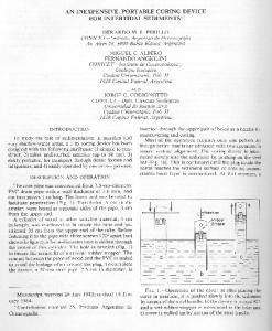
AHI Mean 25.5 SD 22.9 (n=98)
6
4
2
0 0
10
20
30
40
50
60
70
80
90
100
110
120
Apnea Hypopnea Index Figure 1—Distribution of apnea-hypopnea index (AHI) recorded by polysomnography in 98 subjects.
from the analysis due to technical reasons related to the WP_100 device (7 subjects had no recording or PAT signal, and oximetry failure occurred in 1 subject). Six subjects had oximetry failure in ambulatory PSG recordings, and their data were excluded from ODI comparison. One subject lost a substantial portion of the oxygen-saturation signal in the PSG recording but was retained in the ODI comparison (see below). The final study population (n=98, 55 men, 43 women) had a mean age of 60 ± 6.7 years, body mass index of 28.0 ± 4.2 kg/m2, and Epworth Sleepiness Scale score of 6.0 ± 3.5. In all, 21 subjects had a known history of hypertension, and 2 subjects had diabetes (for details see Table 1). The mean PSG AHI of the study population was 25.5 ± 22.9 events per hour. Using PSG AHI 10 as the cut-off value, the prevalence of OSA in this population was 74% (for AHI distribution, see Figure 1). Detailed sleep and breathing characteristics from PSG and WP_100 recordings are shown in Table 2. There was a strong correlation between the AHI, RDI, and ODI assessed by WP_100 and PSG (r = 0.90, 0.88 and 0.92 [n = 92], p < .0001, respectively) (Figure 2). The Bland-Altman plot (Figure 3) revealed a good agreement between the WP_100 and PSG for AHI,
Data Analysis Continuous variables were expressed as means ± SD. Pearson correlation tests were used to test the AHI, RDI, and ODI correlation between the WP_100 and PSG. Bland-Altman plots were used to test the repeatability of AHI, RDI, ODI between WP_100 and PSG. Receiver operating characteristic analysis was performed to validate the WP_100 diagnostic capability. Thresholds of PSG AHI >10, 15, and 20 and RDI >10, 15, and 20 were used as different cut-off points for OSA diagnosis. Areas under the curve were calculated. The agreement between WP_100 automatic sleep-time analysis and PSG-scored total sleep time was compared. All statistical tests were carried out using SPSS 13.0 (SPSS Inc., Chicago, IL), and a p value of .05 or less was considered statistically significant. RESULTS Of the 106 participants, data from 8 subjects were rejected SLEEP, Vol. 29, No. 3, 2006
No. 21
369
Validating a Wrist-Worn Device Using Ambulatory PSG—Zou et al
140
Table 2—Sleep and breathing characteristics comparing Polysomnography and Watch_PAT 100
TST AHI RDI ODI
Monitoring Method Polysomnography WP_100 6.5±1.2 6.3±1.3 25.5±22.9 27.0±18.7 31.6±22.7 30.4±18.7 13.3±15.3 17.7±16.7*
100
WP_100 AHI
Sleep Parameter
120
Data are presented as mean ± SD events per hour, except total sleep time (TST), which is given in hours. WP_100 refers to Watch_PAT 100; AHI, apnea-hypopnea index; RDI, respiratory disturbance index; ODI, oxygen desaturation index. *p < .0001
80
60
40
r=0.90 20
RDI, and ODI (mean difference 1.5±10.2, -1.2±10.9, and 4.4±6.5 [n = 92] events per hour, respectively). The variability of the difference suggested a slight tendency toward higher AHI values detected by the WP_100 at mild to moderate OSA, whereas the opposite was seen in severe cases. The WP_100 tended to overscore ODI in this population (p < .0001). Excluding the outlier did not eliminate the significant ODI difference between the 2 devices (mean 4.1±5.4 [n=91], p < .0001). Receiver-operator characteristic curves (Figure 4) were constructed for WP_100 sensitivity and specificity at different thresholds—PSG AHI and RDI 10, 15, and 20, respectively. The areas under the curve were 0.93, 0.92, and 0.93 for PSG AHI >10, 15, and 20 and 0.88, 0.88, and 0.90 for PSG RDI>10, 15, and 20 (p < .0001, respectively). The mean difference in detected sleep time between the WP_ 100 and PSG was 0.2 ± 1.1 hour. The agreement of the sleep-wake assessment based on 30-second bins between the 2 systems was 82 ± 7% (n = 72).
0 0
20
40
60
80
100
120
140
PSG AHI
140
120
WP_100 RDI
100
80
60
40
r=0.88 20
DISCUSSION This study showed that WP_100, a limited-channel recording technique based on PAT, detected OSA in the unattended home setting with reasonable accuracy. To the best of our knowledge, this is the first study using the WP_100 device for the diagnosis of OSA that addressed a general-population sample with simultaneous unattended home-PSG validation. We propose this procedure as a new standard for validation of simplified recording techniques for the diagnosis of OSA. OSA has been recognized to be a significant and serious public health problem. It is associated with daytime sleepiness,19 cognitive dysfunction,20 poor quality of life,21,22 and increased risk of traffic accidents.23 Moreover, many studies have provided evidence for a strong link between OSA and an increased incidence of hypertension,24 as well as cardiovascular morbidity and mortality.25,26 Current diagnostic guidelines in sleep medicine list PSG as the gold standard for OSA diagnosis because only PSG provides an exact assessment of sleep time and appropriate classification and quantification of OSA based on a calculation of an index defining the number of apneas and hypopneas per hour of actual sleep time. In this context, PSG also provides other important variables, such as mean and minimum oxygen saturation, amount of slow-wave and REM sleep, and the number as well as origin of arousals per hour of sleep. Needless to say, PSG is a cumbersome, complex, and expensive technique performed either in the sleep laboratory or in the unattended home setting. Not only is the prevSLEEP, Vol. 29, No. 3, 2006
0
0
20
40
60
80
100
120
140
PSG RDI 100
WP_100 ODI
80
60
40
20
r=0.92
0
0
20
40
60
80
100
PSG ODI
Figure 2—Scatter plots of (a) apnea-hypopnea index (AHI), (b) respiratory disturbance index (RDI), and (c) oxygen desaturation index (ODI) (n = 92) recorded by polysomnography (PSG) and the Watch_ PAT 100 (WP_100) (r = 0.90, 0.88. 0.92, p < .0001, respectively). 370
Validating a Wrist-Worn Device Using Ambulatory PSG—Zou et al
ROC Curve PSG AHI 10 vs. WP_100 AHI 1.0
Mean = 1.5 (10.2)
20
0.8
Sensitivity
Difference of the methods (WP_100 AHI - PSG AHI)
40
0
-20
0.6
0.4
AUC = 0.93
0.2 -40 0
20
40
60
80
100
120
Mean of the methods (PSG AHI, WP_100 AHI) 0.0 0.0
0.2
0.4
0.6
0.8
1.0
Specificity
Mean = -1.2 (10.9)
ROC Curve PSG RDI 20 vs. WP_100 RDI
20
1.0
0.8
0
Sensitivity
Difference of the methods (WP_100 RDI- PSG RDI)
40
-20
0.6
0.4
-40
0
20
40
60
80
100
120
Mean of the methods (PSG RDI, WP_100 RDI)
AUC = 0.90
Difference of the methods (WP_100 ODI - PSG ODI)
0.2 40
Mean = 4.4 (6.5) P10 [a] and PSG respiratory disturbance index [RDI] > 20 [b] area under the curves [AUC] of 0.93 and 0.90, p < .0001, respectively).
0
-20
alence of OSA in the adult population very high, but numbers also may be anticipated to increase even further as obesity increases dramatically in industrialized countries.27 There is also a growing insight from clinicians in hypertension clinics, stroke units, and cardiology wards that a diagnosis of OSA may affect prognosis and therapeutic strategies. The combination of high prevalence and a complex method of investigation generate a considerable public burden of cost as well as long waiting lists in many sleep laboratories. This dilemma has sparked the development of simpler and po-
-40
0
20
40
60
80
100
Mean of the methods (PSG ODI, WP_100 ODI)
Figure 3—Bland-Altman plots of (a) apnea-hypopnea index (AHI), (b) respiratory disturbance index (RDI), and (c) oxygen desaturation index (ODI) comparisons. Differences were calculated as index Watch_PAT 100 (WP_100) minus index polysomnography (PSG). Values are mean (SD). SLEEP, Vol. 29, No. 3, 2006
371
Validating a Wrist-Worn Device Using Ambulatory PSG—Zou et al
comparator in this type of validation study, and the present study did not run a validation test with attended in-lab PSG. Although other studies have demonstrated similar results in terms of respiratory parameters in comparative studies of unattended home PSG and attended in-lab PSG,2,32,33 there may be discrepancies.34 In our study, the full and complete ODI validation was hampered by oximetry failures among the ambulatory PSG recordings, as described elsewhere.35 In addition, the 2 devices in our study used different oximetry data collection algorithms, and this further complicated the validation.36 As for the study population, it may be argued that we did not address a true population cohort because there was an enrichment of hypertensive subjects and the female participants were almost exclusively postmenopausal. Also, the age span was proportionally narrow, in the region of 60 years, and the body mass index was slightly higher than the population mean. In fact, this may, in part, explain the high prevalence of OSA in our study,37-39 but it is important to remember that symptoms of sleep apnea were not used for selection of subjects. Moreover, correction for the antihypertensive treatment used by a small number of the patients did not substantially influence the results (data not shown). With regard to the recording technique, the high prevalence of sleep-disordered breathing was not entirely unexpected, considering the use of a nasal cannula/pressure together with a thermistor. A recent study showed that the combination of nasal pressure and thermistor could better detect respiratory events than single flow measurement in patients with an AHI less than 50.40 In addition, studies have consistently shown good agreement on respiratory effort-related arousal detection between nasal cannula/pressure recordings and recordings obtained with esophageal manometry.17,41 Some final details may be that it may have been preferable to have the WP_100 device set up by the subject (the user) and that the validation of the WP_100 device had been undertaken in an event-by-event manner rather than as a comparison of indexes in order to provide a fully adjusted validation. It is also important in comparative analyses of this type to, if the function is available, use an appropriate method for determination of the estimated sleep period. Obviously, in order to adequately reflect intended clinical use of a limited-channel device, the sleep period cannot be based on simultaneously acquired PSG data. Instead, the sleep-period determination needs to be based on the automated sleep time estimation, as was done in this study. If the analysis had been based on recorded lights-out to lights-on periods, we could have diluted the calculated index as a result of inadequately included wake episodes in the sleep period. There is a clinical need for simplified recording techniques for the diagnosis of sleep-disordered breathing. Because such devices are intended to diagnose OSA based on standardized PSG criteria, validation process needs to be performed according to certain standard. The current study of the WP_100 was designed to generate such an improved standard by taking a number of potential pitfalls into account. Moreover, this study has demonstrated that nonclassical signals, such as the PAT, may be useful for simplified recording techniques. Although it was not the aim of the present study, such signals were also proved to be useful for quantification of other important events in sleep studies, such as autonomic arousals,42 Cheyne-Stokes breathing,43 or even as surrogates for sleep-stage classification.44 It is concluded that a portable recording technique based on changes in peripheral vascular pulsatile volume in the digital ar-
tentially less-costly methods for diagnostic identification of patients with moderate to severe OSA. Such unattended tools, based on a limited number of diagnostic parameters, have been labelled level 3 (minimum 4 channels including ventilation or airflow) and level 4 (1 to 3 channels or 4 channels but lacking airflow) devices by the American Sleep Disorders Association.5 These simple diagnostic devices record various combinations of respiratory effort and/or flow, oxygen saturation, and heart rate, as well as other indirect parameters for estimation of wakefulness, sleep, body position, and motor activity. Respiratory-output variables are, in general, equally accurate, as compared with those recorded by PSG but not adjusted for the exact sleep time and stage. This difference is an obvious limitation in terms of exact OSA quantification, for instance, when determining cut-off levels for diagnostic classification or therapeutic decisions. The WP_100 device is a portable system that records actigraphy, oximetry, and PAT and derives pulse rate from the PAT signal (level 4). Studies have shown that the PAT signal measures finger pulsatile volume, which changes during sympathetic nervous system activation associated with respiratory events.28-30 The OSA diagnosis of WP_100 has been demonstrated with reasonable reliability and specificity in sleep laboratory cohorts.7-10 Other diagnostic features of WP_100, such as arousal detection31 and sleepwake time assessment,18 have been validated in separate studies. For this reason, WP_100 is considered to provide reasonable accuracy of index calculations of sleep-related respiratory events. The current study attempts to provide an evaluation of this technique by employing a method that addresses several limitations of previous validation studies. First, this study included a general-population sample enriched by a cardiovascular-disease risk group. The study subjects therefore have a broad spectrum and are likely to represent the true target population for simplified OSA diagnostic tools. Most, if not all, previous studies of this type have addressed patient groups with suspected sleep disorders. Investigations of sleep-lab cohorts may systematically favor ruling in a diagnosis of sleep-disordered breathing due to high pretest probability. Second, recordings in the present study were performed in the home setting. Home recordings are evidently less likely to be hampered by changes in environmental factors. These include bed comfort, noise, temperature, sleep partner, dietary intake, and other social activities preceding bedtime that inevitably are induced by studying the patient in a sleep-laboratory setting. Finally, previous validation studies have performed comparative recordings on the same night in the sleep laboratory8,9,31 or on separate nights in the sleep laboratory and the subjects’ home.7,10 Importantly, our study took advantage of simultaneous PSG and WP_100 recordings at home, the environment in which level 3 and 4 devices are mainly used. Both devices were well tolerated, and it was assumed that factors such as night-to-night variability in OSA severity due to changes in body position, sleep length, and sleep-stage distribution, as well as environmental factors, were eliminated by this procedure. Some potential limitations of this study need to be discussed. In terms of the study design, it should be noted that, if a device is primarily intended for OSA diagnostics in a sleep-lab population, it needs to be tested in this population first. This has been the case with the WP_100 device. Once appropriate cut-offs for disease have been validated, the device may be tested separately in the general population. In fact, it may be argued that unattended home PSG recordings (level 2) are inadequate as a “gold standard” SLEEP, Vol. 29, No. 3, 2006
372
Validating a Wrist-Worn Device Using Ambulatory PSG—Zou et al
terial bed adequately identified patients with OSA in a population-based cohort setting. The study has addressed a number of limitations in previous validation studies of simplified recording technique and has resulted in the proposal of a new validation standard of such devices in sleep medicine.
15. 16.
ACKNOWLEDGEMENTS The authors gratefully acknowledge the contributions of Professor Lennart Råstam as a main project initiator of the Skaraborg Hypertension and Diabetes project. We thank the personnel of the Skara Primary Health Care Centre, as well as Helena Axelsson and Paul Murphy, sleep centre Gothenburg, for competent technical assistance. The authors also would like to thank Amir Bar, Itamar Medical, Cesarea, Israel, for the analysis of the WP_100 data.
17. 18. 19.
REFERENCES
20.
1.
21.
2.
3. 4. 5. 6.
7. 8. 9. 10.
11. 12.
13. 14.
Practice parameters for the indications for polysomnography and related procedures. Polysomnography Task Force. American Sleep Disorders Association Standards of Practice Committee. Sleep 1997;20:406-22. Iber C, Redline S, Kaplan Gilpin AM, et al. Polysomnography performed in the unattended home versus the attended laboratory setting—Sleep Heart Health Study methodology. Sleep 2004;27:53640. Redline S, Sanders MH, Lind BK, et al. Methods for obtaining and analyzing unattended polysomnography data for a multicenter study. Sleep Heart Health Research Group. Sleep 1998;21:759-67. Kapur VK, Rapoport DM, Sanders MH, et al. Rates of sensor loss in unattended home polysomnography: the influence of age, gender, obesity, and sleep-disordered breathing. Sleep 2000;23:682-8. Ferber R, Millman R, Coppola M, et al. Portable recording in the assessment of obstructive sleep apnea. ASDA standards of practice. Sleep 1994;17:378-92. Flemons WW, Littner MR, Rowley JA, et al. Home diagnosis of sleep apnea: a systematic review of the literature. An evidence review cosponsored by the American Academy of Sleep Medicine, the American College of Chest Physicians, and the American Thoracic Society. Chest 2003;124:1543-79. Bar A, Pillar G, Dvir I, et al. Evaluation of a portable device based on peripheral arterial tone for unattended home sleep studies. Chest 2003;123:695-703. Ayas NT, Pittman S, MacDonald M, et al. Assessment of a wristworn device in the detection of obstructive sleep apnea. Sleep Med 2003;4:435-42. Penzel T, Kesper K, Pinnow I, et al. Peripheral arterial tonometry, oximetry and actigraphy for ambulatory recording of sleep apnea. Physiol Meas 2004;25:1025-36. Pittman SD, Ayas NT, MacDonald MM, et al. Using a wrist-worn device based on peripheral arterial tonometry to diagnose obstructive sleep apnea: in-laboratory and ambulatory validation. Sleep 2004;27:923-33. Hedner J, Bengtsson-Boström K, Peker Y, et al. Hypertension prevalence in obstructive sleep apnoea and sex: a population-based casecontrol study. Eur Respir J 2006; 27: 1-7. Bog-Hansen E, Lindblad U, Bengtsson K, et al. Risk factor clustering in patients with hypertension and non-insulin-dependent diabetes mellitus. The Skaraborg Hypertension Project. J Intern Med 1998;243:223-32. Johns MW. A new method for measuring daytime sleepiness: the Epworth sleepiness scale. Sleep 1991;14:540-5. Sleep-related breathing disorders in adults: recommendations for syndrome definition and measurement techniques in clinical re-
SLEEP, Vol. 29, No. 3, 2006
22. 23. 24. 25. 26. 27. 28. 29. 30. 31. 32. 33.
34. 35. 36. 373
search. The Report of an American Academy of Sleep Medicine Task Force. Sleep 1999;22:667-89. EEG arousals: scoring rules and examples: a preliminary report from the Sleep Disorders Atlas Task Force of the American Sleep Disorders Association. Sleep 1992;15:173-84. Rechtschaffen A, Kales A. A manual of standardized terminology, techniques and scoring system for sleep stages of human subjects. Bethesda: U.S. Department of Health, Education, and Welfare Public Health Service—National Institutes of Health; 1968. Ayappa I, Norman RG, Krieger AC, et al. Non-Invasive detection of respiratory effort-related arousals (RERAS) by a nasal cannula/ pressure transducer system. Sleep 2000;23:763-71. Hedner J, Pillar G, Pittman SD, et al. A novel adaptive wrist actigraphy algorithm for sleep-wake assessment in sleep apnea patients. Sleep 2004;27:1560-6. Gottlieb DJ, Whitney CW, Bonekat WH, et al. Relation of sleepiness to respiratory disturbance index: the Sleep Heart Health Study. Am J Respir Crit Care Med 1999;159:502-7. Redline S, Strauss ME, Adams N, et al. Neuropsychological function in mild sleep-disordered breathing. Sleep 1997;20:160-7. Finn L, Young T, Palta M, et al. Sleep-disordered breathing and selfreported general health status in the Wisconsin Sleep Cohort Study. Sleep 1998;21:701-6. Baldwin CM, Griffith KA, Nieto FJ, et al. The association of sleepdisordered breathing and sleep symptoms with quality of life in the Sleep Heart Health Study. Sleep 2001;24:96-105. Teran-Santos J, Jimenez-Gomez A, Cordero-Guevara J. The association between sleep apnea and the risk of traffic accidents. Cooperative Group Burgos-Santander. N Engl J Med 1999;340:847-51. Peppard PE, Young T, Palta M, et al. Prospective study of the association between sleep-disordered breathing and hypertension. N Engl J Med 2000;342:1378-84. Shahar E, Whitney CW, Redline S, et al. Sleep-disordered breathing and cardiovascular disease: cross-sectional results of the Sleep Heart Health Study. Am J Respir Crit Care Med 2001;163:19-25. Peker Y, Hedner J, Kraiczi H, et al. Respiratory disturbance index: an independent predictor of mortality in coronary artery disease. Am J Respir Crit Care Med 2000;162:81-6. Young T, Peppard PE, Gottlieb DJ. Epidemiology of obstructive sleep apnea: a population health perspective. Am J Respir Crit Care Med 2002;165:1217-39. Schnall RP, Shlitner A, Sheffy J, et al. Periodic, profound peripheral vasoconstriction--a new marker of obstructive sleep apnea. Sleep 1999;22:939-46. Zou D, Grote L, Eder DN, et al. Obstructive apneic events induce alpha-receptor mediated digital vasoconstriction. Sleep 2004;27:4859. O’Donnell CP, Allan L, Atkinson P, et al. The effect of upper airway obstruction and arousal on peripheral arterial tonometry in obstructive sleep apnea. Am J Respir Crit Care Med 2002;166:965-71. Pillar G, Bar A, Betito M, et al. An automatic ambulatory device for detection of AASM defined arousals from sleep: the WP100. Sleep Med 2003;4:207-12. Fry JM, DiPhillipo MA, Curran K, et al. Full polysomnography in the home. Sleep 1998;21:635-42. Gagnadoux F, Pelletier-Fleury N, Philippe C, et al. Home unattended vs hospital telemonitored polysomnography in suspected obstructive sleep apnea syndrome: a randomized crossover trial. Chest 2002;121:753-8. Portier F, Portmann A, Czernichow P, et al. Evaluation of home versus laboratory polysomnography in the diagnosis of sleep apnea syndrome. Am J Respir Crit Care Med 2000;162:814-8. Mykytyn IJ, Sajkov D, Neill AM, et al. Portable computerized polysomnography in attended and unattended settings. Chest 1999;115:114-22. Davila DG, Richards KC, Marshall BL, et al. Oximeter performance: the influence of acquisition parameters. Chest 2002;122:1654-60. Validating a Wrist-Worn Device Using Ambulatory PSG—Zou et al
37. Worsnop CJ, Naughton MT, Barter CE, et al. The prevalence of obstructive sleep apnea in hypertensives. Am J Respir Crit Care Med 1998;157:111-5. 38. Young T, Finn L, Austin D, et al. Menopausal status and sleep-disordered breathing in the Wisconsin Sleep Cohort Study. Am J Respir Crit Care Med 2003;167:1181-5. 39. Young T, Shahar E, Nieto FJ, et al. Predictors of sleep-disordered breathing in community-dwelling adults: the Sleep Heart Health Study. Arch Intern Med 2002;162:893-900. 40. Teichtahl H, Cunnington D, Cherry G, et al. Scoring polysomnography respiratory events: the utility of nasal pressure and oro-nasal thermal sensor recordings. Sleep Med 2003;4:419-25. 41. Johnson PL, Edwards N, Burgess KR, et al. Detection of increased upper airway resistance during overnight polysomnography. Sleep 2005;28:85-90. 42. Pillar G, Bar A, Shlitner A, et al. Autonomic arousal index: an automated detection based on peripheral arterial tonometry. Sleep 2002;25:543-9. 43. Freimark D, Adler Y, Sheffy J, et al. Oscillations in peripheral arterial tone in congestive heart failure patients: a new marker for Cheyne-Stokes breathing. Cardiology 2002;98:21-4. 44. Dvir I, Adler Y, Freimark D, et al. Evidence for fractal correlation properties in variations of peripheral arterial tone during REM sleep. Am J Physiol Heart Circ Physiol 2002;283:H434-9.
SLEEP, Vol. 29, No. 3, 2006
374
Validating a Wrist-Worn Device Using Ambulatory PSG—Zou et al