EDGE DETECTION AND CLASSIFICATION IN X-RAY IMAGES. APPLICATION TO INTERVENTIONAL 3D VERTEBRA SHAPE RECONSTRUCTION Anne Bilgot*,+, Olivier Le Cadet+, Valérie Perrier+, Laurent Desbat*
(*) Laboratoire TIMC-IMAG, In3S, Faculté de Médecine, Grenoble. (+) Laboratoire LMC-IMAG, Domaine Universitaire, Grenoble. Author for correspondence:
[email protected]
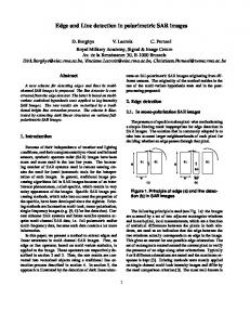
OBJECTIVES Pedicle screw insertion is a delicate surgical intervention, dedicated to restoration of spine stability, in case of disorders such as scoliosis or vertebra fractures. It consists in inserting screws into the pedicles of a vertebra, on which tools are subsequently attached (Fig.1). Roy Camille first performed it in 1963 with open surgery [11]. Since then, it has known reduction of invasiveness with the introduction of percutaneous techniques coupled with fluoroscopy monitoring. Nevertheless, surveys showed that this intervention remains prickly (nervous roots and the spinal cord are very close). Besides, it is known that an intense use of fluoroscopy is damaging in particular for the medical staff. Therefore, several computer-assisted strategies have been
designed to improve the safety of this intervention. One of them consists in the navigation in a pre-operative CT scan: this provides the surgeon with fruitful 3D information [10], but the necessary registration step it induces is rather constraining. Another strategy relies on virtual fluoroscopy; in comparison with the conventional protocol, X-Ray dose is substantially reduced, but 3D information is still lacking [10]. The protocol we are dealing with here comes within this context: in [3,4], Fleute et al. suggest to register a deformable vertebra shape model with two intra-operative orthogonal X-Ray views, so as to provide a 3D model of the surface of the vertebra which is instrumented (see Fig.2). This kind of information indeed turns out to be sufficient for a safe navigation within the vertebra. The generic model they
handle is a statistical model, built, as Cootes and Taylor did [2], by a Principal Component Analysis of the surfaces of a set of manually segmented CT-scans of a type of vertebra. Prior registration, both X-Ray orthogonal views of the vertebra have first to be calibrated, and then processed, in the sense that edges of the vertebra have to be detected (by edges of the vertebra, we refer to projections of external contours of the unknown 3D shape, and thus, on projections, exterior as well as interior contours are of interest). From these detected contours, two back projection beams are built, with which the deformable model is registered through an iterative-closest point algorithm. This method is implemented with a Surgetics Navigation Station (Praxim) and a digital, and so distortion-free, flat panel X-ray detector (PIXIUM 4700, Trixell). As validation of this method, Fleute et al. performed registration experiments for ten different mesh models of vertebrae, which did not belong to the population used to build the statistical model[3]. They simulated contour point projections for both X-Ray views, and obtained a R.M.S. error of 0.62 mm between the reference and the reconstructed meshes. In order to go further into validation, and independently of the influence of the exhaustiveness of the statistical model and the initial position it is given – two difficult problems we do not tackle here - a major issue remains: the automation, as complete as possible, of the edge detection step. This last point constitutes our purpose in this paper. Up to now, the edge detection task was performed manually; one cannot expect a surgeon to do so before an intervention
- it is a time-consuming and boring task. Meanwhile, relying on a completely automatic method is illusory: there are many superimposed and occluded structures in vertebra projections, and the contrast between the vertebra itself and the neighbouring structures can be very low. We thus have turned ourselves towards semi-automatic methods, where we allow the appeal to an expert, in addition to an unsupervised edge detector. As requirements, we would like this detector to be as selective as possible, that is to say that not too much false contour points should be detected. On the other hand, we also need that it extracts sufficient information: for instance, as shown in [1], contenting oneself of the only easiest detectable points in the lateral view can lead to catastrophic reconstruction results.
Fig. 1: Insertion of screws in the pedicle of a vertebra is a prickly intervention, for which transverse information is crucial.
Fig. 2: Illustration of the shape identification method [3]: a vertebra shape model is deformed to as to match the edges detected in two calibrated intra-operative X-Ray images of an unknown vertebra.
METHODS We investigate the use of two different edge detection methods. This section depicts each of them.
1. A wavelet-based edge detector The first tool we use implements a multiscale edge detector, and detects points that are likely to belong to an edge[6]. It relies on a gradient approach, in the sense that edge points are associated to sharp variations of the grey-level intensity in the X-ray image. They are primarily defined as local maxima of the gradient of the intensity along the streamlines associated to the gradient vector field. This definition is then improved by using a multiscale gradient detector, provided by the wavelet transform [7] of the intensity, with an analysing wavelet being taken as the gradient of a smoothing kernel [9]: indeed, in such a case, the vectorial wavelet transform is at each scale proportional to the gradient of the smoothed intensity. In this approach, the location of a local maximum at finest scale belonging to a sufficiently long maxima line across scales provides a significant edge point[8]. Moreover, this framework enables to compute the Lipschitz-regularity of detected edge points, which corresponds to the local regularity of the edge at these points in the transverse direction of the contour, and gives a criterion of classification (for instance, some real vertebra points can be separated from noise false alarms delivered by the detector). In practice, this detector is applied on the image without any pre-processing. It delivers might-be edge points (that can be
gathered into segments according to adjacency criteria). It requires then an interaction with an expert, who is asked to select among segments those that seem to him to be meaningful. At the end of this pruning step, the reconstruction process can be launched, relying on the exterior as well as interior edge points thus isolated.
2. A snake-based edge detector The second tool we assess also relies on a gradient approach. It implements an active contour method, recently developed in [12] and called GVF method (Gradient Vector Flow). It differs from traditional snake methods[5] by the external force applied to the active contour: in GVF method, the field of attraction of an image contour is no more concentrated close to the contour, but is scattered through the image. This makes the evolution of the snake less sensitive to the initial contour, and allows to detect contours of non-simply connected sets. As for previous algorithm, this approach requires an expert interaction. Here it is a pre-processing task, which consists in drawing the initial contour (for instance a polygonal line, roughly following the external contour of the vertebra). The purpose of next section is to draw a comparison between these two edge detectors, by putting them into practice.
RESULTS A previous study was performed on a dry vertebra, using another waveletbased edge detector [1]. In such a case, the X-Ray radiographs are quite easy to process, as no occlusion phenomenon occurs, and indeed, the edge detection
step turned out to enable a satisfactory navigation into the reconstructed vertebra. This work was led only in a qualitative way. The idea in the present study is to get closer from real conditions, while giving better quantitative insight on the reconstruction errors that can be faced when taking the edge detection step into account.
1) Our data In accordance with our validation objectives, we work with a phantom of a synthetic vertebra embedded in a real CT lumbar scan. This phantom is built as follows: we consider a 3D mesh surface, computed from points extracted manually from a CT-scan of a vertebra; as it is difficult to assess with accuracy the error committed during this meshing step, we do not take this preliminary error into account and consider the mesh surface as our target reference. We fill this surface with a model of bone density, getting a model of 3D-vertebra volume. We then compute CT-slices of this synthetic volume: firstly, with no environment, getting a model of a dry vertebra; secondly, with an environment, by substituting, in each slice of a real CT-scan of a vertebra, the real vertebra by our model. We then get a phantom of a vertebra embedded in a realistic environment. The next simulation step consists in simulating orthogonal projections of these two data cubes, getting thus simulated X-Ray images 1) of a dry vertebra, and 2) of an in-vivo vertebra (for this study, we have implemented a very simple simulator of X-ray attenuation phenomenon in parallel geometry). Figure 3 illustrates our simulated data, with an example of a
CT-slice with the synthetic vertebra embedded inside, and the simulated radiographs we will process in the following, in both dry and environment simulated cases. The validation process then goes in two stages, as described in the following.
2) Edge detection results We consider as complete data the two sets of points obtained by projections of the apparent 3D edges of the reference vertebra in the geometry of acquisition. In Fig. 4, they are displayed on the first line. Our experiments consist in applying the two edge detectors to the simulated radiographs of our phantom, and in assessing the amount of edge information that can be provided by these detectors. Note that in these experiments, in the wavelet context, we only process the interaction pruning step in the case of the embedded vertebra phantom, making the a priori assumption that in the dry simulation case, no extra-edges can appear. Results are gathered in Fig. 4. Further work will try and quantify the respective performance of each of them, but by now, a few comments can already be made: 1) GVF algorithm clearly enhances the exterior contour. Meanwhile, interior contour information is lost, whereas it can be recovered when using the wavelet-based algorithm. 2) The interactive pruning step which follows the application of the edge detector when using the waveletbased algorithm and leads to the images shown in Fig. 4, is more constraining for the expert than the drawing of the initial contour needed by the GVF algorithm. In
particular, it requires more anatomical knowledge. Indeed, experiments tend to show that a rough initial contour does not prevent from a correct detection process, whereas a selection of false contour points when pruning can have very bad effects in the subsequent reconstruction. Briefly speaking, the GVF method thus appears of simpler use and less dependant on the expert than the wavelet-based detector. It is very well adapted to exterior contour detection (even if the contour is not convex), but far less adapted to interior contour detection (unless an iterative interactiondetection loop is set up, which is not the case by now). Besides, as regards implementation, one has to mention that the computational cost of the GVF method is higher than the one of the wavelet-based algorithm. Therefore, both approaches have a priori pros and cons. Next section assesses the behaviour of the reconstruction algorithm when coupled with each of them.
3) Reconstruction results In this part, experiments consist in running the reconstruction algorithms on each set of edge points previously detected. We use two kinds of criteria for quantifying each reconstruction result. A first one consists in computing final RMS errors between: 1) the reconstructed surface and the back projection beams (i.e. the error used as minimisation criterion during the registration process), namely the 3D/2D error in Fig. 5. 2) the reconstructed surface and the reference one (the 3D/3D error in Fig.5).
For each error, absolute value (in mm) and relative value, when compared to the largest dimension of the reference vertebra, are given. These measures are global. In contrast, a second criterion consists in computing transverse and frontal slices of the reconstructed surfaces, so as to quantify the “local” reconstruction error in the region of interest: the pedicles. Examples of such slices are displayed in Fig.5. These experiments enable to make the following comments: 1) The errors obtained for the complete-contour case quantify the limits of the reconstruction method. 2) The loss of interior contours (GVF results vs wavelet results) significantly disturbs the subsequent reconstruction. 3) The presence of outliers in the edge map can also have bad effects on the reconstruction. (a false contour on the lateral view in the dry case gives worse reconstruction results than in the realistic case when using the wavelet method), thus confirming the importance of the interaction pruning step. 4) The wavelet-based detection algorithm enables a satisfactory reconstruction result in the realistic phantom case (the error in the pedicle region is less than 2mm (4 pixels)).
CONCLUSION It seems difficult to apply an activecontour based edge detector for the processing of vertebra X-Ray images when detection of interior contours is essential. In contrast, the use of a wavelet-based multiscale edge detector
provides satisfactory results in a synthetic case close from real data, and is thus promising for the application we study – the interventional 3D shape reconstruction of a vertebra. Nevertheless, one must bear in mind that the intervention of an expert with precise anatomical knowledge, although notably lightened by the edge detector pre-processing, remains inescapable. A possible improvement could consist in using as final edge detection result an edge map built simultaneously from the results of both edge detection methods (as the information they supply can complete each other). Another possible one could be the successive coupling of the two edge detection methods together with the reconstruction algorithm: the
idea would be to use the wavelet-based method as described above for performing a first rough surface reconstruction, and then to reproject the contours of this rough surface on the radiographs. This would provide the GVF method with initial contours, both for exterior and interior edges, through an iterative application of the GVF method on successively pointed out initial contours (for instance, once the exterior contour would be dealt with, an expert could run the GVF method on successive initial interior contour selected among interior reprojected contours). One can expect this collaboration between algorithms to supply better reconstruction results.
BIBLIOGRAPHY [1] BARAT C., DUCOTTET C., BILGOT A. DESBAT L. Segmentation of blurred objects using wavelet transform: application to X-Ray images. In Proc.of SPIE, vol. 5266, Wavelet Applications in Industrial Processing, 2004. [2] COOTES T.F., TAYLOR C.J., COOPER D.H., AND GRAHAM J. Training models of shape from sets of examples. In Proc. British Machine Vision Conf., Berlin, Springer, 1992. [3] FLEUTE M. AND LAVALLÉE S. Nonrigid 3-D/2-D Registration of Images Using Statistical Models. In C. Taylor and A. Colchester editors, MICCAI 99. [4] FLEUTE M., LAVALLEE S. AND DESBAT L. Integrated Approach for Matching Statistical Shape
Models with Intra-operative 2D and 3D Data. In T. Dohi and R. Kikinis editors, MICCAI 2002. Springer-Verlag, 2002. [5] KASS M., WITKIN A. AND TERZOPOULOS D. Snakes : Active Contours Models. Int. J. of Computer Vision, vol.1(4), 1987. [6] LE CADET O. Méthodes d'ondelettes pour la segmentation d'images. Applications à l'imagerie médicale et au tatouage d'images. PhD thesis, INPG, 2004.
IEEE Trans. Information Theory 38(2),1992. [9] MALLAT S., ZHONG S. Characterization of Signals from Multiscale Edges. IEEE Trans. Patt. Anal. and Mach. Intell., 14(7), 1992. [10] MERLOZ P. AND HUBERSON C. ET AL. Surgical Navigation for spine : CT virtual imagery versus virtual fluoroscopy. In J. Troccaz and P.Merloz editors, CAMI: tools and applications. Surgetica'2002. Sauramps medical, 2002.
[7] MALLAT S. A wavelet tour of signal processing. Academic Press, 1999.
[11] ROY-CAMILLE R., SAILLANT G. ET AL. Le rachis : aspects fondamentaux, explorations, techniques. Masson, 1995.
[8] MALLAT S. AND HWANG W.L. Singularity detection and processing with wavelets.
[12] XU C. AND PRINCE J.L. Snakes, shapes and Gradient Vector Flow. IEEE Trans. On Im. Proc., vol.7(3), 1998.
Fig. 3: Our phantom. Left: an example of CT-slice where the initial vertebra is replaced by a slice of the reference synthetic vertebra. Middle: the simulated X-Ray images in the dry case. Right: the simulated X-ray images in the realistic case.
Fig 4: Edge detection results for two detectors (wavelet-based or GVF), in comparison with the reference provided by complete contours.
Fig 5: Our reconstruction results. In each case, the reference vertebra is in grey, and the reconstructed vertebra is in white. Position of reconstructed pedicles can be assessed, and global errors are given. The wavelet-based algorithm gives satisfactory results in the realistic case, which is a promising result. In contrast, it seems that GVF-based reconstruction suffers from the loss of information about interior contours.
The authors would like to thank Jean-Marie Nguyen-Xuan-Dang and Sylvain Vallaghe for their fruitful contribution. This work is supported by the Rhône-Alpes Région (AdéMo and Mathématiques pour l’ADéMo grants) and the European Community (IST 1999-12338 MI3 and HPRN-CT 2002-00286 Breaking Complexity grants).