Evaluating Squat Performance with a Single Inertial Measurement Unit Martin O’Reilly*†‡ , Darragh Whelan*†‡ , Charalampos Chanialidis† , Nial Friel† , Eamonn Delahunt ‡ , Tom´as Ward† and Brian Caulfield†‡ † Insight
Centre for Data Analytics of Public Health, Physiotherapy and Population Science University College Dublin Email:
[email protected],
[email protected] *Joint lead authors ‡ School
Abstract—Inertial measurement units (IMUs) may be used during exercise performance to assess form and technique. To maximise practicality and minimise cost a single-sensor system is most desirable. This study sought to investigate whether a single lumbar-worn IMU is capable of identifying seven commonly observed squatting deviations. Twenty-two volunteers (18 males, 4 females, age: 26.09±3.98 years, height: 1.75±0.14m, body mass: 75.2±14.2 kg) performed the squat exercise correctly and with 7 induced deviations. IMU signal features were extracted for each condition. Statistical analysis and leave one subject out classifier evaluation were used to assess the ability of a single sensor to evaluate performance. Binary level classification was able to distinguish between correct and incorrect squatting performance with a sensitivity of 64.41%, specificity of 88.01% and accuracy of 80.45%. Multi-label classification was able to distinguish between specific squat deviations with a sensitivity of 59.65%, specificity of 94.84% and accuracy of 56.55%. These results indicate that a single IMU can successfully discriminate between squatting deviations. A larger data set must be collected and more complex classification techniques developed in order to create a more robust exercise analysis IMU-based system.
I.
I NTRODUCTION
Incorrect exercise performance (i.e. faulty exercise form and technique) may result in ineffective training, inadequate rehabilitation, as well as increasing the likelihood of training induced injuries. This is especially pertinent for athletes who train with free-weights [1]. Training induced injuries are frequently caused by excessive tissue loading as a result of aberrant exercise form and technique [2]. Therefore, feedback on exercise performance is an important consideration to ensure that athletes perform prescribed exercises correctly. Traditionally this feedback has been provided onsite by professional strength and conditioning (S&C) coaches or rehabilitation staff. However, such direct supervision and individualized feedback on exercise performance is not always a possibility, as is the situation when a large number of athletes are training simultaneously [3]. Furthermore, it has also been challenging to provide objective exercise performance data to athletes in this environment with most assessments being subjective in nature. To date marker-based motion analysis systems have been used to provide objective data relative to exercise performance [4]. However, there are a number of limitations with such an approach; set-up is time intensive, the equipment is expensive and the application of markers may hinder normal athletic
movement [2], [5]. Furthermore, this type of analysis is typically performed in specialised research or commercial motion analysis laboratories. These environments may artificially constrict, simplify or influence the movement patterns of those being tested [6]. Therefore, these marker-based systems have not tended to be accepted into routine practice. Recent technological advances support the use of inertial measurement units (IMUs) as a viable option for the assessment and quantification of exercise performance beyond the motion analysis laboratory [2]. These IMUs offer a number of potential advantages over traditional marker-based systems; they are small, inexpensive, easy to set-up and enable the assessment of human movement in an unconstrained environment [7]. Accelerometers and gyroscopes are becoming an increasingly popular method of assessing and quantifying human movement as they are present in many smartphones. This means that these ubiquitous technologies may have the potential to measure human movement and provide feedback relative to the quality of the movement performed [8]. IMUs have been used in a number of different ways from measuring energy expenditure [9] to gait analysis [10] to medical monitoring [11]. These sensors have also been used in the athletic arena in sports such as skiing [12] and golf [13]. Recently the utilization of IMUs as a method of tracking gym and rehabilitation exercises has been investigated. Lin and colleagues [14] evaluated data obtained from IMUs at the hip, knee and ankle during a number of lower limb exercises. Data from the IMUs were used to estimate joint angles; with the authors comparing the IMU derived joint angles to those quantified via a marker-based motion analysis capture system. The authors concluded that these joint angles were accurate when compared to those obtained via the more traditional methodology. However, the quality of the exercise performance was not classified. Pernek and colleagues [8] used accelerometers to assess exercise performance during gym-based resistance type exercises. They assessed movement quality based on the speed of exercise performance. However, different exercise goals may require varying movement speeds and as such, the assessment of movement quality based on speed alone does not offer a holistic way of evaluating exercise technique. Taylor and colleagues [15] attempted to more accurately evaluate exercise performance using IMUs. Five body worn
accelerometers were used to evaluate three lower limb single joint exercises (standing hamstring curl, straight leg raise and reverse hip abduction) in healthy college students. The authors were able to discriminate correct from incorrect exercise performance, with their subsequently developed exercise classifier exhibiting an overall average accuracy of 80% for standing hamstring curl, 65% for reverse hip abduction and 62% for straight leg raise. These results were based on leave-onesubject-out cross-validation (LOSOCV) testing. However, they only recorded data from nine participants and the use of a nonexpert in labelling correct or incorrect exercise performance was a methodological limitation. The same authors built on this work in 2012 [16] and evaluated the use of multi-label classifiers to assess exercise performance in patients with knee osteoarthritis using five IMUs. On this occasion each IMU contained a tri-axial gyroscope as well as an accelerometer. Again their classifiers displayed high accuracy (86%), sensitivity (84%) and specificity (99%) in detecting errors that can occur during the performance of the exercises investigated. However, the overall results and their wider extrapolation are limited by the small participant sample size (n = 8). Furthermore, the exercises utilized were all single joint exercises (standing hamstring curl and straight leg raise) and the number of sensors used may not always be practical. While these exercises may be used in a clinical population during the early stage of rehabilitation they are likely to be inadequate as the rehabilitation progresses or for higher-level conditioning. Velloso and colleagues [3] have also attempted to evaluate the quality of exercises using IMUs. They defined exercise quality as “the adherence to the execution of an activity to its specification”. They evaluated two upper limb single joint exercises (biceps curl and lateral raise). Using a leave-onesubject-out testing protocol they obtained an overall recognition performance of 78.2%. The authors also reported that participants responded favourably to feedback that aided with the correct completion of the exercises. A recent study by Giggins et al [7] suggested that a single IMU may be used to identify poor technique in five of seven single joint exercises investigated (heel slide, straight leg raise, knee extension, hip abduction and hip extension). However, these results were based solely on statistical analysis with the absence of classifier evaluation. A follow up study by the same authors [17] showed that a single IMU worn on the thigh could achieve on average 82% sensitivity, 72% specificity and 83% accuracy in binary classification across the seven exercises and 49% sensitivity, 77% specificity and 61% accuracy in multi-class classification across a subset of four of the exercises. These results were based on LOSOCV testing. A number of studies have demonstrated the viability of multiple IMUs to assess and quantify exercise performance [8], [14]. More recent research has also shown that it may be possible to evaluate these exercises more comprehensively [3], [15], [16], and possibly with a single IMU [7], [17]. This study differs from previous work in the field as it aims to evaluate if a single body-worn IMU is capable of distinguishing between seven levels of performance in a compound exercise (i.e. body weight squat). This may have the potential for applications in the areas of injury screening, S&C and rehabilitation.
II.
M ETHODS
This study was undertaken to determine if a single IMU can discriminate between different levels of squat performance and identify poor exercise technique. Data were acquired from participants as they completed the squat with normal technique for 10 repetitions. IMU data were then acquired while the same exercise was completed for three repetitions with commonly observed deviations from correct technique. A. Participants Twenty two healthy volunteers (18 males, 4 females, age: 26.09±3.98 years, height: 1.75±0.14m, body mass: 75.2±14.2kg) were recruited for the study. No participant had a current or recent musculoskeletal injury that would impair their squat performance. All participants had prior experience with the squat exercise and regularly used it as part of their own training regime for at least one year. Each participant signed a consent form prior to completing the study. The University Human Research Ethics Committee approved the study protocol. B. Exercise Technique and Deviations Participants completed the initial squat with good form as described by the National Strength and Conditioning Association (NSCA) guidelines [p.320-322] [18]. This involved participants holding their chest up and out with the head tilted slightly up. As participants moved down into the squat position they were instructed to allow their hips and knees to flex while keeping their torso to floor angle relatively constant. Furthermore, they were required to keep their heels on the floor and knees aligned over their feet. Participants were required to continue flexing at the hips and knees until their thighs were parallel to the floor. As they moved upward a flat back was to be maintained and they were instructed to keep their chest up and out. Hips and knees were to be extended at the same rate with heels on floor and knees aligned over feet. Participants then extended their hips and knees to reach starting position. The deviations from the aforementioned correct technique that were completed were knee valgus (KVL), knee varus (KVR), weight shift right (WSR), weight shift left (WSL), knees to far forward (KTF), heels elevated (HE) and bent over (BO). These are outlined in table 1. TABLE I: List and description of squat exercise performance. Deviation N KVL KVR WSR WSL KTF HE BO
Explanation Normal squat Knees coming together during downward phase Knees coming apart during downward phase Excessive lean to right hand side during entire squat exercise Excessive lean to left hand side during entire squat exercise Knees ahead of toes during downward phase Heels off ground during entire squat exercise Excessive flexion of hip and torso during entire squat exercise
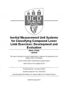
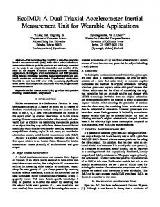
C. Experimental Protocol A pilot study was used to determine an appropriate sampling rate and the ranges for the accelerometer and gyroscope on board the IMU (SHIMMER, Shimmer research, Dublin, Ireland). In the pilot study squat data was collected at 512Hz. A Fourier transform was then used to detect the characteristic frequencies of the signal which were all found to be less than 20Hz. Therefore, a sampling rate of 51.2Hz was deemed appropriate for this study based upon the Nyquist criterion. The Shimmer IMU was configured to stream tri-axial accelerometer (±16G), gyroscope (±500o /s) and magnetometer (±1Ga) data with the sensor ranges chosen also based upon data from the pilot study. The IMU was calibrated for these specific sensor ranges using the Shimmer 9DoF Calibration. When participants arrived to the laboratory the testing protocol was explained to them. Following this they completed a ten minute warm-up on an exercise bike maintaining a power output of 100W at 75-85 revolutions per minute. Next the IMU was secured on the participant at the level of the 5th lumbar vertebra using an elasticated strap. This sensor placement was selected based on clinical judgement as to the location that would most likely identify deviations and is shown below in Figure 1. The orientation and location of the IMU was consistent for all study participants. Participants were then instructed on how to complete the squat with good form and biomechanical alignment as outlined in the NSCA guidelines as explained in section B. They completed ten repetitions with this good form. Once the squat had been completed with normal technique the participant was instructed to complete the exercise with the deviations specified in table 1. They completed three repetitions of each deviation. Verbal instructions and a demonstration were provided to all participants and they were allowed a trial to ensure they were comfortable completing the deviations. All squats were completed using body weight only. A Chartered Physiotherapist was present throughout all data collection to ensure the squat had been completed as instructed. D. Data Analysis Data were low-pass filtered at fc =20 Hz using a Butterworth filter of order n=8 in order to remove high frequency noise and ensure all data analysed related to each participants movement as confirmed using the Fourier transform during the pilot study. For each repetition of the exercise a total of fifteen features were extracted from the IMU to allow for statistical analysis. These were maximum, minimum and range of the acceleration (accel) signals in X, Y and Z planes and maximum and minimum angular velocity (gyro) in X, Y and Z planes. Initially a repeated measures t test was considered as an appropriate comparison between the eight squat conditions. However, it was shown using a normal quantiles plot that the difference between the means of any two conditions did not follow the Gaussian distribution (Figure 2) and thus the data is not normally distributed. Therefore, the non-parametric pairwise Wilcoxon signed-rank test was used to analyse whether there was a difference in the IMU parameters between the various squat techniques. A P value