Hierarchical clustering of brain activity during human nonrapid eye movement sleep Mélanie Bolya,1, Vincent Perlbargb,1, Guillaume Marrelecb, Manuel Schabusa,2, Steven Laureysa, Julien Doyonc, Mélanie Pélégrini-Issacb, Pierre Maqueta,3, and Habib Benalib a Cyclotron Research Centre, University of Liège, 4000 Liège, Belgium; bInstitut National de la Santé et de la Recherche Médicale–Université Pierre et Marie Curie Paris 06, Unité Mixte de Recherche-S 678, Laboratoire d’Imagerie Fonctionnelle, 75634 Cedex 13 Paris, France; and cFunctional Neuroimaging Unit, Montreal Geriatrics Institute, Montreal, QC, Canada H3W 1W5
Consciousness is reduced during nonrapid eye movement (NREM) sleep due to changes in brain function that are still poorly understood. Here, we tested the hypothesis that impaired consciousness during NREM sleep is associated with an increased modularity of brain activity. Cerebral connectivity was quantified in restingstate functional magnetic resonance imaging times series acquired in 13 healthy volunteers during wakefulness and NREM sleep. The analysis revealed a modification of the hierarchical organization of large-scale networks into smaller independent modules during NREM sleep, independently from EEG markers of the slow oscillation. Such modifications in brain connectivity, possibly driven by sleep ultraslow oscillations, could hinder the brain’s ability to integrate information and account for decreased consciousness during NREM sleep. complexity
| integration
D
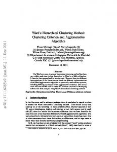
uring nonrapid eye movement (NREM) sleep, we are less aware of ourselves and our environment and, if we awaken, are less able to recollect any mental representation than during full-blown wakefulness (1). The mechanisms underpinning the reduction in conscious content during NREM sleep are still uncertain. Consciousness has been associated with the ability of a system to integrate information (2), which could be altered during NREM sleep. Here, in contrast to previous work (3, 4), we quantified changes in information integration from wakefulness to NREM sleep in large-scale brain networks and computed both their total integration and their degree of functional clustering. Functional clustering estimates how integration is hierarchically organized within and across the constituent parts of a system. It has been proposed as an empirically tractable measure for complexity of brain integration (5), which is considered a better estimate of the capacity to integrate information than total integration (6). We assessed brain functional connectivity on functional MRI (fMRI) data, which reflect the slow dynamics of local field potentials rather than instantaneous neural activities (7). Data were collected in a single nocturnal session in 13 participants who maintained periods of steady NREM sleep. At awakening, none of the subjects could recall any mental conscious content since sleep onset. From this dataset, we extracted for each subject two subsets of consecutive volumes recorded, respectively, during wakefulness and NREM sleep. Six spatially independent patterns, which we refer to as networks (Fig. 1A), were identified at the group level on wakefulness data, using a data-driven method (independent component analysis). These networks [visual (VIS), motor (MOT), default mode (DM), dorsal attentional (dATT), executive control (EC), and salience (SAL)] were previously identified in many studies investigating restingstate fMRI correlations in the literature (8–10). Network composition was very similar in data obtained during NREM sleep compared with wakefulness, in terms of within-networks areas distribution and Euclidian distance between networks (SI Results and Fig. S1). The six networks consistently identified during wakefulness were used only to select regions of interest (ROIs) www.pnas.org/cgi/doi/10.1073/pnas.1111133109
for further analyses. In total, 77 ROIs were selected around the local maxima of these networks (Table S1). To quantify the amount of functional interactions within and between these networks across vigilance states, we computed a hierarchical measure of integration between brain regions during both NREM sleep and wakefulness. First, an average ROI activity time course was extracted and an averaged correlation matrix was computed for each network on NREM sleep data. On the basis of the resulting similarity tree, each network was further parsed in anatomically and physiologically meaningful assemblies of bilateral homologous areas (Fig. S2). We then considered brain connectivity at three nested levels: brain, networks, and assemblies of areas (Fig. 1B). The brain was divided into six networks and each network into three to seven assemblies of two to five areas. Finally, we probed the hierarchical structure of brain integration by computing integration at whole-brain and network levels and derived their respective functional clustering ratio (FCR), the proportion of interactions within each subsystem, relative to between them. Differences in FCR between wakefulness and NREM were tested in a Bayesian framework and inferences were conducted at a probability of difference >0.95. Results Compared with wakefulness, NREM sleep was characterized by a diffuse increase in FCR values, both at the whole-brain level and in each and every brain network (Fig. 1C and Table 1). Rather than the mere breakdown of functional connectivity (11), this result reveals a profound modification of the hierarchical organization of functional integration. Total brain integration increased during NREM sleep relative to wakefulness (Table 2) due to an increase in both within- and between-network integration. Within each network, a fairly consistent change consisted of an increase in within-assembly integration (Iws), observed in all networks except the MOT network (Table 3). In contrast, during NREM sleep, the integration between assemblies (Ibs) increased (VIS and ATT), remained stable (EC), or decreased (MOT, DM, and SAL; Table 4). However, in all networks, the change in within-assembly integration was proportionally larger than that in between-assembly integration, resulting in a consistent increase in FCR. Complementary analyses revealed that this (fixed-effects) group result of an increase in FCR during NREM sleep compared with wakefulness was reproducible across indi-
Author contributions: M.S. and P.M. designed research; M.B. and M.S. performed research; V.P., G.M., and H.B. contributed new reagents/analytic tools; M.B., V.P., G.M., M.P.-I., P.M., and H.B. analyzed data; and M.B., V.P., G.M., S.L., J.D., M.P.-I., P.M., and H.B. wrote the paper. The authors declare no conflict of interest. This article is a PNAS Direct Submission. 1
M.B. and V.P. contributed equally to this work.
2
Present address: Department of Psychology, University of Salzburg, 5020 Salzburg, Austria.
3
To whom correspondence should be addressed. E-mail:
[email protected].
This article contains supporting information online at www.pnas.org/lookup/suppl/doi:10. 1073/pnas.1111133109/-/DCSupplemental.
PNAS Early Edition | 1 of 6
NEUROSCIENCE
Edited by Marcus E. Raichle, Washington University in St. Louis, St. Louis, MO, and approved February 13, 2012 (received for review July 19, 2011)
Fig. 1. (A) The six networks extracted during wakefulness. (B) Levels of brain hierarchical integration. (C) Increases in functional clustering ratio in the brain and the six networks (all significant with a probability >0.95). Networks: dATT, dorsal attentional; DM, default mode; EC, executive control; MOT, sensorimotor; SAL, salience; VIS, visual.
viduals, both at the brain level and for each network (Table S2). Additional results obtained using graph theory tools also confirmed that brain activity modularity was increased in NREM sleep compared with wakefulness (Fig. 2). These findings indicate that over and above changes in individual network integration or in total brain integration, an essential difference in brain function between wakefulness and NREM sleep consists of
increased functional clustering of brain activity into assemblies of homologous areas. Within each network, we then quantified how much information was exchanged between each node and the rest of the network. We observed a moderate reordering of the importance of the nodes in each network (Fig. 3). Although many areas changed their within-network rank from wakefulness to NREM sleep, the hierarchical organization of the networks was relatively preserved with the transition between states.
Table 1. Functional clustering ratios (FCR) computed within whole brain and within each network during wakefulness and NREM sleep Wakefulness FCR = Iws/Ibs
Mean
NREM sleep–wakefulness SD
Brain level vs. networks Brain 1.50 0.03 System level vs. assemblies MOT 4.72 0.30 VIS 2.92 0.27 DM 0.93 0.06 dATT 1.35 0.12 EC 0.54 0.04 SAL 1.37 0.07
Table 2. Total brain integration computed within whole brain and within each network during wakefulness and NREM sleep
Sleep
Variations prob > 0.95
Mean
SD
(+)
1.70
0.03
(+) (+) (+) (+) (+) (+)
5.95 6.24 1.24 1.93 1.03 3.57
0.37 0.37 0.06 0.09 0.05 0.18
The central column in Tables 1 and 2 indicates the variations with a probability >0.95.
2 of 6 | www.pnas.org/cgi/doi/10.1073/pnas.1111133109
Wakefulness Itot
Mean
NREM sleep–wakefulness SD
Brain level vs. networks Brain 5.45 0.08 System level vs. assemblies MOT 0.83 0.02 VIS 0.32 0.02 DM 0.56 0.02 dATT 0.32 0.02 EC 0.40 0.02 SAL 0.84 0.03
Sleep
Variations prob > 0.95
Mean
SD
(+)
6.37
0.07
(−) (+) (=) (+) (+) (−)
0.76 0.74 0.57 0.63 0.55 0.73
0.02 0.02 0.02 0.02 0.02 0.02
Boly et al.
Wakefulness Iws
Mean
NREM sleep–wakefulness SD
Brain level vs. networks Brain 3.27 0.06 System level vs. assemblies MOT 0.69 0.02 VIS 0.24 0.01 DM 0.27 0.01 dATT 0.18 0.01 EC 0.14 0.01 SAL 0.49 0.02
Neurobiological Interpretation of Total-, Within-, and Between-System Integration and Functional Clustering Ratio. Total integration
Sleep
Variations prob > 0.95
Mean
SD
(+)
3.98
0.05
(=) (+) (+) (+) (+) (+)
0.66 0.64 0.31 0.42 0.28 0.57
0.02 0.02 0.01 0.01 0.01 0.02
Because the slow oscillation is a fundamental rhythm of NREM sleep potentially associated with a breakdown in cerebral connectivity (11), slow-wave activity (SWA) was regressed to integration measures. Total brain integration was negatively correlated to SWA during NREM sleep (Tables 4 and 5 and Fig. S3). However, there was no significant relation between FCR and SWA (Table 6). Discussion Content of Consciousness During NREM Sleep. The current work is
based on the theoretical prediction that consciousness is related to the ability of the brain to integrate information. This ability is related to the mathematical construct of complexity, which we approached by computing the functional clustering ratio. To test this hypothesis, we contrasted two polygraphically identified states of vigilance that substantially differ in mental content, namely resting wakefulness and NREM sleep. The reduction of mental content during NREM sleep was confirmed during debriefing after awakening and we are confident that the mental content differed between wakefulness and sleep in our volunteers. Importantly, we do not claim that NREM sleep is associated with a total obliteration of conscious mental representations. The brain remains responsive to external stimuli during NREM sleep (12, 13) and dreams have been reported after NREM sleep awakenings (14). However, the aim of the current paper was not to characterize the neural correlates of these mental representations but rather to contrast brain function during normal wakefulness to that observed during a state of impoverished conscious content such as NREM sleep.
Fig. 2. Brain connectivity modularity indexes (group mean result ± SDs) during wakefulness and during NREM sleep. The modularity index, calculated by using the Louvain algorithm, quantifies the degree to which a system may be subdivided into clearly delineated clusters.
Boly et al.
assesses the sum of information (in the Shannon sense) exchanged between the main functional brain networks and within each of them. Our results show that total integration is larger during NREM sleep than during resting wakefulness. This is an unexpected result. Indeed, although previous studies investigating resting-state fMRI connectivity during states of altered consciousness produced conflicting results (3, 4), they previously mainly revealed a decrease in connectivity. The present results also contrast with previous findings of a globally decreased brain connectivity that has been reported in other altered states of consciousness such as in coma (15) or during anesthesia (ref. 16, in a study performed using the same brain integration quantification technique). The central point of this paper is to show that beyond these changes in total brain integration, functional interactions are hierarchically modified throughout the brain during NREM sleep, relative to wakefulness. The increase in total brain integration results from a combined increase in both within- and between-network integration. In turn, such changes in integration within each network are due to modifications of the integration both within and between their constituent systems (i.e., their assemblies). In all networks during NREM sleep, the within-system integration becomes proportionally larger than between-system integration, resulting in a consistent increase in FCR. Increased functional clustering of brain activity in small independent modules appears as a general phenomenon and reflects deep, hierarchical modifications in information flow during NREM sleep. Theoretical considerations predict that increased clustering of brain activity should be associated with a decreased ability to integrate information (6). Information integration should decrease for modular compared with more homogeneously interconnected systems because integrated information is predicted to be maximal for systems that are both highly connected and not decomposable in individual subsystems. Integrated information indeed refers to the information generated by the causal interactions in a whole system, over and above the information generated by the parts (17). Increased functional modularity potentially results in a decrease in information integration at the systemic level (2) and, according to the tested hypothesis, this result could then account for the decrease in consciousness during NREM sleep, despite preserved total information processing in the brain. According to this theory, the ability of a system to integrate information appears to be a function not only of the total amount of connectivity in the system, but also of the complexity of the interactions leading to the observed dynamics (5). Functional clustering has been proposed as one empirically tractable measure for the complexity of brain integration, which is supposed to characterize conscious processes (5). Our measure of brain activity functional clustering, defined by the ratio of between- and within-subsystems integration, quantifies the degree of functional segregation of a given system into subsystems. An increase of functional clustering ratio means that the architecture of the system is modified toward a greater proportion of exchanges within subsystems, rather than at the system level—i.e., that the subsystems (networks or assemblies in each network) become more independent from one another. The fact that we observe this phenomenon both at the brain and at the networks level suggests a pervasive presence of this phenomenon during NREM sleep. Additional analyses using classical graph partitioning tools confirmed our result of an increase in brain modularity during NREM sleep compared with wakefulness (Fig. 2). However, to be reliable, such graph theoretical tools have to be applied on a relatively large number of regions. We thus could not apply this same technique to fine network substructure characterization as provided by the use of FCR. PNAS Early Edition | 3 of 6
NEUROSCIENCE
Table 3. Within-subsystems integration values computed for whole brain and for each network during wakefulness and NREM sleep
Fig. 3. Within-network key node density maps (quantifying the integration between each ROI and the rest of the network) during wakefulness and NREM sleep. The values were sorted for the two conditions to allow one to compare the relative importance of different nodes in each network. Values are displayed on a scatter plot to allow comparison between wakefulness and NREM sleep. The lengths of ellipsoid axes correspond to condition-specific group results SDs. Color codes correspond to the areas for which the rank in the hierarchy is the same (black), higher (blue), or lower (red) during NREM sleep compared with wakefulness. Please refer to Table S1 for ROI coordinates and name abbreviations.
Altogether, these findings are in line with a recent modeling work, showing that reaching high information integration values in a given system requires a subtle balance between functional specialization and integration between the elements constituting the system (6). Following this model, increased clustering of brain activity would result in a decrease in the information integration ability of the system, despite preserved total information processing in the brain. Theoretical predictions indeed state that not only the richness of connectivity, but also its spatial organization matter and that a system that is strongly modular is unlikely to generate high levels of integrated information (6) that has been related to the level of consciousness (2). In this view, investigating general properties of the system, like its modularity, is more relevant to the assessment of the ability of a system to integrate information than merely assessing the amount of information present within this system. Changes in the Relative Importance of Key Nodes Within Networks. In
addition to an increased clustering of brain activity, NREM sleep was also associated to a reordering of brain areas in each network— typically placing the network node presenting the highest number of connections during wakefulness at a lowest rank of the hierarchy of areas participating in within-network integration. This result is in line with the proposed role of key integration nodes for normal 4 of 6 | www.pnas.org/cgi/doi/10.1073/pnas.1111133109
consciousness (18). However, this modification was not present in all networks (e.g., VIS or DM), and the hierarchical architecture of the networks seemed relatively preserved in NREM sleep compared with wakefulness. Nevertheless, it is possible that these qualitative modifications of within-networks interactions could be
Table 4. Between-subsystems integration values computed at the whole brain level (between networks) and within each network (between assemblies) during wakefulness and NREM sleep Wakefulness Ibs
Mean
NREM sleep–wakefulness SD
Brain level vs. networks Brain 2.18 0.04 System level vs. assemblies MOT 0.15 0.01 VIS 0.08 0.01 DM 0.29 0.01 dATT 0.14 0.01 EC 0.25 0.01 SAL 0.36 0.02
Sleep
Variations prob > 0.95
Mean
SD
(+)
2.39
0.03
(−) (+) (−) (+) (=) (−)
0.11 0.10 0.25 0.22 0.27 0.16
0.01 0.01 0.01 0.01 0.01 0.01
Boly et al.
Itot vs. SWA
R
Brain level vs. networks Brain −0.42 Network level vs. assemblies MOT −0.25 VIS −0.07 DM −0.32 dATT −0.43 EC −0.36 SAL −0.43
P value (two-tailed) 0.0037* 0.0938 0.6439 0.0301* 0.0029* 0.0140* 0.0029*
*Significant correlation with a P value