To identify the tumor's location rel coordinate system, fiducial markers are head and the bespoke software to id location in preoperative CT images developed. 3.
Paper271 2013 13th International Conference on Control, Automation and Systems (ICCAS 2013) Oct. 20-23, 2013 in Kimdaejung Convention Center, Gwangju, Korea
Image-guided Positioning Robot for Single-port Brain Surgery Robotic Manipulator Sungmin Seung, Pengxin Liu, Sukho Park, Jong-Oh Park* and Seong Young Ko* Chonnam National University, School of Mechanical Engineering, Gwangju, 500-757, Korea (Tel : +82-62-530-1679; E-mail: {jop, sko}@jnu.ac.kr) * Corresponding author Abstract: The paper presents an image-guided positioning robot for a single-port brain surgery robotic manipulator. Due to its limited workspace, the robotic manipulator that performs a fine operation needs to be precisely and stably positioned near the target area. For this purpose, an accurate guidance of a positioning robot to the target position using CT images is developed. The developed brain surgery robot system consists of a single-port robotic manipulator, a positioning robot, an optical tracking system, a brain phantom, and an image model obtained from the pre-operative CT data of the brain phantom. The positioning robot supports the single-port robotic manipulator and positions it near a surgical site. To guide the surgical manipulator into a variety of positions and orientations, a 6-DOF serial robot is introduced as the positioning robot. The coordinate transformations among the image model, the brain phantom and the positioning robot and the single-port robotic manipulator are obtained to guide the positioning robot and to perform the registration between CT image of the phantom and the real phantom. The simple algorithm to specify the target location of the positioning robot in CT coordinate system is proposed. Using the coordinate transformations and the desired target location and orientation, it is possible to guide the positioning robot into the desired target position and orientation. The experimental result shows the targeting error of the proposed method is sufficiently small. Keywords: robotic manipulator, single-port brain surgery, image-guided surgery manipulator. The system provides an immersive operating environment for surgeons by providing both high quality stereo visualization and a man-machine interface that transfers directly the surgeon's hand motion to the motion of surgical tool tips inside the patient's body [4]. Next is the NeuroMate, the first USA FDA-approved, image-guided and robotic-assisted system, which is used for stereotactic procedures in neurosurgery. It has been successfully used in frameless operations for movement disorder surgery. Its registration is performed using an ultrasound or X-rays images. The external positioning robot moves a probe autonomously along a predefined position before locking and powering off. The probe is then manually inserted into the brain [5]. The path finder is an image-guided frameless 6-DOF robot that provides a stable and accurate tool positioning for neurosurgery in the context of ROBOCAST project [6]. Fiducial markers are attached to the patient’s skin before acquiring the preoperative CT data for a registration. Inspired by these surgical robotic systems, we developed a stable and accurate positioning robotic system which is guided automatically using CT data of a brain. We calculate the relative position between a patient and a surgical robot manipulator through coordinate transformation. The remainder of this paper is constructed as follows. Section II introduces an overview of the proposed image-guided robotic system.
1. INTRODUCTION Recently, various types of surgical robotic systems have been developed to achieve minimal invasiveness and to improve the maneuverability of surgical tools. Regarding a brain tumor surgery, many surgical treatment methods including an endoscopic resection of brain tumors using a slender conduit [1] have been proposed. For the endoscopic resection of brain tumor, we have developed a single-port multi-robotic arm manipulator system, which is composed of two small robotic arms and one stereo-endoscope in a long slender housing [2]. The robotic arms have been designed so that its workspace can cover the normal brain tumor size while minimizing its mechanical size and maximizing the tool’s accuracy. Since its workspace is limited, a positioning robotic system, which positions and supports the manipulator having small arms near the lesion’s position, is needed. Utilization of small robotic arms and external positioning robot makes it possible to achieve a large workspace and an accurate tool motion, at the same time [3]. There are a large number of existing surgical robotic systems which include the positioning robot system to support surgical manipulators. First, da Vinci system developed by Intuitive Surgical Inc., U.S.A is one of the successful surgery robots for a minimal invasive laparoscopic surgery. This system consists of 3 slave robots and each one has a 7-degree-of-freedom (DOF)
978-89-93215-05-2 95560/13/$15 ⓒICROS
623
Next, the control method for image-bbased guidance is described in Section III. Experimentss and results are explained in Section IV. Finally, Secttion V concludes the paper with some list of the future woork.
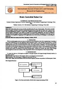
2. OVERVIEW OF PROPOSE ED SYSTEM Since our single-port surgical manipulator m has optimized workspace for the tumor size, it needs a positioning robotic system for moving it near the brain tumor. Therefore, the whole surgery sysstem consists of a surgery robot system, which includess a small robotic manipulator and an external positioninng robot carrying the manipulator, an optical tracking syystem to trace the positions of a patient and the robots, the t patient that is replaced by a brain phantom in this worrk, and at last, the preoperative medical images that is converted into a virtual models as shown in Fig. 1.
Fig. 2 The single-port robootic manipulator for a brain tumor removval procedure 2.2 Positioning Robot System A positioning robot supports the single-port robotic i near a surgical site. To manipulator and positions it guide the surgical manipulatoor into various positions and orientations, a 6-DOF seriaal robot is introduced. In addition, we propose a metthod to calculate a trocar’s position using the patient's medical m imaging i.e. CT data to guide the manipulator. The positioning robot is chosen as a 6-DOF articulaated serial industrial robot Tx-90 manufactured by Stääubli international AG. Its repeatability is 0.03mm and its i reach is 925mm. Fig. 3 shows the overall surgical s setup including the single-port robotic manipulattor, the positioning robot on a flange of which the manipulator is mounted, a patient and the optical tracking system.
Fig. 1 The concept of an image-guided robotic system for single-port brain surgerry
2.1 Single-port Robotic Manipulator As shown in Fig.1, the single-port robbotic manipulator is inserted into slender hole in the braiin and performs a surgical tumor removal procedure. Thuus, the single-port robotic manipulator is designed so ass to have several small robotic arms that have multipple DOFs in the limited workspace. The manipulator is also safe and precise, and able to provide a sufficientt force and torque for grasping and incision of the soft tissue. As shown in Fig. 2, the developed manipulator conssists of a gripping end-effector, a suction/injection endd-effector and a stereo-endoscope in the trocar as explainned in [2].
Fig. 3 The image-guided poositioning robot system for a single-port robootic manipulator m 2.3 Optical Tracking System To register the tumor’s loccation to the robotic system and to trace the position of thhe patient’s head relative to the robotic manipulator, we attach 3D markers on the patient’s head and the robotiic manipulator to define the dynamic reference frames. To detect the frames, a three-dimensional (3D) opticcal tracking system (OTS), Polaris Spectra, manufacturedd by Northern Digital Inc. is adopted. The Polaris Spectrra is selected since it uses passive-type probes to measuure 6-DOF frames.
624
To identify the tumor’s location rellative the patient coordinate system, fiducial markers aree attached on the head and the bespoke software to iddentify the tumor location in preoperative CT imagess has also been developed.
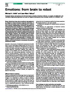
3. Image-Guided Control Method M 3.1 Coordinate Transformations Fig. 4 shows coordinate transformaations among an image model, a brain phantom, a possitioning robot, a single-port robotic manipulator, and maaster devices. The registration between a CT image of thhe brain phantom and a real brain phantom is carriedd out first using point-to-point matching to find CTTOTS thhe transformation matrix from the coordinate system of thhe CT image {CT} to the coordinate system of OTS {OT TS}, and then by OTS measuring TP the transformation maatrix from OTS to the patient coordinate system {P}. Siince we can also measure OTSTRO the transformation mattrix of the robotic manipulator’s base relative to OTS, it is i possible to find the tumor’s location relative to the robbotic manipulator. In addition, CTTGR a transformation matrix from the c system coordinate system of CT image to the coordinate of 3-D graphics is defined to visualizze the 3D image properly. Using these transformatioon matrices, the positioning robot can be positioned properly if the targeting position/orientation is defined relative to {CT}.
vector from the entry pointt to tumor position is not sufficient to define a 3×3 rootational matrix, we define one more point P3 to inddicate up-direction. Fig. 5 visualizes the proposed method to define the targeting frame {Ot} (position and orrientation), along which the positioning robot will be inseerted into the trocar. First of all, the differrence between P1 and P2 is obtained as in (1) and then its normalized vector is is defined as x-axis x of the targeting frame calculated. {Ot}. To find z-axis, thee difference between P3 and P2 is obtained as in (2) and thhen its normalized vector is also calculated. = =
-
(1) (2)
and are not orthogonal, o however, y-axis Since is defined as that is calcuulated by a cross product of and as in (3), and z-aaxis is defined by that is calculated by a cross product of and as in (4).
= =
(3) (4)
The targeting rotational maatrix and the transformation matrix are defined as in (5) annd (6), respectively. = =
(5) 0
1
(6)
Fig. 4 The coordinate transformations for image-based guidance.
3.2 Pre-operative Robot Planning Meethod We define a desired targeting positioon and orientation of the positioning robot using a patient’s CT image. The targeting position is defined as the entrry point P2 of the trocar in CT image of the brain phantoom. The targeting orientation is defined as the vector from m a trocar’s entry point P2 to a tumor’s center point P1. Since, however, the
t desired targeting position Fig. 5 The method to define the and orientation of thhe positioning robot
4. EXPERIMENT T AND RESULT Fig. 6 shows the experim mental setup to evaluate the targeting accuracy. In order to protect the robotic arms
625
from crushing to the surrounding, a thin and flexible needle is attached instead of the roboticc arms. We attach 3D marker to a 5mm grid paper to defiine the coordinate system of the real phantom. The plate with w grid paper is also CT-scanned to define the taargeting position precisely. The positioning robot system is mooved in order to reach the target position using the propoosed image-based guiding method. We have calculateed the measured position error from the target positioon to the actual position of the tip of the positioning robot. Targeting RMS error of the image-guided positioning robot is measured 2.2mm. The targeting error includes a robot calibration error, a model registrationn error and OTS error.
ACKNOWLE EDGEMENT This study was supporteed by the business of IT original technology devellopment (Project No. : KI001802) in Ministry of Knnowledge Economy, and by Leading Foreign Researcch Institute Recruitment Program (No. 2012-0267440) through the National Research Foundation of Koorea (NRF) funded by the Ministry of Education, Science and Technology (MEST).
REFERE ENCES E A. H. Mintz et al., [1] A. B. Kassam, J. A. Engh, “Completely endosccopic resection of intraparenchymal brainn tumors,” Journal of Neurosurgery, vol. 110, noo. 1, pp. 116-123, 2009. [2] S. M. Seung, H. S. Choi, C W. Y. Kim et al., "Development of manipulator including exchange-type multi-artticulated end-effector for single port surgical robot.." Robotics and Biomimetics (ROBIO), pp. 425-430, 20011. [3] J. Arata, Y. Tada, H. Koozuka et al., “Neurosurgical robotic system for brain tumor removal,” International Journal of Computer Assisted Radiology and Surgery, vol. 6, no. 3, pp. 375-385, 2011. [4] G. S. Guthart, and J. Salisbury, Jr., "The IntuitiveTM telesurgery system: overvview and application." IEEE International Conferennce on Robotics and Automation, pp. 618-621 vol.1, 2000. [5] P. S. Morgan, T. Carteer, S. Davis et al., “The application accuracy of the Pathfinder neurosurgical robot,” International Conngress Series, vol. 1256, pp. 561-567, 2003. [6] E. De Momi, P. Cerveri,, and G. Ferrigno, "ROBOt and sensors integration foor computer assisted surgery and therapy (ICT-2007- 215190-ROBOCAST)." 2 pp. 1-6.
Fig. 6 Targeting setup: (a) The red cirrcles indicate 3D reference frame’s marker, (b)(c) The grreen dots indicate target positions.
5. CONCLUSION This paper proposes an image-baseed guidance of a positioning robot for a single-port robbotic manipulator. Our brain surgery robotic system consists of a single-port robotic manipulator, a posiitioning robot, an optical tracking system, a patient, andd a virtual patient model obtained from the pre-operative CT C images. The coordinate transformation am mong the frames attached to the aforementioned compoonents is used to guide the positioning robot to the targetting frame, which is defined in patient’s CT image. mage-guided force In the future, we will develop an im feedback algorithm for the singgle-port robotic manipulation.
626