Proceedings of the 6th WSEAS International Conference on Applied Computer Science, Hangzhou, China, April 15-17, 2007
Introducing Automated Melanoma Detection in a Topic Map Based Image Retrieval System A. PAPASTERGIOU, A. HATZIGAIDAS, Z. ZAHARIS, G. TRYFON, K. MOUSTAKAS, D. IOANNIDIS Department of Electronics, Alexander Technological Educational Institute of Thessaloniki, Sindos, 57400 Thessaloniki, Macedonia, GREECE
[email protected],
[email protected],
[email protected],
[email protected],
[email protected],
[email protected]
Abstract: Early diagnosis is the most reliable solution for an effective treatment of melanoma. There is an ongoing research effort to develop computer aided imaging tools and functional content-based image retrieval systems as diagnostic support to dermatologists. Following this spirit, a Topic Map based management and retrieval system for melanoma images has been developed. Currently, this research work focuses on developing ‘ABCD’ calculation program for Automated Melanoma Detection and employing it in the TM-based system. This paper introduces the design and methodology towards this direction. Key-words: Melanoma, Dermatoscopy, Topic maps, ABCD, Image processing, Segmentation
1 Introduction The increasing incidence of melanoma cases worldwide resulted to an indispensable need to support dermatologists with effective diagnostic tools and methods, towards early diagnosis of melanoma [8, 15]. Dermoscopy (dermatoscopy, epiluminescence microscopy) has been established as a non invasive method for improving the early diagnosis of melanoma [2,4,7,13,16]. The dermoscopic diagnosis of melanoma is based on various analytic approaches and algorithms that have been set forth in the last few years [7]. The ABCD rule of dermatoscopy is a well established standard used in dermatoscopy analysis for classification of dermatological images to benign, suspicious or melanoma [9, 14, 33]. ABCD stands for the following features: A (asymmetry), B (border), C (colour), D (Diameter or Differential structures) [33]. Additionally, there has been a great deal on scientific research aimed to provide improved and reliable diagnostic support to dermatologists by means of computer-based digital image analysis systems [5,6,18,27,29,31]. Computerised methods could increase diagnostic accuracy for dermatologists and enable storing of images with diagnostic information for further investigations or creation of new methods of diagnosis [11,12,17,21,32].
Furthermore, the increasing amount of digital dermoscopic images that are produced in medical institutions results to a proportional research interest to develop new approaches for building advanced image repositories for more efficient information storage and management, that could be used for diagnostic and teaching support [20, 28,34]. Recently several research efforts focus on employing Web and Semantic Web technologies in medical image databases, in to achieve reusability and shareability of information as well as enhanced mechanisms for storage, access and retrieval of digital medical images [1,3,25]. In this background, a topic map-based information management and retrieval system for possible melanoma images, had been developed during the research work that is conducted in our Laboratory [22,23,24]. This research work is currently focusing on developing an ‘ABCD’ calculation program for Automated Melanoma Detection and employing it into the TM-based system. A detailed design technique and methodology towards this direction is presented in the following sections.
2 Employing ‘ABCD’ application in the TM-based system In the framework of this research, a topic map based information retrieval system for handling TM-encoded dermatoscopic melanoma images and
452
Proceedings of the 6th WSEAS International Conference on Applied Computer Science, Hangzhou, China, April 15-17, 2007
related medical data has already been formulated (Fig. 1). The main goal of this research was to explore potential benefits of encoding information using topic map technology, in order to create an effective diagnostic support tool for dermatologists in the sensitive field of malignant melanoma cases [22].
Fig. 1: Representation of TM-based system The core of the proposed system is the Enterprise Topic Map Server (ETMS) that administrates TM and coordinates communication issues with client applications and MySQL database. Communication issues and exchange of TM are established via customized Web Services (Yellow WS) [22]. On the client, software tool (Yellow-TM) is enabling authoring and visualization of TM, as well as definition of TM Schema and expression of rules and queries [24]. Yellow-TM has been used in order to construct a functional repository based on the topic map model that contains melanoma dermatoscopic images and related clinical data [23]. Ontomel application is provided for end-users that simply want to navigate in TM information and they don’t have an implicit knowledge of the topic map model [22]. More specifically, simple windowing and form-based graphical user interfaces (GUIs) have been implemented to
facilitate the needs of end-users (Fig 1). Registered users log into the system and subsequently interact with it. Dermatological image, related data like ABCD parameters, patients’ and clinical data can be uploaded in a simple and perceivable way. ETMS will encode all input data according to TM structure and save them in the TM-based database. Moreover, enhanced navigational and retrieval functionalities are provided via GUIs that minimise users’ input requirements and offer an efficient working environment. User may locate a patient from a predefined list, and correlate all related images and clinical information. Additionally, the predefined rules and queries that have been established in the system can be used for more sophisticated access and retrieve of information [22]. At present, the proposed system is technically and functionally evaluated. Initial results present satisfactory overall effectiveness of the system. Nevertheless, a main demand that seem to be indispensable in effectively diagnosing is the automatic detection of ABCD parameters. Currently, this research focuses on creating an ‘ABCD’ program for automated calculation of ABCD values and incorporating it into the TMbased system. ABCD application will be part of ETMS Server. When a user uploads an image to the system, ABCD parameters could be automatically estimated and attached to the specific dermatological image. ‘ABCD’ program, according to similar programs [21, 29, 31], should perform the following steps: segmentation of image in order to separate the tumor area from the surrounding normal skin, feature extraction for calculation of ABCD values return of the estimated diagnostic values to the user. Subsequently, ETMS will encode these values and input data related to this image, as TM structures and will save them in the systems’ database. In the following subsections image segmentation and feature extraction issues towards the implementation of ‘ABCD’ calculation program are outlined.
3 Introducing ‘ABCD’ application 3.1 Image Segmentation Issues According to the design analysis, the first step towards implementation of ‘ABCD program is the
453
Proceedings of the 6th WSEAS International Conference on Applied Computer Science, Hangzhou, China, April 15-17, 2007
definition of tumor area covered by melanoma and its extraction from the normal skin [10, 29,30,35]. The KMCC algorithm is used to segment the initial image so as to distinguish the area of the melanoma from the rest of the skin [19]. In the KMCC algorithm a spatial distance is inserted in the distance between the vectorial spaces that are considered for clustering [19]. Assuming, the vectorial spaces V (n-dimension) and S (m-dimension). Every point to be registered to one of the clusters as well as every center Κi, has as characteristic a n-dimensional vector and an mdimensional vector, that represent the projection of the point into the vectorial spaces V and S respectively. The distance of a point with vectors v1 and s1 from a center Κ with characteristic vectors v and s is defined from the following equation: D = v1 − v + w s1 − s (1) The parameter w is called connectivity factor and defines the degree in which each vectorial space S contributes to the final value of function (1). For the case of an image the vectorial space V is the three dimensional color space, while the S space is the two dimensional image space. In that sense the generalized distance of an image point, with color coordinates (r1,g1,b1) and spatial coordinates (x1,y1), from a center Κ with coordinates (r,g,b) and (x,y) results from the following equation: D=
(r1 − r )2 + (g 1 − g )2 + (b1 − b )2 +
λ A
(x1 − x )2 + ( y1 − y )2
1 Nj
∑s
(4)
s∈Ω j
where v are the color coordinates, s the spatial, Νj the number of points that have already been registered to the cluster j, Ωj the set of all points V S that belong to the team j and j − new , j − new the new color and spatial centers respectively. The KMCC algorithm outperforms the regular k-means approach for the processed images since it tends to generate connected regions as well as to support a tunable connectivity factor λ that can be tuned according to the context of the processed image [19]. Exams of the estimated area of the possible melanoma are illustrated in Fig 2.
Fig. 2: Extraction of melanoma area
+
3.2 Feature extraction (2)
In equation (2) the connectivity factor λ is divided with the constant Α which stands for the number of points that have been already registered in the corresponding cluster Ωj (centered at K), where j is the index of the cluster. The update of the centers of the KMCC is updated in a sense similar to the standard K-means with the only difference that in each team-cluster correspond two centers that are separately calculated using the following formulae: 1 V j −new = v (3) N j v∈Ω j
∑
S j − new =
Assuming now that the area of the possible melanoma is estimated, let us work on the set W of the points of this area. The ‘ABCD’ calculation program will be based on the ABCD dermoscopic classification rule. The features that has been decided to be extracted from the images, are A, B, C and D values that correspond to the ABCD rule [33]. The ABCD rule has a point based system, where the physician assigns a score to each of the four features. The four features are: A (asymmetry), B (border), C (colour), D (Differential structures). The Total Dermatoscopy Score (TDS) is defined by TDS = A*1.3 + B*0.1 + C*0.5 + D*0.5, with a maximum score of 8.9. A high ABCD score means that the lesion is more likely to be a malignant melanoma (TDS > 5.45) [33,13,17]. The following paragraphs present a brief description of the ABCD rule, as well as the proposed approaches for
454
Proceedings of the 6th WSEAS International Conference on Applied Computer Science, Hangzhou, China, April 15-17, 2007
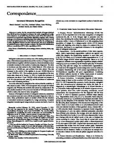
automatic estimation of ABCD values. For testing and evaluating the proposed automatic estimation procedures, dermatological images from a Tutorial CD have been used [26]. These images have defined ‘ABCD values’ as well as TDS scores. Thus, the estimated values of the proposed system can be compared with the default values. Rule A: Using principal components analysis (PCA) the two principal directions are estimated as illustrated in Figure 3 [10, 30]. Then any other state-of-the-art algorithm can be applied to estimate the asymmetry of the region. In the proposed approach the Stoltz method is adopted where the image is tested for asymmetry in terms of the two principal axis of the region [33]. A score of 0 is given when the two axes don’t have asymmetry properties, otherwise 1 or 2 points is given when asymmetry is present in one or two axes, respectively [13, 33].
colours inside the lesion gives 1 point to each for the calculation of the C score (0-6 points) [14,17, 33]. The implementation of this rule is the next step towards the development of the ABCD calculation program. Rule D: The patterns and structure of the lesion are considered. These characteristics include network, structureless areas, dots, globules, and streaks (0-5 points) [13,33]. In some literature diameter of the lesion is estimated for D. If the diameter is more than 6 mm it can be stated as suspicious [9,14,17,33]. For reasons of simplicity, in the present approach Rule D estimates diameter. The diameter of the region is estimated in pixel coordinates simply by calculating the largest distance between the contour points of the region, which stands for the diameter. To summarize, Asymmetry, Border and Diameter values are automatic estimated, while the realization of automatic definition of Color and Differential structure parameters is the current proceed of this work.
3.3 Implementation issues A preliminary attempt to create a functional ‘ABCD’ application using C language and to incorporate it in the TM-based system, is under progression (Fig. 4).
Fig. 3: Estimation of asymmetry For example for the image of Fig. 3, the ‘A score’ that stands for Asymmetry and is automatic estimated, is 0. Default ‘A value’ from the CD is 0 too. Rule B: Irregularities of lesions may indicate melanoma. Thus, regarding Rule B, the image is divided into 8 equi-angular areas using as center the center of mass O of the region [13, 33]. Then the contour points of region are identified and if the distribution of the radii of the polar coordinates of the contour points exhibit large variation for one of the eight areas the B value is increased by one. This happens for all eighths until the final value of B is estimated that should be between 0 and 8.For example for the Image of Fig 3, ‘B value’ is estimated 0, which is the same as the “B value” in the CD. Rule C: The presence of up to six colours (white, red, light brown, dark brown, blue gray, black), are considered. Determination of presence of 6 basic
Fig. 4: Representing expected functionalities
455
Proceedings of the 6th WSEAS International Conference on Applied Computer Science, Hangzhou, China, April 15-17, 2007
A simple GUI will allow user to upload a dermatoscopic image from his computer to the ETM Server. Then the ‘ABDC’ application will automatically define ABCD values. After the segmentation process and the ABCD calculation of the program, the user will receive the lesion diagnosis. The system must return the results to the user, by representing them in a simple numerical output. Ιn the background, ETMS will encode the estimated ABCD values; the uploaded image and all related input data as TM structures, and save them in the MySQL database.
4 Conclusion This paper presented the design and methodology towards the creation of the ‘ABCD’ calculation program for automatic melanoma detection. This work is part of an ongoing research focused on creating a semantic web-based melanoma image management and retrieval system, for diagnostic support in the sensitive field of melanoma cases, using topic map model. In the proposed approach, the KMCC algorithm [19], is used for image segmentation and extraction of melanoma area from the normal skin. The ‘ABCD’ calculation program is based on the ABCD rule for classification of dermatological images to benign, suspicious and melanoma. In conclusion, it should pointed out that the implementation of the ABCD rule may result to different degrees of diagnostic accuracy, depending on the complexity of the algorithm used. Consequently, it is expected that the algorithm will have a great deal of influence towards future development. Future proceeds of this work include realisation of Rule C and D for differential structure, as they have been described during the design procedure, completion of GUI of ‘ABCD’ program and incorporation in the TM-based retrieval system for melanoma images. Moreover, it is anticipated to evaluate the program using a large number of images and compare the estimated ABCD values with accurate result from expert dermatologists is essential. Successful comparison would warranty, a reliable and effective support tool for early diagnosis of melanoma.
References: [1] Aghbari Z. and Makinouchi A., Semantic approach to image database classification and retrieval, NII Journal, No 7, 2003, pp 1-9, http://www.nii.ac.jp/journal/pdf/07/07-01.pdf
[2] Argenziano G, Soyer HP., Dermoscopy of pigmented skin lesions—a valuable tool for early diagnosis of melanoma, Lancet Oncol, Vol 2, 2002 , pp 443–449 [3] Berners-Lee,T., Hendler J. and Lassila O., The Semantic Web, Scientific American, May 2001, http://www.sciam.com/2001/0501issue/0501be r ners-lee.html [4] Binder M, Kittler H, Seeber A et al., Epiluminescence microscopy based classification of pigmented skin lesions using computerized image analysis and an artificial neural network, Melanoma Res, Vol 8,1998,pp 261–266 [5] Blum A., Luedtke H., Ellwanger U., Schwabe R., Rassner G. & Garbe C., Digital image analysis for diagnosis of cutaneous melanoma. Development of a highly effective computer algorithm based on analysis of 837 melanocytic lesions , British Journal of Dermatology, Vol 151, No 5, 2004, pp 1029– 1038 [6] Cascinelli N, Ferrario M, Bufalino R, Zurrida S, Galimberti V, Mascheroni L, Bartoli C, Clemente C., Results obtained by using a computerised image analysis system designed as an aid to diagnosis of cutaneous melanoma, Melanoma Res, Vol 2,1992, pp 167–170 [7] Consensus Net Meeting on Dermoscopy http://www.dermoscopy.org/default.asp [8] Diepgen TL, Mahler V., The epidemiolsogy of skin cancer, Br J Dermatol, Vol 146, 2002, pp 1–6 [9] Feldmann R, Fellenz C, Gschnait F., The ABCD rule in dermatoscopy: analysis of 500 melanocytic lesions, Hautarzt , Vol 49, 1998, pp 473–476 [10] Gonzalez R C, Woods R E., Digital Image Processing: Second Edition, Prentice Hall, inc. 2002 [11] Green A, Martin N, Pfitzner J, O’Rourke M, Knight N., Computer image analysis in the diagnosis of melanoma, J Am Acad Dermatol, Vol 31, 1994, pp 958–964. [12] Hall PN, Claridge E, Morris Smith JD., Computer screening for early detection of melanoma—is there a future?, Br J Dermatol, Vol 132, 1995, pp 325–38. Atlas of Dermoscopy [13] Interactive http://www.dermoscopy.org/atlas/default.asp RH., Dermoscopy: Alternative [14] Johr Melanocytic Algorithms—The ABCD Rule of Dermatoscopy, Clinics in Dermatology ,Vol 20, 2002, pp 240–247
456
Proceedings of the 6th WSEAS International Conference on Applied Computer Science, Hangzhou, China, April 15-17, 2007
[15] Koh H., Cutaneous melanoma, N Engl J Med, Vol 325,1991, pp 171–182 [16] Mayer J., Systematic review of the diagnostic accuracy of dermatoscopy in detecting malignant melanoma, Med J Aust, Vol 167, 1997, pp 206–210 skin cancer reviewed, [17] Melanoma http://melanoma.blogsome.com/ [18] Menzies SW, Cutaneous melanoma: making a clinical diagnosis, present and future, Dermatologic Therapy ,Vol 19, No 1, 2006, pp 32–39 [19] Mezaris V., Kompatsiaris I. and. Strintzis M.G, Still Image Segmentation Tools for Object-based Multimedia Applications, International Journal of Pattern Recognition and Artificial Intelligence, Vol. 18, No. 4, 2004, pp. 701-725 [20] Muller H., Michoux N., Bandon D. and Geissbuhler A., A Review of Content- Based Image Retrieval Systems in Medical Applications - Clinical Benefits and Future Directions, International Journal of Medical Informatics, Vol 73, 2004, pp 1–30 [21] Oka H., Hashimoto M., Iyatomi H. and Tanaka M., Internet-based program for automatic discrimination of dermoscopic images between melanoma and Clark nevi, British Journal of Dermatology, No.150, 5, 2004., p.1041 [22] Papastergiou A., Grammatikopoulos G., Hatzigaidas A. & Tryfon G., Presenting a 3tier System for Managing Melanoma Images with Topic Maps, WSEAS Trans. on Information Science and Applications, Issue 4, Vol. 4, 2007, pp. 820-826 [23] Papastergiou A., Grammatikopoulos G., Hatzigaidas A., Zaharis Z., Lazaridis P., Kampitaki D., Tryfon G. & Ioannidis D. Applying topic map technology to create a knowledge base of melanoma images for diagnostic support, WSEAS Trans. on Information Science and Applications, Issue 2, Vol. 4, 2007, pp. 406-412 A., Hatzigaidas A., [24] Papastergiou Grammatikopoulos G., Zaharis Z., Lazaridis P., Kampitaki D. & Tryfon G. Introducing an advanced Topic Map software tool towards the deployment of a TM-based system for managing melanoma cases images, WSEAS Trans. on Information Science and Applications, Issue 3, Vol. 4, 2007, pp. 452459 [25] Paslaru Bontas E., Tietz S. , Schrade T., Experiences Using Semantic Web Technologies to Realize an Information
Retrieval System for Pathology, Berliner XML Tage, 2004, pp 82-93 [26] Quizz Of Dermatoscopy, Pigmented skin tumors, Dr. Raoul Triller, Hopital FrancoBritannique - Levallois - FranceLa RochePosay, Laboratoire Pharmaceutique [27] Rosado B, Menzies S, Harbauer A et al. , Accuracy of computer diagnosis of melanoma: a quantitative meta-analysis, Arch Dermatol, Vol 139, 2003, pp 361–367 [28] Rui Y., Huang T. S., and Chang S. F., Image Retrieval: Current Techniques, Promising Directions, and Open Issues, Journal of Visual Communication and Image Representation, Vol. 10,1999, pp. 39-62 [29] Sboner A, Eccher C, Blanzieri E, Bauer P, Cristofolini M, Zumiani G, Forti S., A multiple classifier system for early melanoma diagnosis, Artificial Intelligence in Medicine, Vol 27, 2003, pp 29-44 [30] Schmid Ph., Segmentation & symmetry measure for image analysis: application to digital dermatoscopy, Thesis no. 2045, Swiss Federal Institute of Technology at Lausanne (EPFL), Signal Processing Laboratory (LTS); 1999, http://ltswww.epfl.ch/pub_files/schmid/ [31] Schmid-Saugeon P., Guillod J. and Thiran JP, Towards a computer-aided diagnosis system for pigmented skin lesions, Computerized Medical Imaging and Graphics, Vol. 27, 2003, pp. 65–78 [32] Stoecker W.V. & Moss R.H., Digital imaging in dermatology, Computerized Medical Imaging and Graphics, Vol 16, No 3, 1992, pp145-150 [33] Stolz W., Braun-Falco O., Bilek Landthanler, M., Conneta, A. B. and Riemann, A. ,ABCD rule of dermatoscopy: a new practical method for early recognition of malignant melanoma, Eur J. Dermatol., Vol. 4, 1994, pp. 521-527 [34] Tang LHY, Hanka R, Ip HHS, .A review of intelligent content based indexing and browsing of medical images, Health Informatics Journal , 5,1999, pp 40-49 [35] Xu L, Jackowski M, Goshtasby A, Roseman D, Bines S, Yu C, Dhawan A, Huntley A., Segmentation of skin cancer images, Image and Vision Computing ,Vol 17,1999, pp 65-74 [36] Zsolt BA., Dermoscopy (epiluminescence microscopy) of pigmented skin lesions, Dermatol Clin , Vol 15, 1997, pp 79–95
457