International Journal of Computer Information Systems and Industrial Management Applications. ISSN 2150-7988 Volume 8 (2016) pp. 115-124 © MIR Labs, www.mirlabs.net/ijcisim/index.html
Multi-Agent Segmentation using Region Growing and Contour Detection: Syntetic evaluation in MR Images with 3D CAD Reconstruction Abdelhafid NACHOUR1, Latifa OUZIZI2, Youssef AOURA3 ENSAM, Ecole Nationale Superieure d’Arts et Meiters, Moulay Ismail University Meknes, Morocco
[email protected], 2l.ouzizi @ensam-umi.ac.ma, 3y.aoura @ensam-umi.ac.ma
Abstract: Computer assisted surgery navigation takes full advantage of progress in engineering disciplines. Developed models increase the accuracy of replacement technique, especially in hip surgery to reduce the risk of component malpositioning. This paper presents a 3D model reconstruction from contour extracted through a proposed multi-agent segmentation (MAS) approach. We first describe parallel agents’ behaviors for extracting the object of interest from MR Images. The proposed algorithm is formulated by combining region growing and contour detection ensuring an overall segmentation. The 3D CAD model is generated using MATLAB code implemented as per the MAS method and gives us good result of reconstruction in most of the cases. The comparison of the proposed method with the traditional approach is made in terms of run times segmentation and edge detection accuracy. Keywords: Virtual Surgery; Multi-agent Systems; Segmentation; 3D reconstruction; CAD model; Human Femur.
I. Introduction A virtual surgery simulation significantly increases the likelihood of obtaining satisfactory results. The simulation refers to replicating a model or a process in a computer [1]. Habitually the starting point is a set of 3D models generated from medical images, usually computed tomography (CT) image or Magnetic Resonance Images (MRI). The establishment of total prostheses assisted by computer is a new technique since the first implantation is dated in 1997 [2]. the principle is to replace damaged joint by resurfacing the bone end of the thigh and hip bone, capping them with metal, to ensure support, flexibility and motion, without pain. The 3D models are used as reference in the virtual surgery to determine the patient-specific implant geometry [3]. These models have recently been applied to various applications [4]-[7]. The required models decrease the preoperative workup time and increase the accuracy of model preparation
and subsequent surgery. This 3D models are reconstructed from MR Image that produce high image quality of human bone. 3D Image-based reconstruction techniques can be divised in three major parts, such as depth-map-based approaches, volume-based approaches, and surface-based approaches [8]. Following image acquisition, the preoperative traitements in the 3D reconstruction consist of: (i) Segmenting images into objects, (ii) assessment of objects of interest, (iii) set of cross-sectional objects of interest contours. Image segmentation refers to the technique that partitions a digital image into set of segments typically used to identify regions of interest (Regions of Interest, ROI) or other relevant information in digital images [9] based on criteria such as similarity and homogeneity. According to Cocquerez, [10], the choice of a technique is linked to the nature of the image and the treatments after this segmentation. The existing image segmentation algorithms can be classified into four categories [11]: (1) Local filtering approaches, (2) Snake and Balloon methods, (3) Region growing and merging techniques, and (4) Global optimization approaches based on energy functions or Bayesian and MDL (Minimum Description Length) criteria. Region growing method has been widely used for image segmentation [12], and in particular medical image applications. It is a region-based segmentation in which pixels are segmented by grouping similar neighboring pixels of seed points [13]. For example, if a similarity measure of the two adjacent pixels is greater than a threshold, these pixels are similar and thus are grouped together. The grouping of neighboring pixels continues until no similar pixels remain. However, two major problems plaguing the traditional region growing algorithms: first the difficulties to select the appropriate initial seed automatically, second, the noises and regions with holes form [14]. For the first problem, automatic Dynamic Publishers, Inc., USA
116 segmentation algorithm by integrating color-edge extraction and seeded region growing is done in [15]. The authors use an Edge detection algorithm conducted on the image to obtain the major geometric structures as an intermediate to select the initial seeds. Approximate center point of the lesion region is taken as the initial seed in the automatic seed point selection algorithm proposed in [16]. For the second problem, the usual practice is to remove the noise by Gaussian filtering [16], [17] or median filtering [18], [19] applied before segmentation. However, this tool often causes two problems: edge blur [20] and over segmentation [21]. Over segmentation is the process by which the objects of interest are themselves segmented or fractured into regions. Based on the existing algorithms in optimization theory, there are two main approaches to attacking the problem [22]: using random operators and employing multi-individual (agents) based algorithms. The field of multi-agent systems (MAS) arose during the late 80s when several researchers started to work with mobile robots performing coordinated task [23]. So far, it is currently a very active field of research for many types of applications [24]-[27] and disciplines where the medical interest has increased considerably. The MASs are distributed applications consisting of relatively independent modules called agents, which sometimes employ artificial intelligence techniques to accomplish complex operations [28],[29]. The Multi-agent are used as a useful approach in medical practice, especially, in real-time applications. Various applications of MASs have been proposed in image segmentation where a distributed agent makes possible to apply more advanced algorithms and to perform demanding tasks quickly. Liu et al. [30] and Rodin et al. [31] present a parallel image processing system based on simple reactive agents. Agents act according to a perception–action model without problem solving or deliberation. Bovenkamp [32] elaborate a high-level knowledge-based control over lowlevel image segmentation algorithms. The agents dynamically adapt segmentation algorithms based on knowledge about global constraints, contextual knowledge, local image information and personal beliefs. Settache et al. [33] use multi-agent system to share the result of a quad three algorithm used to identify regions primitive, and a Shen filter used to determine edges in the MRI brain part’s detection. Bellet et al. [34] present incremental processes of region growing and edge detection where the cooperation
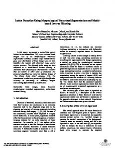
NACHOUR et al… between two types of agent is dynamic and allows to transmit informations when it becomes necessary for a taking of decisions. This cooperative approach increases the segmentation’s quality by confronting the information provided from different algorithms. Yanai et al. [35] use a MAS to extract primitives information like lines, edges or regions using different types of algorithms. Each agent is located on the image and builds a set of coherent primitives. Then, the agents interact to negotiate their local primitives. In this paper, we develope a multi agent segmentation approach for 3D CAD model reconstruction of human femur from MR Images. We first present a parallel agents’ behaviors for extracting the object of interest by combining region growing and contour detection ensuring an overall segmentation (Fig1). This is similar to the convergence of individuals in multi-agent optimization algorithms [36]-[38], in which each agent starts from different initial seed but they are all supposed to converge to the best possible solution. We use two reactive agent equipped with ability to define pixels with the same primitive which discern regions and edges: ‘‘Region’’ agents to solve the particular difficulty of region growing algorithm and ‘‘Edge’’ agents for overall segmentation. Each agent is responsible for the detection of exactly one type of image object and can be communicate with each agent doing its image interpretation in parallel with others agents. The results of the segmentation step are used for the proposed 3D reconstruction of the bony elements. The 3D CAD model is generated using MATLAB code implemented as per the MAS method and gives us good result of reconstruction in most of cases. The paper is organized as follow: in the next section, the proposed method for 3D image recostruction is presented. Section III presents the results of a 3D CAD model of human femur. Finally, section IV is devoted for conclusion.
II. Proposed 3D reconstruction The approch adopted in this paper has three steps. At first, the MR Images are exported from DICOM format(Digital Imaging in Medicine and Communications) to JPEG type with dimensions of 200×550 pixels. The second step focuses on segmentation technique using multi-agent system to extract the edge of the femur and the process of 3D CAD model reconstruction is developed in third step.
Multi-Agent Segmentation using Region Growing and Contour Detection
117
Figure 1. Operational MAS steps. (1) Thresholding operation produces 11 thresholds value for MR Image, (2) Controlller agent initiates the region growing algorithm with 10 agents, and (3) Edge detection after each end message where the last agent represent the image borders. A. MR Image multi-agent segmentation One of the major problems encountered in medical image segmentation is to separate the regions and edges corresponding to object of interest, from the regions that correspond to the background. The proposed MAS is constituted by the agents and their environment which contains the images. Each pixel of image is characterized by a gray level and boolean value which defines if the pixel has already been explored by an agent. We use three types of agents initialized automatically: Agent named “controller” contains all information required to initialize and operate others agents. Agents named “region” responsible to segment homogeneous regions. Agents named “edge”, represent region’s boundaries. Each agent is further responsible for one processing task and cooperates with other agents to come to a consistent overall image segmentation. JADE (Java Agent Development Framework) created by TILAB laboratory and described by Bellifemine et al. [39] is used to build the system. In the following sub-sections, we define each agent as well as their interactions. 1) controller agent: In the first operational step, “controller” agent is responsible for initializing each “region” agent. For instance, if there are n class region in the image, n “region” agents should be created. In this step, unique “region” agent is defined for each class. In order to obtain a completely automatic segmentation, seed pixels that initiate region growing algorithm, are determined the first step using thresholding operation in histogram processing [40]. After each end message from “region” agents, “controller” agent activates the “edge” agent to create the boundaries of the last region. 2)
Region agent:
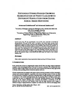
The “region” agent receives a seed pixel and moves to its neighbors and iteratively merges pixels into sets, according to homogeneity criteria (Fig.2).
Figure 2. Region growing algorithm: (a) represent seed pixel in red (b) (c) region growing process by adding homogenious neighbors pixel and (d) represent segmented region. Homogeneity is defined according to the local statistics of the window formed for each image pixel [41]. To determine whether the pixel neighbors should be added to the region, homogeneity is calculated using two parameters: standard deviation and discontinuities (gradient norm). The standard deviation describes the brightnesse within a region with n pixels (n=9 in our case), and the discontinuity indicates an abrupt change in gray level of pixel neighbors. If a similarity measure |H(P)-H(Pn)| of the two adjacent pixels P and Pn is less than a pre-defined threshold ε, these pixels are similar and thus are grouped together. The grouping of neighboring pixels continues until no similar pixels remain. We consider Iij the intensity of the pixel Pij at the position (i,j) in image SMN where (M, N) is the image dimensions, 0