Original Article Reducing Interpolation Artifacts for Mutual Information Based Image Registration Hossein Soleimani, Mohammadali Khosravifard Department of Electrical and Computer Engineering, Isfahan University of Technology, 84156-83111 Isfahan, Iran
ABSTRACT Medical image registration methods which use mutual information as similarity measure have been improved in recent decades. Mutual information is a basic concept of information theory which indicates the dependency of two random variables (or two images). In order to evaluate the mutual information of two images, their joint probability distribution is required. Several interpolation methods, such as Partial Volume (PV) and bilinear, are used to estimate joint probability distribution. Both of these two methods yield some artifacts on mutual information function. Hanning window‑ Partial Volume and Generalized Partial Volume methods are introduced to remove such artifacts. In this paper, we show that the acceptable performance of these methods is not due to their kernel function. It’s because of the number of pixels which incorporate in interpolation. Since using more pixels requires more complex and time consuming interpolation process, we propose a new interpolation method which uses only four pixels (the same as PV and bilinear interpolations) and removes most of the artifacts. Experimental results of the registration of Computed Tomography images show superiority of the proposed scheme.
Key words: Artifact, image registration, interpolation, mutual information
INTRODUCTION Image registration is the alignment of two or more different images of a view or an object such that their similar pixels coincide to each other. These images might be acquired by several sensors or by a single sensor in different viewpoints, moments or other different conditions. The goal of image registration is to find a transform function by which the floating image is aligned to the reference image. Every image registration algorithm requires three basic ingredients: 1) a spatial transformation model which determines the set of possible solutions, 2) an objective similarity measure which estimates the quality of each potential solution, and 3) an optimization algorithm which looks for the best solution.[1,2] There are numerous medical imaging modalities that show the anatomy or morphology. Especially, computed tomography (CT) and magnetic resonance (MR) imaging of the head for diagnosis and surgical issues, provide significant information for surgeons. To obtain more complete information about the patient, monitoring tumor growth and comparison of the patient’s data with anatomical information, registration between these modalities is necessary.[2,3] Multimodality registration is of great importance in many medical applications. Over the years, many different
methods are advised for this task. A complete survey of these methods and classification of approaches can be found in.[4,5] Image registration methods are classified to 1) intensity and 2) feature based methods. Feature based methods use some features (edge, surface, and line etc), which requires segmentation and feature selection. Intensity based methods, which are more popular in medical image registration,[6] employ the intensity of pixels and no feature is needed. Mutual information is an information-theoretic concept which has been widely used as the similarity measure in intensity based methods. It shows high accuracy and robustness with intensity variation and noise.[7,8] Mutual information is also employed in other image processing applications such as template matching[9] and object tracking.[10] The most important and critical step in calculating the mutual information of two random variables (or two images) is to find their joint probability distribution. Since the acquired images are actually samples of continuous two-dimensional signals, calculation of their joint probability distribution cannot be perfect. Thus, an interpolation method should be employed for estimating
Address for correspondence: Mr. Hossein Soleimani, Department of Electrical and Computer Engineering, Isfahan University of technology, 84156-83111 Isfahan, Iran. E-mail:
[email protected]
Vol 1 | Issue 3 | Sep-Dec 2011
177 Journal of Medical Signals & Sensors
Soleimani and Khosravifard: Reducing Interpolation Artifacts for Mutual Information Based Image Registration
In order to remove such artifacts, Hanning windowPartial Volume (HPV) and generalized partial volume (GPV) interpolation methods are proposed.[5,13] With these methods, interpolation procedure is performed by using Hanning windowed sinc and Spline as kernel functions instead of boxcar function used in PV interpolation. In,[5] it is shown that the performance of these methods is better than that of PV. But this comparison is questionable, since in HPV and GPV, 16 pixels are involved in the interpolation, while for PV, only 4 pixels are involved. As a result, one cannot conclude that the superiority of HPV and GPV is due to their appropriate kernel functions. In contrast, in this paper, we show that the importance of the number of involved pixels in the performance of registration is more than the selected kernel function. Also, we propose a new interpolation method which uses only four pixels with better performance with respect to PV and bilinear interpolations. Since the resulted mutual information function is smoother, the registration is more accurate. The paper is organized as follows. In Section 2, the problem of generated artifacts in mutual information function is stated. In Section 3, different interpolation methods are explained. The main idea of the paper is also discussed in Section 3. Finally, in Section 4, performance of PV, bilinear, and proposed methods are compared.
ARTIFACTS IN MUTUAL INFORMATION FUNCTION Mutual information is one of the basic concepts of information theory which indicates the dependency of two random variables. Initially, it was used in medical image registration by Viola and Wells in 1995.[14] It is well-known that two random variables, A and B, with marginal probability distributions pA ( a) and pB ( b) and joint probability distribution pA, B ( a, b) are independent iff pA, B ( a, b) = pA ( a). pB ( b). Mutual information I ( A, B) measures the dependency of A and B through the Kullback-Leibler distance of the distributions pA, B ( a, b) and pA ( a). pB ( b),[15] i.e. 178 Journal of Medical Signals & Sensors
a, b
pA, B ( a, b) pA ( a). pB ( b)
)
(1)
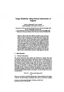
Probability distributions of images are easily derived by normalizing the correspondent histograms. Mutual information takes its maximum value when the underlying random variables (or images) are absolutely dependent (one of them is a function of the other one). In such case, the joint probability matrix (or joint histogram matrix) is diagonal. Reversely, if the images are independent, then the mutual information takes its minimum value, i.e. zero. In order to align two images, a transformation function T should be applied to the floating one. Transforming a grid point of floating image usually gives a point in the reference image which does not coincide with a grid point. Thus, an interpolation method is required to estimate the joint histogram of the images. The prevalent interpolation method in image processing is bilinear interpolation, which uses four nearest pixels of the transformed coordinate in the reference image. Joint histogram is easily obtained by a simple counting procedure.[11] With this method, the resulted mutual information function is not smooth and the optimization algorithm may converge to a wrong solution. The situation is more critical when the images are noisy. In order to overcome this problem, PV interpolation method was proposed. But it has been shown in[12] that if the underlying images have the same pixel size, PV and bilinear interpolation yield some local optima (artifacts) on the surface of mutual information function. A typical pattern of mutual information function in terms of displacement (translation) in horizontal and vertical directions is shown in Figure 1. It can be seen that the surface has some local minimums (res. maximums) for bilinear (res. PV) interpolation method.
INTERPOLATION METHODS AND PROPOSED ALGORITHM In this section, first we describe some interpolation
5.5 5.0 4.5 4.0 3.5 3.0 60
Mutual Information
The joint probability distribution can be derived by using Partial Volume (PV) or bilinear interpolation methods. [11] When the underlying images are acquired by a single sensor, and hence the size of their pixels is equal, mutual information (as a function of translation) shows some local optima at integer values.[12] These artifacts may cause the optimization algorithm to converge to wrong solutions. This will degrade the accuracy of the registration.
I ( A, B) = ∑ pA, B ( a, b).log(
Mutual Information
the joint probability distribution which influences the quality of image registration. Therefore, selection of a reliable interpolation method for estimating the joint probability distribution is of great importance.
40
20
Y-displacement
00
(a)
6 5 4 3
2 50 60 40 40 30 20 10 20 X-displacement Y-displacement
00
(b)
50 30 40 10 20 X-displacement
Figure 1: Typical patterns of local extremes of mutual information resulting from two interpolation methods. (a) PV; (b) Bilinear
Vol 1 | Issue 3 | Sep-Dec 2011
Soleimani and Khosravifard: Reducing Interpolation Artifacts for Mutual Information Based Image Registration
methods such as bilinear, PV, and its modifications. We investigate the reason behind the acceptable performance of GPV and HPV methods. Also, an interpolation scheme is proposed.
Bilinear Interpolation As mentioned in the previous section, transforming coordinate of a pixel of floating image F gives a new coordinate in the reference image R, which may not be a grid point. Hence, the intensity of such point must be somehow interpolated. In bilinear interpolation, the intensity is estimated by computing a weighted average of the intensity of four nearest pixels in the reference image. If the point p with coordinate s in the floating image is transformed (by transformation T ) to point q with coordinate TS in the reference image [Figure 2], the intensity of the reference image in coordinate TS is given by 4
R(Ts ) = ∑ wi R( ni ) i =1
w1 = (1 − d1 )(1 − d2 ) w3 = (1 − d1 )d2
w2 = d1 (1 − d2 ) w4 = d1d2
(2)
Where, ni , i=1,2,3,4 denote the coordinate of four nearest grid points to point q in the reference image. Note that we 4
have ∑ wi = 1.
which is known as Boxcar function. In,[9] Chen and Varshney proposed GPV interpolation method. They replaced Boxcar function with a third order Spline, i.e., x3 2 2 2 −x +3 (2 − x)3 f GPV ( x) = 6 0
1≤ x < 2
(5)
otherwise
In this method, sixteen pixels are used to calculate wi’s and update the joint histogram. As a result, for each pixel of floating image, sixteen bins are updated in the joint histogram. Note that the support of this function is the interval (0,2). In,[8] HPV interpolation method was proposed by replacing the boxcar function with Hanning windowed sinc function 1 + cos(2 πx / 4) f HPV ( x) = 4 0
0≤x