Texture Classification in Bioindicator Images Processing Martina Mudrov´a, Petra Slav´ıkov´a, and Aleˇs Proch´azka
Abstract The section deals with classification of microscope images of Picea Abies stomas. There is an assumption that a stoma character strongly depends on the level of air pollution, so that stoma can stand for an important environmental bioindicator. According to the level of stoma incrustation it is possible to distinguish several classes of stoma structures. A proposal of an algorithm enabling the automatic recognition of a stoma incrustation level is a main goal of this study. There are two principles discussed in the paper: The first principle is based on gradient methods while the second one uses a wavelet transform. Possibilities of application of mentioned attitudes were investigated and the classification criteria distinguishing the stoma character were suggested, as well. The resulting algorithm was verified for a set of four hundred real images and results achieved were compared with an expert’s sensual classification. Selected methods of image preprocessing as noise reduction, brightness correction and resampling are studied, as well.
1 Introduction Image processing represents a widely spread interdisciplinary research area with many applications in various disciplines including engineering, medicine and technology [8, 46, 21, 43]. The section is devoted to the application of selected mathePh.D. Martina Mudrov´a Department of Computing and Control Engineering, Institute of Chemical Technology Prague, Technick´a 1905, Prague 6, Czech Republic, e-mail:
[email protected] MSc. Petra Slav´ıkov´a Department of Computing and Control Engineering, Institute of Chemical Technology Prague, Technick´a 1905, Prague 6, Czech Republic, e-mail:
[email protected] Prof. Aleˇs Proch´azka Department of Computing and Control Engineering, Institute of Chemical Technology Prague, Technick´a 1905, Prague 6, Czech Republic, e-mail:
[email protected]
1
2
Martina Mudrov´a, Petra Slav´ıkov´a, and Aleˇs Proch´azka
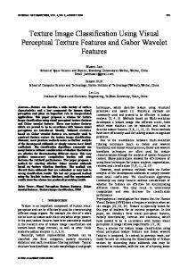
Fig. 1 Dust particles concentration over the Czech Republic observed during a selected day and time measured by ground measuring stations and extrapolated over the whole region pointing to the most polluted areas
matical methods in environmental engineering to detect and to analyze the quality of vegetation which can be substantially affected by air pollution and dust particles concentration. The air pollution is regularly observed by ground measuring stations in most countries. Figure 1 presents the situation in the Czech Republic with 103 observation points precisely defined by their longitude and latitude. Fig. 1 presents these surface observations interpolated to the whole Czech Republic. In this case, the spline two dimensional interpolation has been used allowing for evaluation of a given variable at the chosen grid points. This method allows the estimation of air pollution in places without measuring stations and detection of air pollution sources as well. Remote satellite sensing represents another method used for air pollution detection. Real data presented in Fig. 2 epresent images taken by satellites operated by NOAA (The National Oceanic and Atmospheric Administration). All these satellites fly on elliptical or circular orbits around the planet earth, the earth’s centre being the focal or central point and they belong to the so-called polar orbiting satel-
Latitude
(a) SATELLITE CHANNEL 1
(b) SATELLITE CHANNEL 2
51
51
50
50
49
49 12
14
16 Longitude
18
12
14
16 Longitude
18
Fig. 2 Satellite images of the Czech Republic from the NOAA satellite simultaneously observed by the scanning radiometer AVHRR using different spectral ranges with results obtained from its (a) channel 1 and (b) channel 2
Texture Classification in Bioindicator Images Processing
3
Fig. 3 Selected examples of electron-microscope images of stomas of Picea Abies presenting different stages of the affect of air pollution with the level of stoma incrustation increasing from the top to the bottom
lites which fly in rather low altitudes, typically at 850 km height. Polar orbiters provide excellent pictures of all parts of the earth including the polar regions. The main apparatus of NOAA satellites is the scanning radiometer AVHRR (Advanced Very High Resolution Radiometer). This is a five-channel apparatus covering spectral ranges (1) 0.58-0.68µ m, (2) 0.725-1.1µ m, (3) 3.55-3.93µ m, (4)10.3-11.3µ m, (5) 11.5-12.5µ m. The first two channels work with the reflected sun radiation only in red and close infrared region, the last two ones are fully heat radiation channels, and channel 3 is a mixed one. Figure 2 presents satellite images of the Czech Republic from the NOAA satellite simultaneously observed in two different spectral ranges. Their difference can be used as a measure of dust particles concentration owing to their different reflection providing an alternative to the ground observations in this way. Figure 3 presents the effect of air pollution to vegetation samples taken from selected parts of the Czech Republic. The figure shows electron-microscope images of stomas of Picea Abies. This tree is considered to be an important bioindicator of air pollution in a given area owing to the great appearance of this biotope within the Czech Republic and Europe generally. The stoma epidermis has usually miscellaneous structure but in the case of polluted air it covers with epicuticular vax to protect itself against negative influences of pollutants, and stoma epidermis incrusts. The level of incrustation can be used as a factor for air pollution assessment. Stoma
4
Martina Mudrov´a, Petra Slav´ıkov´a, and Aleˇs Proch´azka
Table 1 List of monitored localities 1 - Svratouch 2 - B´ysˇ´t u Pardubic 3 - Trnov´a
ˇ 4 - Repice u Strakonic ´ 5 - Borˇsice-Ujezd 6 - Velk´a Losenice
quality can be investigated using its microscope images and the level of incrustation can be divided into several different categories. In connection with the experimental results five classes [28] are used in the following text. As the class assignment can be easily affected by an expert’s personality in the case of human visual evaluation, a requirement of independent fair-minded images classification appears. The section presents analysis of the extensive set of electron-microscope images from several regions of the Czech Republic summarized in Table 1. The location of these areas is presented in Fig. 4. Mathematical methods for processing of time-series and images are very close. The section is devoted to methods of two-dimensional digital signal processing forming the basic tool for automated description, classification or evaluation of images [29], their segmentation [38], compression, pattern recognition [3, 37, 1, 2, 23, 15, 41] and image enhancement [34]. These methods include both time-frequency [5] and time-scale [9, 20, 18, 35, 19, 10, 16] signal analysis procedures. There is a large amount of books and papers devoted both to image processing theory and to the description and to the development of mathematical and algorithm background [44, 6, 9, 12, 40, 45, 47]. Publications devoted to the application of selected methods in various branches are widely spread as well. The goal of this contribution is in the development of an automatic algorithm which can recognize the level of stoma changes [33] by means of methods of texture classification. Essential algorithmic procedures discussed further cover topics presented in Fig. 5 and including image preprocessing methods, segmentation procedures and classification algorithms. The following study is devoted to the discussion of two basic principles including (i) the application of gradient methods and (ii) the use of methods based on the wavelet transform. These methods are preceded by selected image preprocessing
2 3 1 6
Fig. 4 Localities within the Czech Republic from where vegetation samples were collected
4 5
Texture Classification in Bioindicator Images Processing
5
Input Image
PRE-PROCESSING
SEGMENTATION
Resampling
Image Segmentation STOMA SELECTION
Median Filtering
Segment Criteria Evaluation
Brightness Correction
Segment Classification
Output Classified Image
Fig. 5 The block diagram presenting the fundamental steps used for classification of microscopic images including their preprocessing, feature extraction and classification
procedures for image enhancement and noise reduction. The whole algorithm proposed based upon the structure presented in Fig. 5 is presented in the final part of the paper. Results achieved [39] are based upon the analysis of the set of about four hundred images collected from 6 localities in the Czech Republic with their location summarized in Table 1 and presented in Fig. 4.
2 Image Preprocessing The set of about four hundred real images was processed in this study. As images were observed under different conditions thanks to various stoma size and character, the image preprocessing was unavoidable to eliminate unexpected effects. Images were unified into the same resolution, cropped to the same sizes and their energy was normalized. The application of non-linear digital filters turns out to be suitable before their feature extraction and image classification. Median filter was used as a proper method in this case.
2.1 Image Resizing In the first step it was necessary to unify image resolution. Various methods of the two-dimensional interpolation [17, 42] can be used in the process of image resampling. The method of the nearest neighbor, bilinear, bicubic and spline interpolation were tested and their results compared. An interpolation method based on the discrete two-dimensional Fourier transform [11] have been studied in this connection
6
Martina Mudrov´a, Petra Slav´ıkov´a, and Aleˇs Proch´azka
as well. In the final version, the bilinear method has been selected. Together with the change of resolution it was necessary to crop images into the same size to enable comparative data processing and results.
2.2 Energy Normalization Unification of level of brightness was a second step of image preprocessing as images were taken under various light condition. The energy E of each image x of size M x N evaluated by relation M
E=∑
N
∑ xi,2 j
(1)
i=1 j=1
was normalized for all images with the mean value of image pixels set to 0 and the standard deviation set to 1 at the same step.
2.3 Median Filtering During the following processing the necessity of lowpass filtering appears. It is possible to notice the white spots in the lowest image in the Fig. 3. These spots are considered to be indicators of stoma structure recovery. They appear only in some images of class 5. Their presence affects the wrong classification of these stomas so they should be removed. It is possible to use various methods of noise rejection [4]. Median filtering is considered to be a suitable method used to decrease the effect of this shoot-typed noise. Filter size selection depends on image resolution and the size of disturbing areas. Filter size of 3 × 3 pixels has been satisfactory in this case.
3 Texture Analysis Methods As it is not trivial to delimitate the stoma border within the image, methods of texture classification were applied. There are various attitudes to solution of a general problem of texture analysis mentioned in [26, 13]. Presented paper is focussed to the application of gradient methods and edge detection and to the possibilities of wavelet transform use for analysis of given images. Various edge detectors were investigated as well as various wavelet functions. The principle of application the selected method to the given image set included (i) the split of each image into several areas of the same size and (ii) separate processing of each area. The subimage size was selected so that it would be easy to use the wavelet transform, i.e. dimensions of them should be a power of 2. Then it
Texture Classification in Bioindicator Images Processing
7
Fig. 6 Selected templates of textures found in given images. Each row corresponds to one class, so that a level No. 1 is in the first row and level No. 5 is in a bottom.
is possible to assign various weights according to image area site as the stoma was usually located in the center of image, as well. As the definition of stoma classification [28] is uncertain and it is given by several phrases only, a database of cuts was created at first and they were investigated by an experienced expert to obtain his classification of each of them. In the case that his classification was not obviously evaluated, the cut was excluded from the database. Clearly evaluated cuts solved in the next processing as the templates for criteria values determination. A selected set of these templates is presented in Fig. 6. Each row includes typical representatives of the same class.
3.1 Gradient Methods and Edge Detection Gradient methods mentioned e.g. in [12, 32] belong to the basic tool of image processing. As they discover changes of brightness function, edge detection provides their primary use. The principle of gradient methods is based on the convolution of an image x of size M x N and a convolution mask h according to relation M−1 N−1
y(i, j) =
∑ ∑ x(m, n)h(i − m, j − n)
(2)
m=0 n=0
Resulting image y can be thresholded to obtain binary image only in which the pixels with a value ”one” indicate a part with the highest level of brightness changes these pixels represent edges in the original image. The shape and size of the convolution mask h depends upon the degree of derivation used, its direction and method.
8
Martina Mudrov´a, Petra Slav´ıkov´a, and Aleˇs Proch´azka
Robinson, Prewitt, Kirsch and Sobel convolution masks were applied to the set of image segments with a size 128 x 128 pixels with known level 1 - 5 of stoma incrustation. Selected example of a set of such image segments is presented in Fig. 6. As the operators mentioned above have directional behavior, resulted image was obtained as a sum of convolutions in each direction. Otsu’s algorithm [30] was used for the threshold selection during the process of conversion into a binary image. Number of white pixels G in each segment was evaluated and its correlation with a class number was compared - see Fig. 7. It is obvious that with increasing level of incrustation the fine structure of stoma is gradually disappearing and the number of edge pixels G is decreasing. More sophisticated Canny method [7] for edge detection was used as well. Application of Canny’s method provides the best correlation with a segment class (see Fig. 7) so a criterion Kg could be established. It splits segments’ classes according to percentage of white pixels - see Table 2. Segments with values Kg > 17.5 are considered to be class 1 while segments with Kg < 12.2 are supposed to be class 5.
(a) Prewitt
(b) Sobel
5000
5000 G
7500
G
7500
2500
2500
0
0 1
2
3 Class
4
5
1
2
(c) Kirsch
3 Class
4
5
(d) Canny 3000 G
G
4000
2000
2000 1000 0
0 1
2
3 Class
4
5
1
2
3 Class
4
5
Fig. 7 Results of application of Prewitt (a), Sobel (b), Kirsch (c) and Canny (d) methods to the selected set of image cuts with a known classification. Number of pixels representing edges G by given method is drawn in dependence upon stoma class. Mean values and standard deviations are marked, as well.
3.2 Wavelet Transform Use for Image Classification The discrete wavelet transform (DWT) [9, 45] is studied for more than 20 years already with its mathematical roots going to last several centuries. There is a large
Texture Classification in Bioindicator Images Processing
9
Table 2 Image Texture Classification Using Gradient Method Border values of gradient criterium Criterion
1−2
2−3
3−4
4−5
Kg [%]
17.5
16.2
14.0
12.2
amount of papers concerning its application in the area of image segmentation, noise reduction, classification and others [4, 6]. The basic idea is based on the use of a special set of wavelet and scaling functions to decompose the original one-dimensional continuous signal x into its detail and approximation coefficients. The the basic idea of 1-dimensional wavelet transform based on initial wavelet function dilation and translation can be mathematically described by relation DW T {x(k)} ≡ X(m, n) = 2− 2
m
∑ x(k)ψ (2−m k − n)
(3)
k
The Mallat’s pyramidal scheme [25, 40] presented in Fig. 8 describes another attitude for wavelet transform understanding. It describes the DWT as a set of band filters which are applied subsequently in several levels to obtain approximation coefficients. A wavelet and proper scaling function use guarantees the possibility of perfect reconstruction of the original signal.
Signal reconstruction in the 1st level
Scale
Signal decomposition
Fig. 8 Mallat’s pyramid scheme of signal decomposition. The principle of the reconstruction in the first level is denoted, as well
n Wavelet coefficients
10
Martina Mudrov´a, Petra Slav´ıkov´a, and Aleˇs Proch´azka
Fig. 9 Wavelet transform of a selected image cut. Decomposition to the second level is shown
Thanks to these properties the DWT can be simply extended into two dimensions and in this form it can be used as a powerful tool in image processing. The wavelet decomposition at each level can be performed separately in the rows and columns of the image. The combination of DWT application in both directories is provides two-dimensional wavelet coefficients passing on into the next decomposition level. Fig. 9 presents an example of wavelet decomposition of selected real image segment into 2 levels. Various wavelet functions were used for processing of the given set of real image segments (with the same size of 128 x 128 pixels, again). The proper criteria for stoma structure class evaluation were searched. Selected example of wavelet decomposition of presented set of image segment is shown in Figures 10 and 11 where the sum CW of absolute values of wavelet coefficients ci, j is explored in dependence upon the decomposition level. Coefficients belonging to the same class segments are drawn in the separated graphs with increasing class level from the left to the right. A set of ten selected segments of the same class were decomposed into the third level and the sum of their wavelet coefficient in their absolute values were explored. Again it is possible to notice the decreasing level of these sums with increasing segment’s class. This fact can be simply explained by vanishing fine stoma structure with increasing segment class, so that the details in the wavelet decomposition subsequently disappears.
300
CW
200
100
0 1
2
31
2
31 2 31 Level of Decomposition
2
31
2
3
Fig. 10 A sum of wavelet coefficients in their absolute values in the three levels of decomposition. Daubechies 1 wavelet was used for decomposition of a selected set of 10 cuts with known classification. Class level is increasing from the left graph (class No. 1) to the right one (class No. 5)
Texture Classification in Bioindicator Images Processing
11
150
CW
100
50
0 1
2
31
2
31 2 31 Level of Decomposition
2
31
2
3
Fig. 11 A sum of wavelet coefficients in their absolute values in the three levels of decomposition. Daubechies 8 wavelet was used for decomposition of a selected set of 10 cuts with known classification. Class level is increasing from the left graph (class No. 1) to the right one (class No. 5)
According to achieving results, the following three border criteria were suggested: The criterion Ke according the Eq. (4) evaluates a sum of wavelet coefficients of the first level decomposition using Daubechies 1 wavelet function. The criterion Ks1 given by Eq. (5) is suggested as a difference of sum of Daubechies 8 wavelet coefficients the second and the first level. The coefficients are summarized in their absolute values. Similarly, the criterion Ks2 defined according to Eq. (6) is counted using the Daubechies 8 wavelet coefficients coming from the third and the second level. M
N
Ke = ∑ ∑ |ci, j |
(4)
i=1 j=1
M
Ks1 = ∑
N
∑ |c2i, j level | − |c1i, j level | st
nd
(5)
i=1 j=1 M
Ks2 = ∑
N
∑ |c3i, j level | − |ci,2 j level | rd
nd
(6)
i=1 j=1
The Table 3 presents border values of mentioned criteria solving for classification of a given image segment. Table 3 Image Texture Classification Using Wavelet Method Border values of wavelet criteria Criterion
1−2
2−3
3−4
4−5
Ke Ks1 Ks2
187 115 -32
171 98 17.7
160 79 10
131 72 0
12
Martina Mudrov´a, Petra Slav´ıkov´a, and Aleˇs Proch´azka
Table 4 Comparison of automatic and sensual stoma evaluation of the set of four hundred images Differences between Automatic and Sensual Classification Criterion
Kg
Ks1
Ks2
Ke
Difference of ±1 class (%) Difference of ±2 class (%) Difference of ±3 class (%)
95.40 4.60 0.00
92.33 6.65 1.02
81.33 17.90 0.77
87.98 11.25 0.77
4 Results According to suggested criteria the resulting algorithm was proposed - see Fig. 5 and it was verified for more than four hundred real images. As the real images differ in value of brightness and they have the various level of resolution, these parameters had to been unified at first. The influence of noise was reduced by the application of median filter with size 3 × 3 pixels. The image segments were evaluated by a selected criterion while the segments belonging to stoma only were considered. Achieved results were compared with an experts’ sensual image evaluation. The difference of one class was omitted. Comparison of automatic and individual results is presented in Table 4. A selected example of segment classification is presented in Fig. 12. It would be also interesting to compare an achieving results with another air pollution indicators. The Table 5 could solve for such a comparison.
Fig. 12 Result of a selected image classification.
4
4
3
3
4
4
3
1
2
4
4
3
2
2
4
4
4
5
4
4
Texture Classification in Bioindicator Images Processing
13
Table 5 Mean Level of Stomas’ Class in the monitored localities according to suggested algorithm No. of Locality Criterion Kg Ks1 Ks2 Ke
1 3.3 ± 0.9 3.2 ± 0.7 3.3 ± 0.6 2.5 ± 0.7
2 3.5 ± 0.9 3.5 ± 0.6 2.6 ± 0.6 2.8 ± 0.8
3 3.4 ± 0.6 3.3 ± 0.5 2.6 ± 0.5 2.5 ± 0.5
4 3.3 ± 0.6 3.2 ± 0.5 2.2 ± 0.5 2.5 ± 0.7
5 3.1 ± 0.8 3.1 ± 0.6 2.2 ± 0.5 2.5 ± 0.6
6 3.5 ± 0.7 3.4 ± 0.6 2.5 ± 0.6 2.6 ± 0.6
5 Conclusion The paper presents the problem of the affect of air pollution to the quality of vegetation. In the general sense this problem is a global one and it can be extrapolated to the study of environment pollution and its affect to the environment quality both on the earth’s continents and inside oceans and seas. Algorithmic tools proposed in the paper are devoted to the analysis of the quality of vegetation derived from microscopic images of tree needles in the different areas of the Czech Republic. Results obtained by the wavelet transform are compared with those obtained by general gradient methods. Further research will be devoted to the study of the most appropriate methods for analysis of textures closely related to the quality of the vegetation. More detail study will be devoted to the correlation between air pollution sources and changes of vegetation.
Acknowledgement This work has been supported by the Ministry of Education of the Czech Republic (program No. MSM 6046137306). This support is very gratefully acknowledged.
References 1. Arivazhagan, S., Ganesan, L.: Texture Classification Using Wavelet Transform. Pattern Recogn. Lett. 24(9-10), 1513–1521 (2003) 2. Arivazhagan, S., Ganesan, L.: Texture Segmentation Using Wavelet Transform. Pattern Recogn. Lett. 24(16), 3197–3203 (2003) 3. Bishop, C.M.: Neural Networks for Pattern Recognition. Oxford University Press (1995) 4. Boashash, B.: Time-Frequency Signal Analysis and Processing - A Comprehensive Reference, Elsevier Science, Oxford, UK, 2003 5. Bracewell, R.N.: Fourier Analysis and Imaging. Kluwer Academic Press (2003) 6. Burger, W. and Burge, M. J. Digital Image Processing, Springer, USA, 2008
14
Martina Mudrov´a, Petra Slav´ıkov´a, and Aleˇs Proch´azka
7. Canny, J., F., A computational approach to edge detection. IEEE Transactions on Pattern Analysis and Machine Intelligence, 8, 1986, pp. 679-698. 8. Chellapa, R.: Digital Image Processing. IEEE Computer Society Press, Los Alamitos (1992) 9. Daubechies, I.: Orthonormal Bases of Compactly Supported Wavelets. Comm. Pure and Applied Math, 41, Nov. 1998, pp. 909-996 10. Debnath, L.: Wavelets and Signal Processing. Birkhauser Boston, Boston, U.S.A. (2003) 11. Frayer, D.: Interpolation by the FFT Revised-an Experimental Investigation. IEEE Trans. Acoustics, Speech and Signal Processing, 37(5), 1989, pp. 665-675 12. Gonzales, R.C. and Woods, R.E.: Digital Image Processing, 2nd ed. Prentice Hall, New Yersey, USA, 2002 13. Haralick, R.M., Shanmugan, K. and Dinstein, I.: Texture Features for Image Classification. IEEE Trans. on Syst. Man Cybern., 3(6), 1992, pp. 610-612 14. Huang, K. and Aviyente, S.: Wavelet Selection for Image Classification. IEEE Trans. on Image Processing, 17(9), Sept. 2008, pp. 1709-1720 15. Jafari-Khouzani, K., Soltanian-Zadeh, H.: Rotation-Invariant Multiresolution Texture Analysis Using Radon and Wavelet Transforms. IEEE Trans. on Image processing 14(6), 783–795 (2005) 16. Jafari-Khouzani, K., Soltanian-Zadeh, H.: Rotation-invariant multiresolution texture analysis using Radon and wavelet transforms. IEEE Transaction on Image Processing 14(6), 783–795 (2005) 17. Keys, R.: Cubic Convolution Interpolation for Digital Image Processing. IEEE Trans. Acoustics, Speech and Signal Processing, 29(6), 1981, pp. 1153-1160 18. Kingsbury, N.G.: Complex Wavelets for Shift Invariant Analysis and Filtering of Signals. Journal of Applied and Computational Harmonic Analysis 10(3), 234–253 (2001) 19. Selesnick, I.W., Baraniuk, R.G. and Kingsbury, N.G.: The Dual-Tree Complex Wavelet Transform. IEEE Signal Processing Magazine 22, 123–151 (2005) 20. Kingsbury, N.G., Mugarey, J.F.A.: Wavelet Transforms in Image Processing. In: A. Proch´azka, J. Uhl´ıˇr, P.J.W. Rayner, N.G. Kingsbury (eds.) Signal Analysis and Prediction, Applied and Numerical Harmonic Analysis, chap. 2. Birkhauser, Boston, U.S.A. (1998) 21. Klette, R., Zamperoni, P.: Handbook of Image Processing Operators. John Wiley & Sons, New York (1994) 22. Laine, A. and Fan, J.: Texture Classification by Wavelet Packet Signature. IEEE Trans. on Pattern Analysis and Machine Intelligence, 15(11), 1993, pp. 1186-1190 23. Li, S., Shawe-Taylor, J.: Comparison and fusion of multiresolution features for texture classification. Pattern Recogn. Lett. 25 (2004) 24. Lindeberg, T.: Edge Detection and Ridge Detection with Automatic Scale Selection. Internation Journal of Computer Vision, 30(2), 1998, pp. 117-154 25. Mallat, S.:A Theory of Multiresolution Signal Decomposition: The Wavelet Representation. IEEE Trans. on Pattern Analysis and Machine Intelligence, 11(7), 1989, pp. 674-693 26. Mirmehdi, M., Xie X. and Suri, J. Handbook of Texture Analysis, World Scientific Publishing, New Jersey, USA, 2008 27. Moosmann, F. and Nowak, E. anod Jurie, E.: Randomized Clustering Forests for Image Classification. IEEE Trans. on Pattern Analysis and Machine Intelligence, 30(9), Sept. 2008, pp. 1632-1646 28. N´ahl´ık, J., Cudl´ın, P., Kaˇsov´a, E., Barton´ıcˇ kov´a, R.: Studium morfologie stomat´aln´ıho vosku ˇ pomoc ERM. ˇ smrku ztepil´eho na vybranch lokalit´ach CR Stav a Perspektivy ekologick´eho v´yskumu horsk´ych lesn´ych ekosyst´emov, 2001, Slovensk republika, Polana, SR 2001 29. Nixon, M., Aguado, A.: Feature Extraction & Image Processing. NewNes Elsevier (2004) 30. Otsu, N., A Threshold Selection Method from Gray-Level Histograms IEEE Transactions on Systems, Man, and Cybernetics, 9, No. 1, 1979, pp. 62-66. 31. Parker, A.J., Kenyon, R.V. and Troxel, D.E.: Comparison of Interpolating Methods for Image Resampling. IEEE Trans. on Medical Imaging, 2(1), 2007, pp. 31-39 32. Petrou, M. and Bosdogianni, P. Image Processing, The Fundamentals, John Wiley and Sons, New York, USA, 2000
Texture Classification in Bioindicator Images Processing
15
33. Proch´azka, A. and Gavlasov´a, A., and Volka, K.: Wavelet Transform in Image Recognition. In: International conference ELMAR05, Zadar. Croatia. IEEE (2005) 34. Proch´azka, A. and Pt´acˇ ek, J.: Wavelet Transform Application in Biomedical Image Recovery and Enhancement. In: The 8th Multi-Conference Systemics, Cybernetics and Informatic, Orlando, USA, 6, pp. 82–87. IEEE (2004) ˇ 35. Pt´acˇ ek, J., Sindel´ aˇrov´a, I., Proch´azka, A., Smith, J.: Wavelet Transforms In Signal And Image Resolution Enhancement. In: International Conference Algoritmy 2002, Podbanske. STU (2002) 36. Randen, T. and Husoy, J.H.: Filtering for Texture Classification: A Comparative study. IEEE Trans. on Patern Analysis and Machine Inteligence, 21(4), 1999, pp. 291-310 37. Randen, T., Husoy, J.H.: Filtering for Texture Classification: A Comparative Study. IEEE Trans. on PAMI 21(4), 291–310 (2000) 38. Shaffrey, C.W.: Multiscale Techniques for Image Segmentation, Classification and Retrieval. Ph.D. thesis, University of Cambridge, Department of Engineering (2003) 39. Slav´ıkov´a, P., Mudrov´a, M. and Proch´azka, A.: Automatic Bionindicator Images Evaluation. Intelligent Engineering Systems, 2010. INES 2010. 40. Strang, G., and Nguyene, T., Wavelets and Filter Banks, Wellesley-Cambridge Press, USA, 1996 41. Stringer, S.M.: Invariant Object Recognition in the Visual System with Novel Views of 3D Objects. Neural Computing (14), 2585–2596 (2002) 42. Unser, M.: Texture Classification and Segmentation using Wavelet Frames. IEEE Trans. on Image Processing, 4(11), 1995, pp. 1549-1560 43. Van der Heijden, F.: Image Based Measurement Systems. John Wiley & Sons, New York (1994) 44. Vaseghi, S.V.: Advanced Digital Signal Processing and Noise Reduction. John Wiley & Sons Ltd (2006) 45. Vetterli, M. and Kovacevic, Wavelets and Subband Coding, Prentice Hall New Yersey, 1995 46. Watkins, C., Sadun, A., Marenka, S.: Modern Image Processing: Warping, Morphing, and Classical Techniques. Academic Press, Ltd., London (1993) 47. Weibao, Zou and Yan, Li: Image Classification Using Wavelet Coefficients in Low-pass Bands. Neural Networks, 2007. IJCNN 2007, pp.114–118 48. Ziou, D. and Tabbone, S.: Edge Detection Techniques: An Overview. Internation Journal of Pattern Recognition and Image Analysis, 8(8), 1998, pp. 537-559 49. Zhang, W. and Berholm, F.: Multi-scale Blur Estimation and Edge Type Classification for Scene Analysis. Internation Journal of Computer Vision, 24(3), 1997, pp. 219-250
Index
air pollution bioindicators, 3 ground observation, 2 satelite observation, 3
Interpolation two dimensional methods use, 2, 5
gradient methods edge detectors application Canny, 8 Kirsch, 8 Prewitt, 8 Robinson, 8 Sobel, 8
noise removing, 6
image processing application, 3
median filter application, 5, 6
stoma classification, 3 texture classification, 4 wavelet transform application, 10 wavelet function selection, 10
17