Seizure 26 (2015) 56–64
Contents lists available at ScienceDirect
Seizure journal homepage: www.elsevier.com/locate/yseiz
Review
Wavelet-based EEG processing for computer-aided seizure detection and epilepsy diagnosis Oliver Faust a, U. Rajendra Acharya b,*, Hojjat Adeli c,d,e,f, Amir Adeli g a
School of Science and Engineering, Habib University, Karachi, Pakistan Department of Electronics and Computer Engineering, Ngee Ann Polytechnic, Singapore 599489, Singapore c Department of Neuroscience, The Ohio State University, Columbus, OH, USA d Department of Biomedical Engineering, The Ohio State University, Columbus, OH, USA e Department of Biomedical Informatics, The Ohio State University, Columbus, OH, USA f Department Electrical and Computer Engineering, The Ohio State University, Columbus, OH, USA g Department of Neurology, The Ohio State University, 470 Hitchcock Hall, 2070 Neil Avenue, Columbus, OH 43210, USA b
A R T I C L E I N F O
A B S T R A C T
Article history: Received 25 October 2014 Received in revised form 15 January 2015 Accepted 18 January 2015
Electroencephalography (EEG) is an important tool for studying the human brain activity and epileptic processes in particular. EEG signals provide important information about epileptogenic networks that must be analyzed and understood before the initiation of therapeutic procedures. Very small variations in EEG signals depict a definite type of brain abnormality. The challenge is to design and develop signal processing algorithms which extract this subtle information and use it for diagnosis, monitoring and treatment of patients with epilepsy. This paper presents a review of wavelet techniques for computeraided seizure detection and epilepsy diagnosis with an emphasis on research reported during the past decade. A multiparadigm approach based on the integration of wavelets, nonlinear dynamics and chaos theory, and neural networks advanced by Adeli and associates is the most effective method for automated EEG-based diagnosis of epilepsy. ß 2015 British Epilepsy Association. Published by Elsevier Ltd. All rights reserved.
Keywords: Continuous Wavelet Transform Discrete Wavelet Transform Electroencephalogram Epilepsy
1. Introduction Scientific work on mechanizing the detection of epileptic seizures began around 1970 [1]. However, it took about 30 years to develop the algorithms to turn these ideas into physical problem solutions [2]. Various Electroencephalography (EEG) analysis and classification methods use the fact that the information processing in the brain is reflected in the EEG as dynamical changes of the electrical activity in time, frequency, and space. Various frequency and nonlinear methods have been used to understand the mechanisms behind the information processing [3,4]. Among time–frequency analysis methods the Wavelet Transform (WT) stands out in terms of algorithmic elegance and efficiency. WT captures the subtle changes in the EEG signal well. These minute variations are difficult to spot using the naked eye in the EEG signal. Therefore, this review is dedicated to WT-based EEG processing and computer-aided seizure detection and epilepsy diagnosis.
* Corresponding author. Tel.: +65 64606135; fax: +65 64671730. E-mail address:
[email protected] (U.R. Acharya).
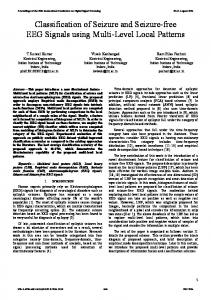
Fig. 1 shows sample normal, interictal and ictal (epileptic) EEG signals from a database hosted by the University of Bonn [5]. The signals in the database were obtained from five normal subjects and five epileptic patients. The EEG data is classified into three categories: control, interictal, and ictal/epileptic. The interictal EEG signals were obtained from the hippocampal formations during seizure free intervals. The ictal signals were recorded from the lateral and basal regions of the neocortex. The EEG signals were recorded using a 128-channel amplifier system digitized with a sampling frequency of 173.61 Hz, and filtered using a band pass filter of 0.53–40 Hz.
2. Computer aided seizure detection A trend in healthcare is shifting from clinician-centric care to patient-centric care where the patient becomes an active participant in her care management. Automated Computer-Aided Diagnosis of epilepsy which is significantly more challenging than computer-aided seizure detection was advanced with the seminal work of Adeli et al. [6] for diagnosis of the absence seizure more than a decade ago. This review aim to summarize the research reported since then. A CAD system can help neurologists make the
http://dx.doi.org/10.1016/j.seizure.2015.01.012 1059-1311/ß 2015 British Epilepsy Association. Published by Elsevier Ltd. All rights reserved.
O. Faust et al. / Seizure 26 (2015) 56–64
57
provides a smooth representation, unlike windowed representation in the short time Fourier transform. Hence, one can capture very minute details, sudden changes and similarities in the EEG signals. WT acts like a mathematical microscope, because it has the capability to analyze EEG signals at different scales [12]. Researchers have proposed unique wavelets. Fig. 3 shows examples of mother wavelets used for EEG processing: (a) Daubechies (db), (b) Morlet, (c) Biorthog-onal (bior), (d) Orthogonal Cubic Spline (ocs), (e) Mexican Hat (MH), (f) Haar, (g) Complex Gaussian (CG), and (h) Coiflet (coif) wavelet. The following two sections introduce the application of WT in computer aided seizure detection. 3. Signal preprocessing/denoising
Fig. 1. Sample EEG signals.
diagnosis more efficiently and accurately. Fig. 2 presents a block diagram for a computer-aided seizure detection and epilepsy diagnosis system. It consists of an offline and an online system. The offline system consists of the design steps necessary to create and test the CAD algorithm structure. This algorithm is then used in the online system by the neurologist as a decision support system. WT is used for signal preprocessing/denoising and for feature extraction. WT was developed in the 1980s as a powerful signal processing technique to overcome the shortcomings of other methods such as the Fourier transform [11]. Since then it has been used in a variety of signal processing applications [8,9]. WT
Raw EEG signals suffer from poor spatial resolution, low signalto-noise ratio and artifacts [7]. Preprocessing is the denoising step which aims to improve the signal-to-noise ratio of the EEG. The WT is now a well-known tool for removing noise from the signal. Multi-resolution analysis provides information about the signal in different frequency bands. The wavelet decomposition of a noisy signal concentrates intrinsic signal information in a few wavelet coefficients having large absolute values without modifying the random distribution of noise. Therefore, denoising can be achieved by thresholding the wavelet coefficients. The WT gives a time-variant decomposition, an advantage over techniques such as Wiener filtering. With a time-variant decomposition it is possible to choose different filtering settings (i.e. wavelet coefficients) for different time ranges. Hence, it is possible to create event-related filter responses. With time-invariant approaches, such as Wiener filtering, it is not possible to find a unique implementation that is suitable for all event related potentials [10]. 4. Wavelet analysis for signal feature extraction In addition to denoising, wavelets can be used for feature extraction. The mother wavelet is shifted by a small interval in the x-axis and correlation coefficients are computed. This procedure is repeated for various scaling factors (dilations) in the y-axis.
Fig. 2. Block diagram for a computer-aided seizure detection and epilepsy diagnosis system.
O. Faust et al. / Seizure 26 (2015) 56–64
58
(a) db Wavelet
(c) Biorthogonal Wavelet
(e) Mexican Hat Wavelet
(g) Gaussian Wavelet
(b) Morlet Wavelet
(d) Spline Wavelet
(f) Haar Wavelet
(h) Coiflet Wavelet
Fig. 3. Examples of wavelets used for EEG processing.
There are two types of wavelet analysis: Continuous Wavelet Transforms (CWT) and Discrete Wavelet Transform (DWT) [12]. The CWT coefficients are evaluated for a continuous variation (infinitesimal increments) of both translation and dilation factors. DWT processes the input signals with finite impulse response filters. 4.1. Continuous time wavelet analysis (CWT) Duration and location of the abnormality can be detected using dilation and translation factors. Fig. 4 shows scalogram plots of the normal, ictal, and interictal EEG signals shown in Fig. 1, where
x-axis represents time, y-axis is the scale factor (reciprocal of frequency) and pixel intensity represents the magnitude of the correlation. Scalogram plots are unique and the sudden changes in the epileptic EEG signal can be observed through changes in the color. There is continuous variation in the color in the plot depicting the randomness as well chaotic nature of the EEG signal. Fig. 4(b) shows repeating patterns compared with Fig. 4(a) which appears as random. Table 1 summarizes published research on EEG signal feature extraction using CWT in terms of goal, wavelet used (W), number of signal classes (No) and results evaluation. The EEG data used has five subgroups Z, O, N, F, and S. Subgroups Z and O correspond to
O. Faust et al. / Seizure 26 (2015) 56–64
59
Fig. 4. Scalogram plots of the signals shown in Fig. 1 using CWT.
the EEG signal acquired from normal subjects with eyes closed and open respectively. N and F subgroups indicate the interictal (seizure free) EEG signals. S group denotes the seizure EEGs. No = 2 indicates two classes of normal (O) and seizure (S) classes; No = 3
means three classes of normal (O), interictal (F) and seizure (S) classes; No = 4 indicates four classes of normal (O), seizure-free intervals of five patients from hippocampal formation of opposite hemisphere (N), seizure-free intervals of five patients from
Table 1 Summary of published research on EEG signal feature extraction using CWT in terms of goal, wavelet used (W), number of signal classes (No), and results evaluation. Author
Year
Goal
W
No
Result evaluation
Gadhoumi et al. [74] Acharya et al. [29]
2013 2013
Seizure prediction CAD of epilepsy
Morse MH
2 3
Gadhoumi et al. [75]
2012
Morse
2
Indic and Narayanan [76]
2011
Morlet
–
–
Abibullaev et al. [77] Pravin Kumar et al. [78] Arab et al. [79]
2010 2010 2010
bior bior4.4 bior3.3
2 3 3
ANN, accuracy ANN, accuracy sensitivity specificity ANN, accuracy
Abibullaev et al. [80] Meier et al. [81] Chiu et al. [82] Subasi et al. [83] Gigola et al. [84] Rosso et al. [85] Latka et al. [86]
2010 2008 2006 2005 2004 2003 2003
Discrimination of pre-ictal and interictal states Study of EEG before and during mesial temporal lobe seizures CAD of epilepsy Seizure detection Feature extraction and de-noising topographic brain mapping and epilepsy classification Seizure detection Seizure detection Online seizure detection CAD of epilepsy Seizure prediction Seizure analysis Seizure analysis
Nonstandard classifier Various standard classifier, accuracy sensitivity specificity Nonstandard classifier, sensitivity
bior1.5 Symlet Nr Morlet db4 Ocs MH
3 2 4 2 – 3 –
ANN, SVM, ANN, ANN, – –
accuracy accuracy accuracy accuracy sensitivity specificity
O. Faust et al. / Seizure 26 (2015) 56–64
60
epileptogenic zone (F), and seizure (S) classes, and No = 5 indicates five classes: normal with eyes open (O), normal with eyes closed (Z), seizure-free intervals of five patients from hippocampal formation of opposite hemisphere (N), seizure-free intervals of five patients from epileptogenic zone (F), and seizure (S) classes. 4.2. Discrete Wavelet Transform (DWT) The DWT algorithm decomposes a given signal into approximation and detail coefficients to obtain a first level of decomposition. The approximation coefficients in every level are further decomposed into next level of approximation and detail coefficients [12]. The features extracted from the detailed coefficients at various levels or different frequency bands can reveal the characteristics of the time series and can be used in automated seizure detection systems [13,14]. Leung et al. used an absolute slope method for DWT-based EEG denoising and feature extraction [16]. Is¸ik and Sezer used DWT-based denoising and feature
extraction prior to application of Artificial Neural Networks (ANN) for epilepsy diagnosis [18]. Table 2 summarizes published research on EEG signal feature extraction using CWT in terms of goal, wavelet used (W), number of signal classes (N) and results evaluation. Table 3 shows 40 studies used DWT and 21 studies used CWT. A classification accuracy of 99.7% was reported using DWT [24] and a classification accuracy of 96% was reported using CWT [28]. CWT provides a pictorial representation of the signal. However, classification algorithms often require two-dimensional (2D) feature vectors. DWT coefficients can be used directly as features. It turns out that the DWT coefficients represent the subtle changes in the EEG signal very well. It can be seen from Tables 1 and 2 that various studies have been conducted using wavelet features for automated seizure detection. The detailed coefficients of the DWT at various levels have the signatures of the normal, interictal and ictal EEG classes. These features can be used for automated detection of seizure. In CWT, various 2D features like texture, fractal dimension, entropies,
Table 2 Summary of published research on EEG signal feature extraction using DWT in terms of goal, wavelet used (W), number of signal classes (No), and results evaluation (Var indicates that different mother wavelets were used). Year
Goal
W
No
Result evaluation
Nunes et al. [35]
2014
EEG signal classification
Var
4
Chen [87]
2013
Nr
4
Xie and Krishnan [33]
2013
Haar
2
SVM k-NN FLD
Acharya et al. [46]
2012
Seizure detection with dual-tree complex DWT Seizure detection and epilepsy diagnosis Seizure diagnosis
OPF 10-fold stratified cross validation accuracy sensitivity PPV Various standard classifier, accuracy
Bior
3
Various classifiers, accuracy,
Acharya et al. [24]
2012
Acharya et al. [27]
Author
sensitivity, specificity Various standard classifier, accuracy, sensitivity, specificity Various standard classifiers, accuracy sensitivity specificity PPV k-NN, accuracy ANN, accuracy, sensitivity, specificity Sensitivity, specificity, ROC PNN, accuracy, sensitivity, specificity ANN, accuracy, sensitivity, specificity SVM, accuracy ANN, accuracy, sensitivity, specificity – Threshold Threshold, sensitivity, specificity Modified ANN, parametric and sensitivity analysis ANN, specificity, selectivity ANN, sensitivity, specificity, ROC ANN, accuracy Spiking neural network, accuracy – – ANN, accuracy, sensitivity, specificity – Mixture of experts, sensitivity, specificity Adaptive neuro-fuzzy inference system, sensitivity, specificity – – ANN and fuzzy logic, sensitivity, specificity ANN, sensitivity, specificity –
db10
3
2011
CAD of epilepsy based on Wavelet packet transform (WPT) CAD of epilepsy
Nr
3
Guo et al. [31] Guo et al. [41] Zandi et al. [38] Gandhi et al. [88] Guo et al. [43] Lima et al. [89] ¨ beyli [90] U Magosso et al. [91] Ocak [96] Indiradevi et al. [92] Ghosh-Dastidar et al. [93]
2011 2010 2010 2010 2010 2009 2009 2009 2008 2008 2008
Seizure detection Dual tree complex DWT Real-time seizure detection with WPT Expert model for epilepsy detection Seizure detection EEG signal classification EEG signal classification Seizure analysis Seizure detection Seizure detection CAD of epilepsy
db4 db6 db6 db4 db4 db4 db2 db4 Nr db4 db4
3 2 2 2 2 2 3 – 2 2 3
Patnaik and Manyam [94] ¨ beyli [95] U Ocak [96] Ghosh-Dastidar and Adeli [97] Pereyra et al. [98] Adeli et al. [99] Ghosh-Dastidar et al. [23] Ouyang et al. [100] Subasi [39] Subasi [101]
2008 2008 2008 2007 2007 2007 2007 2007 2007 2007
Epileptic EEG detection EEG signal classification CAD of epilepsy based on WPT CAD of epilepsy Studying the dynamics of EEG CAD of epilepsy CAD of epilepsy Seizure analysis Seizure detection Seizure detection
Nr db2 db2 db4 Ocs db4 db4 db4 db4 db4
3 3 4 3 – 3 3 – 2 2
Figliola et al. [102] Rosso et al. [103] Subasi [104] Subasi [105] Rosso et al. [106]
2007 2006 2006 2005 2005
Ocs Ocs db4 db4 Ocs
– – 2 2 –
¨ beyli [107] Gu¨ler and U
2005
Seizure analysis Seizure analysis Seizure detection Seizure detection Gain insights into the dynamics of neural activity EEG signals classification
db2
5
Guler and Ubeyli [108] Rosso et al. [109]
2005 2005
db1 Ocs
2 –
Subasi and Erc¸elebi [32] Khan and Gotman [110] Adeli et al. [6]
2005 2003 2003
Epilepsy detection Study of children with and without childhood absence epilepsy Seizure detection Seizure detection CAD of epilepsy
Adaptive neuro-fuzzy inference system, accuracy, sensitivity, specificity ANN, accuracy, specificity, sensitivity Statistical analysis
db4 db4 db4
2 – –
PNN, accuracy, sensitivity, specificity ROC Basic statistical methods –
O. Faust et al. / Seizure 26 (2015) 56–64 Table 3 Summary of the research in terms of number of signal classes and type of wavelet transform used. Measures Number Number Number Number Number Number
of of of of of of
studies studies studies studies studies studies
with with with with with
no classes 2 classes 3 classes 4 classes 5 classes
DWT
CWT
Total
40 9 15 12 3 1
21 9 6 5 1 0
81 19 22 17 4 1
(22.5%) (37.5%) (30%) (7.5%) (2.5%)
(42.8%) (28.5%) (23.8%) (4.7%) (0%)
higher order spectra features can be extracted from the scalogram and used for seizure detection. 5. Nonlinear dynamics and chaos theory Adeli and associates advanced the idea of a multi-paradigm approach for automated EEG-based diagnosis of epilepsy through adroit integration of wavelets, a signal processing technique, nonlinear dynamics [6,14,23,54,100,102] and chaos theory [19], and neural networks, a pattern recognition and classification method [15,17,19–22,26,50]. Employing nonlinear dynamics and chaos theory researchers have extracted various nonlinear features such as entropies [24], energy [25], correlation dimension [47], fractal dimension [47,27], Lyapunov exponent [47], Higher Order Spectra (HOS) [28,25] from both detailed and approximate coefficients of the WT and used them for signal classification and seizure detection and epilepsy diagnosis [29,30,31]. 6. Classification Examples of classification algorithms used for seizure detection and epilepsy diagnosis are: k-Nearest Neighbor algorithm (k-NN) [31], Probabilistic Neural Network (PNN) [32], Fisher’s linear discriminant (FLD) [33], Support Vector Machine (SVM) [34], Optimum Path Forest (OPF) [35], Principal Component Analysis (PCA) [36], and Enhanced Probabilistic Neural Network (EPNN) [37]. The offline classification results are assessed by accuracy, sensitivity specificity, Positive Predictive Value (PPV) and Receiver Operating Characteristic (ROC) [38]. This assessment is used to select the best classification algorithm. The following features are used for the two class classification of epilepsy: Approximate Entropy (ApEn) on DWT coefficients [40,41], relative wavelet energy [42], line length feature [43], Principal Component Analysis (PCA), Independent Component Analysis (ICA) and Linear Discriminant Analysis (LDA) on DWT coefficients [44] and wavelet packet entropy [45]. In the three class classification of epilepsy the following features are used: nonlinear features extracted from DWT coefficients [23,24], HOS cumulants extracted from Wavelet Packet Decomposition (WPD) coefficients [27], wavelet coefficients using WPT, and extracted eigenvalues from the resultant wavelet coefficients using PCA [46], ICA on the DWT coefficients [29] and HOS and texture features from scalogram plots [28]. 7. Discussion Table 3 summarizes the research in terms of number of signal classes and type of wavelet transform used. It shows that DWT has been used in research more often than CWT. Table 4 summarizes the research in terms of the type of wavelet used. When the researchers did not report the type of wavelet used it is noted as Not Reported (nr). Table 4 shows that db is the most commonly
61
Table 4 Summary of the research in terms of the type of wavelet used. Wavelet db4 db/=4 Morlet ocs bior nr MH Morse coif Haar Biphasic (bip) rbio6.8 Symlet 5 CG ocs
DWT
CWT
Total
19 7 0 5 0 4 0 0 1 1 1 1 0 0 0
0 1 7 1 4 1 3 2 0 0 0 0 1 1 1
19 8 7 6 4 5 3 2 1 1 1 1 1 1 1
used wavelet for DWT research. In previous papers authors [43– 49] explored various types of wavelet functions for automated classification. The highest classification accuracy was obtained using db4. Table 4 shows a summary of the research in terms of the type of wavelet used. It indicates that db4 is the most widely used wavelet for seizure detection. It appears that db4 is the most suitable wavelet for seizure detection. Physiological signal analysis is challenging because the origins of the observable physical quantities are largely unknown [24]. Nowhere is this statement more true than for EEG signal processing. The analysis of EEG signals can lead toward a better understanding of the electrical activity of the brain. The research has moved from automated seizure detection toward automated epileptic background detection and epilepsy diagnosis. This was only possible through the development of increasingly sophisticated algorithms. Following this trend, our understanding will grow and we will have the ability to diagnose epilepsy even when there is no epileptic background. The work on epilepsy has opened the door for new and exciting research on other brain disorders [51–53]. Recently, Adeli and associates have developed novel algorithms for automated EEGbased diagnosis of other neurological and psychiatric disorders such as the Alzheimer’s disease [54–59], Autism Spectrum Disorder [60–62], Attention Deficit Hyperactivity Disorder (ADHD) [63–65] and Major Depressive Disorder [66,67]. Further, EEG signal analysis has been used to detect alcohol related changes in the electrical activity of the brain [68,69]. Similar EEG signal analysis techniques have been used to study drug abuse-induced changes on the electrical activity of the brain [67]. Research also will advance in other types of brain signals. Recently, using Magneto-Encephalogram (MEG) signals, Ahmadlou et al. studied the differences of complexity of functional connectivity network, a global property of the brain, between Mild Cognitive Impairment (MCI) and normal elderly subjects during a working memory task. They measured the brain networks’ complexities by Graph Index Complexity and Efficiency Complexity computed in theta and alpha bands. Their results show Efficiency Complexity at theta band can be used as an index for assessing working memory deficits and potentially as a biomarker for the diagnosis of MCI [68]. Another promising application of automated EEG processing is brain computer interfaces [72,73]. This technology can be used for rehabilitation and mobility improvements for paralyzed patients. All the aforementioned approaches and systems were based on only one physiological measurement, i.e. EEG. However, a single source of physiological information may not deliver a clear picture of the patient health. Therefore, future CAD systems will be based
O. Faust et al. / Seizure 26 (2015) 56–64
62
on a range of physiological measurements, such as Electrocardiogram (ECG) [74–76]. 8. Conclusion In this review authors explored the importance of WT for EEGbased computer aided seizure detection and epilepsy diagnosis. The paper began by investigating the rational behind computerized EEG processing. Relevant information about diseases, such as epilepsy, is hidden within the main structure of the EEG signal. For seizure detection and prediction a good understanding of both time and frequency location of the abnormalities is necessary. While WT makes this information more accessible, additional feature extraction steps are necessary to refine and distil information from the wavelet coefficients. Only with this refinement it is possible to build reliable CAD systems. Extensive review of the literature established that WT is the method of choice for EEG-based seizure detection, no other signal processing method featured so prominently. It was found more scientific work has been carried out using the DWT than the CWT. This review also discussed the measurements used to assess the quality of wavelet-based EEG analysis systems. The rationale behind this interest comes from the fact that only rigorously assessed methods will continue to be relevant in future. Conflict of interest We declare that we have no conflict of interest. Appendix A. Acronyms ADHD ApEn ANN ApEn bior bip CAD CG coif CWT db DBS DWT ECG EEG FLD FT HOS ICA k-NN LDA MCI MEG MH nr ocs OPF PCA PPV PNN ROC SVM TMS WPD WPT WT
Attention Deficit Hyperactivity Disorder Approximate Entropy Artificial Neural Network Approximate Entropy Biorthogonal Biphasic Computer-Aided Diagnosis Complex Gaussian Coiflet Wavelet Continuous Wavelet Transforms Daubechies Deep Brain Stimulation Discrete Wavelet Transform Electrocardiogram Electroencephalography Fisher’s linear discriminant Fourier Transform Higher Order Spectra Independent Component Analysis k-Nearest Neighbor algorithm Linear Discriminant Analysis Mild Cognitive Impairment Magneto-Encephalogram Mexican Hat Not Reported Orthogonal Cubic Spline Optimum Path Forest Principal Component Analysis Positive Predictive Value Probabilistic Neural Network Receiver Operating Characteristic Support Vector Machine Transcranial Magnetic Stimulation Wavelet Packet Decomposition Wavelet Packet Transform Wavelet Transform
References [1] Viglione SS, Walsh GO, Proceedings: Epileptic seizure prediction. Electroencephalography and clinical neurophysiology 1975; 39 (4): 435–436.
[2] Litt B, Echauz J, Prediction of epileptic seizures. The Lancet Neurology 2002; 1 (1): 22–30. [3] Rosso OA, Martin MT, Plastino A. Brain electrical activity analysis using wavelet-based informational tools. Physica A 2002;313(3):587–608. [4] Osorio I, Frei MG. Real-time detection, quantification, warning, and control of epileptic seizures: the foundations for a scientific epileptology. Epilepsy Behav 2009;16(3):391–6. [5] Andrzejak RG, Lehnertz K, Mormann F, Rieke C, David P, Elger CE. Indications of nonlinear deterministic and finite-dimensional structures in time series of brain electrical activity: dependence on recording region and brain state. Phys Rev E 2001;64(6):061907. [6] Adeli H, Zhou Z, Dadmehr N. Analysis of EEG records in an epileptic patient using wavelet transform. J Neurosci Methods 2003;123(1):69–87. [7] Temko A, Boylan G, Marnane W, Lightbody G. Robust neonatal EEG seizure detection through adaptive background modeling. Int J Neural Syst 2013;23(4): 1350018. [8] Addison PS. Wavelet transforms and the ECG: a review. Physiol Meas 2005;26(5):R155. [9] Kodogiannis VS, Amina M, Petrounias I. A clustering-based fuzzy-wavelet neural network model for short-term load forecasting. Int J Neural Syst 2013;23(5):1350024. 19 pp.. [10] Quiroga RQ, Garcia H. Single-trial event-related potentials with wavelet denoising. Clin Neurophysiol 2003;114(2):376–90. [11] Goupillaud P, Grossmann A, Morlet J. Cycle-octave and related transforms in seismic signal analysis. Geoexploration 1984;23(1):85–102. [12] Meyer Y. Wavelets and applications. Paris: Masson; 1992. [13] Ghosh-dastidar S, Adeli H. Spiking neural networks. Int J Neural Syst 2009;19(4):295–308. [14] Ghosh-Dastidar S, Adeli H. A new supervised learning algorithm for multiple spiking neural networks with application in epilepsy and seizure detection. Neural Netw 2009;22(10):1419–31. [15] Acir N, Oztura I, Kuntalp M, Baklan B, Guzelis C. Automatic detection of epileptiform events in EEG by a three-stage procedure based on artificial neural networks. IEEE Trans Biomed Eng 2005;52(1):30–40. [16] Leung H, Schindler K, Chan AYY, Lau AYL, Leung KL, Ng EHS, et al. Waveletdenoising of electroencephalogram and the absolute slope method: a new tool to improve electroencephalographic localization and lateralization. Clin Neurophysiol 2009;120(7):1273–81. [17] Zhang Y, Ge H. Freeway travel time prediction using Takagi-Sugeno-Kang fuzzy neural network. Comput-Aided Civil Infrastruct Eng 2013;28(8):594– 603. [18] Is¸ik H, Sezer E. Diagnosis of epilepsy from electroencephalography signals using multilayer perceptron and Elman artificial neural networks and wavelet transform. J Med Syst 2012;36(1):1–13. [19] Rigatos GG. Adaptive fuzzy control for differentially flat MIMO nonlinear dynamical systems. Integr Comput-Aided Eng 2013;20(2):111–26. [20] Cen Z, Wei J, Jiang R. A grey-box neural network-based model identification and fault estimation scheme for nonlinear dynamic systems. Int J Neural Syst 2013;23(6):1350025 [17 pages]. [21] Boutalis Y, Christodoulou M, Theodoridis D. Indirect adaptive control of nonlinear systems based on bilinear neuro-fuzzy approximation. Int J Neural Syst 2013;23(5):1350022. 18 pp.. [22] Zhang C, Wang H, Wang H, Wu M. EG-based expert system using complexity measures and probability density function control in alpha sub-band. Integr Comput-Aided Eng 2013;20(4):391–405. [23] Ghosh-Dastidar S, Adeli H, Dadmehr N. Mixed-band wavelet-chaos-neural network methodology for epilepsy and epileptic seizure detection. IEEE Trans Biomed Eng 2007;54(9):1545–51. [24] Acharya UR, Sree SV, Ang PCA, Yanti R, Suri JS. Application of non-linear and wavelet based features for the automated identification of epileptic EEG signals. Int J Neural Syst 2012;22(2):1250002. [25] Martis RJ, Acharya UR, Lim CM, Mandana KM, Ray AK, Chakraborty C. Application of higher order cumulant features for cardiac health diagnosis using ECG signals. Int J Neural Syst 2013;23(4):1350014. [26] Hsu WY. Single-trial motor imagery classification using asymmetry ratio, phase relation, wavelet-based fractal, and their selected combination. Int J Neural Syst 2013;23(2):1350007. 13 pp.. [27] Acharya UR, Sree SV, Suri JS. Automatic detection of epileptic EEG signals using higher order cumulant features. Int J Neural Syst 2011;21(5):403–14. [28] Acharya UR, Yanti R, Zheng JW, Krishnan MMR, Tan JH, Martis RJ, et al. Automated diagnosis of epilepsy using CWT, HOS and texture parameters. Int J Neural Syst 2013;23(3):1350009. [29] Acharya UR, Yanti R, Swapna G, Sree VS, Martis RJ, Suri JS. Automated diagnosis of epileptic electroencephalogram using independent component analysis and discrete wavelet transform for different electroencephalogram durations. Proc Inst Mech Eng Part H 2013;227(3):234–44. [30] Selesnick IW, Baraniuk RG, Kingsbury NC. The dual-tree complex wavelet transform. IEEE Signal Process Mag 2005;22(6):123–51. [31] Guo L, Rivero D, Dorado J, Munteanu CR, Pazos A. Automatic feature extraction using genetic programming: an application to epileptic EEG classification. Expert Syst Appl 2011;38(8):10425–36. [32] Subasi A, Erc¸elebi E. Classification of EEG signals using neural network and logistic regression. Comput Methods Programs Biomed 2005;78(2):87–99. [33] Xie S, Krishnan S. Wavelet-based sparse functional linear model with applications to EEGs seizure detection and epilepsy diagnosis. Med Biol Eng Comput 2013;51(1/2):49–60.
O. Faust et al. / Seizure 26 (2015) 56–64 [34] Li D, Xu L, Goodman E, Xu Y, Wu Y. Integrating a statistical background– foreground extraction algorithm and SVM classifier for pedestrian detection and tracking. Integr Comput-Aided Eng 2013;20(3):201–16. [35] Nunes TM, Coelho ALV, Lima CAM, Papa JP, de Albuquerque VHC. EEG signal classification for epilepsy diagnosis via optimum path forest – a systematic assessment. Neurocomputing 2014;136:103–23. [36] Meraoumia A, Chitroub S, Bouridane A. 2D and 3D Palmprint Information, PCA and HMM for an improved person recognition performance. Integr Comput-Aided Eng 2013;20(3):303–19. [37] Ahmadlou M, Adeli H. Enhanced probabilistic neural network with local decision circles: a robust classifier. Integr Comput-Aided Eng 2010;17(3): 197–210. [38] Zandi AS, Javidan M, Dumont GA, Tafreshi R. Automated real-time epileptic seizure detection in scalp EEG recordings using an algorithm based on wavelet packet transform. IEEE Trans Biomed Eng 2010;57(7):1639–51. [39] Subasi A. EEG signal classification using wavelet feature extraction and a mixture of expert model. Expert Syst Appl 2007;32(4):1084–93. [40] Ocak H. Automatic detection of epileptic seizures in EEG using discrete wavelet transform and approximate entropy. Expert Syst Appl 2009;36(2): 2027–36. [41] Guo L, Rivero D, Pazos A. Epileptic seizure detection using multiwavelet transform based approximate entropy and artificial neural networks. J Neurosci Methods 2010;193(1):156–63. [42] Chiu AWL, Daniel S, Khosravani H, Carlen PL, Bardakjian BL. Prediction of seizure onset in an in-vitro hippocampal slice model of epilepsy using Gaussian-based and wavelet-based artificial neural networks. Ann Biomed Eng 2005;33(6):798–810. [43] Guo L, Rivero D, Dorado J, Rabunal JR, Pazos A. Automatic epileptic seizure detection in EEGs based on line length feature and artificial neural networks. J Neurosci Methods 2010;191(1):101–9. [44] Subasi A, Ismail Gursoy M. EEG signal classification using PCA, ICA, LDA and support vector machines. Expert Syst Appl 2010;37(12):8659–66. [45] Wang D, Miao D, Xie C. Best basis-based wavelet packet entropy feature extraction and hierarchical EEG classification for epileptic detection. Expert Syst Appl 2011;38(11):14314–20. [46] Acharya UR, Vinitha Sree S, Alvin APC, Suri JS. Use of principal component analysis for automatic classification of epileptic EEG activities in wavelet framework. Expert Syst Appl 2012;39(10):9072–8. [47] Faust O, Bairy MG. Nonlinear analysis of physiological signals: a review. J Mech Med Biol 2012;12(4). [48] Ahmadlou M, Adeli H. Functional community analysis of brain: a new approach for EEG-based investigation of the brain pathology. Neuroimage 2011;58(2):401–8. [49] Faust O, Alvin APC, Puthankattil SD, Jospeh PK. Depression diagnosis support system based on EEG signal entropies. J Mech Med Biol 2013;14(3):1450035. 20 pp.. [50] Ahmadlou M, Adeli H. Visibility graph similarity: a new measure of generalized synchronization in coupled dynamic systems. Physica D 2012;241(4): 326–32. [51] Adeli H, Ghosh-Dastidar S, Dadmehr N. A spatio-temporal wavelet-chaos methodology for EEG-based diagnosis of Alzheimer’s disease. Neurosci Lett 2008;444(2):190–4. [52] Ahmadlou M, Adeli H, Adeli A. New diagnostic EEG markers of the Alzheimers disease using visibility graph. J Neural Transm 2010;117(9):1099–109. [53] Ahmadlou M, Adeli H, Adeli A. Fractality and a wavelet-chaos-methodology for EEG-based diagnosis of Alzheimer disease. Alzheimer Dis Assoc Disord 2011;25(1):85–92. [54] Sankari Z, Adeli H. Probabilistic neural networks for diagnosis of Alzheimer’s disease using conventional and wavelet coherence. J Neurosci Methods 2011;197(1):165–70. [55] Sankari Z, Adeli H, Adeli A. Intrahemispheric, interhemispheric, and distal EEG coherence in Alzheimers disease. Clin Neurophysiol 2011;122(5):897– 906. [56] Sankari Z, Adeli H, Adeli A. Wavelet coherence model for diagnosis of Alzheimer disease. Clin EEG Neurosci 2012;43(4):268–78. [57] Ahmadlou M, Adeli H, Adeli A. Fractality and a wavelet-chaos-neural network methodology for EEG-based diagnosis of autistic spectrum disorder. J Clin Neurophysiol 2010;27(5):328–33. [58] Ahmadlou M, Adeli H, Adeli A. Improved visibility graph fractality with application for the diagnosis of autism spectrum disorder. Physica A 2012;391(20):4720–6. [59] Ahmadlou M, Adeli H, Adeli A. Fuzzy synchronization likelihood-wavelet methodology for diagnosis of autism spectrum disorder. J Neurosci Methods 2012;211(2):203–9. [60] Ahmadlou M, Adeli H. Wavelet-synchronization methodology: a new approach for EEG-based diagnosis of ADHD. Clin EEG Neurosci 2010;41(1):1– 10. [61] Ahmadlou M, Adeli H. Fuzzy synchronization likelihood with application to attention-deficit/hyperactivity disorder. Clin EEG Neurosci 2011;42(1):6–13. [62] Ahmadlou M, Adeli H, Adeli A. Graph theoretical analysis of organization of functional brain networks in ADHD. Clin EEG Neurosci 2012;43(1):5–13. [63] Ahmadlou M, Adeli H, Adeli A. Fractality analysis of frontal brain in major depressive disorder. Int J Psychophysiol 2012;85(2):206–11. [64] Ahmadlou M, Adeli H, Adeli A. Spatiotemporal analysis of relative convergence of EEGs reveals differences between brain dynamics of depressive women and men. Clin EEG Neurosci 2013;85:206–11.
63
[65] Faust O, Yanti R, Yu W. Automated detection of alcohol related changes in electroencephalograph signals. J Med Imag Health Inform 2013;3(2):333–9. [66] Faust O, Acharya UR, Allen AR, Lin CM. Analysis of EEG signals during epileptic and alcoholic states using AR modeling techniques. IRBM 2008;29(1):44–52. [67] Coullaut-Valera R, Arbaiza I, Bajo R, Arru´e R, Lo´pez ME, Coullaut-Valera J, et al. Drug polyconsumption is associated with increased synchronization of brain electrical-activity at rest and in a counting task. Int J Neural Syst 2014;24(1):1450005. [68] Ahmadlou M, Adeli A, Bajo R, Adeli H. Complexity of functional connectivity networks in mild cognitive impairment subjects during a working memory task. Clin Neurophysiol 2014;125(4):694–702. [69] Ortiz-Rosario A, Adeli H. Brain–computer interface technologies: from signal to action. Rev Neurosci 2013;24(5):537–52. [70] Ortiz-Rosario A, Berrios-Torres I, Adeli H, Buford JA. Combined corticospinal and reticulospinal effects on upper limb muscles. Neurosci Lett 2014;561:30–4. [71] Sankari Z, Adeli H. ‘Heartsaver’: a mobile cardiac monitoring system for autodetection of atrial fibrillation, myocardial infarction, and atrio-ventricular block. Comput Biol Med 2011;41(4):211–20. [72] Martis RJ, Acharya UR, Adeli H. Current methods in electrocardiogram characterization. Comput Biol Med 2014;48:133–49. [73] Martis RJ, Acharya UR, Adeli H, Prasad H, Tan JH, Chua KC, et al. Computer aided diagnosis of atrial arrhythmia using dimensionality reduction methods on transform domain representation. Biomed Signal Process Control 2014;13: 295–305. [74] Gadhoumi K, Lina JM, Gotman J. Seizure prediction in patients with mesial temporal lobe epilepsy using EEG measures of state similarity. Clin Neurophysiol 2013;124(9):1745–54. [75] Gadhoumi K, Lina JM, Gotman J. Discriminating preictal and interictal states in patients with temporal lobe epilepsy using wavelet analysis of intracerebral EEG. Clin Neurophysiol 2012;123(10):1906–16. [76] Indic P, Narayanan J. Wavelet based algorithm for the estimation of frequency flow from electroencephalogram data during epileptic seizure. Clin Neurophysiol 2011;122(4):680–6. [77] Abibullaev B, Seo HD, Kim MS. Epileptic spike detection using continuous wavelet transforms and artificial neural networks. Int J Wavelets Multiresolut Inf Process 2010;8(1):33–48. [78] Pravin Kumar S, Sriraam N, Benakop PG, Jinaga BC. Entropies based detection of epileptic seizures with artificial neural network classifiers. Expert Syst Appl 2010;37(4):3284–91. [79] Arab MR, Suratgar AA, Ashtiani AR. Electroencephalogram signals processing for topographic brain mapping and epilepsies classification. Comput Biol Med 2010;40(9):733–9. [80] Abibullaev B, Kim MS, Seo HD. Seizure detection in temporal lobe epileptic EEGs using the best basis wavelet functions. J Med Syst 2010;34(4):755–65. [81] Meier R, Dittrich H, Schulze-Bonhage A, Aertsen A. Detecting epileptic seizures in long-term human EEG: a new approach to automatic online and real-time detection and classification of polymorphic seizure patterns. J Clin Neurophysiol 2008;25(3):119–31. [82] Chiu AWL, Kang EE, Derchansky M, Carlen PL, Bardakjian BL. Online prediction of onsets of seizure-like events in hippocampal neural networks using wavelet artificial neural networks. Ann Biomed Eng 2006;34(2):282–94. [83] Subasi A, Alkan A, Koklukaya E, Kiymik MK. Wavelet neural network classification of EEG signals by using AR model with MLE preprocessing. Neural Netw 2005;18(7):985–97. [84] Gigola S, Ortiz F, D’attellis CE, Silva W, Kochen S. Prediction of epileptic seizures using accumulated energy in a multiresolution framework. J Neurosci Methods 2004;138(1):107–11. [85] Rosso OA, Blanco S, Rabinowicz A. Wavelet analysis of generalized tonic– clonic epileptic seizures. Signal Process 2003;83(6):1275–89. [86] Latka M, Was Z, Kozik A, West BJ. Wavelet analysis of epileptic spikes. Phys Rev E 2003;67(5):052902. [87] Chen G. Automatic EEG seizure detection using dual-tree complex wavelet – Fourier features. Expert Syst Appl 2014;41(5):2391–4. [88] Gandhi T, Panigrahi BK, Bhatia M, Anand S. Expert model for detection of epileptic activity in EEG signature. Expert Systems with Applications 2010;37(4):3513–20. [89] Lima CAM, Coelho ALV, Chagas S. Automatic EEG signal classification for epilepsy diagnosis with relevance vector machines. Expert Syst Appl 2009;36(6): 10054–59. ¨ beyli ED. Combined neural network model employing wavelet coefficients [90] U for EEG signals classification. Digital Signal Process 2009;19(2):297–308. [91] Magosso E, Ursino M, Zaniboni A, Gardella E. A wavelet-based energetic approach for the analysis of biomedical signals: application to the electroencephalogram and electro-oculogram. Appl Math Comput 2009;207(1):42–62. [92] Indiradevi KP, Elias E, Sathidevi PS, Dinesh Nayak S, Radhakrishnan K. A multi-level wavelet approach for automatic detection of epileptic spikes in the electroencephalogram. Comput Biol Med 2008;38(7):805–16. [93] Ghosh-Dastidar S, Adeli H, Dadmehr N. Principal component analysis-enhanced cosine radial basis function neural network for robust epilepsy and seizure detection. IEEE Trans Biomed Eng 2008;55(2):512–8. [94] Patnaik LM, Manyam OK. Epileptic EEG detection using neural networks and post-classification. Comput Methods Programs Biomed 2008;91(2):100–9. ¨ beyli ED. Wavelet/mixture of experts network structure for EEG signals [95] U classification. Expert Syst Appl 2008;34(3):1954–62. [96] Ocak H. Optimal classification of epileptic seizures in EEG using wavelet analysis and genetic algorithm. Signal Process 2008;88(7):1858–67.
64
O. Faust et al. / Seizure 26 (2015) 56–64
[97] Ghosh-Dastidar S, Adeli H. Improved spiking neural networks for EEG classification and epilepsy and seizure detection. Integr Comput-Aided Eng 2007;14(3):187–212. [98] Pereyra ME, Lamberti PW, Rosso OA. Wavelet Jensen–Shannon divergence as a tool for studying the dynamics of frequency band components in EEG epileptic seizures. Physica A 2007;379(1):122–32. [99] Adeli H, Ghosh-Dastidar S, Dadmehr N. A wavelet-chaos methodology for analysis of EEGs and EEG subbands to detect seizure and epilepsy. IEEE Trans Biomed Eng 2007;54(2):205–11. [100] Ouyang G, Li X, Li Y, Guan X. Application of wavelet-based similarity analysis to epileptic seizures prediction. Comput Biol Med 2007;37(4):430–7. [101] Subasi A. Application of adaptive neuro-fuzzy inference system for epileptic seizure detection using wavelet feature extraction. Comput Biol Med 2007;37(2):227–44. [102] Figliola A, Serrano E, Rosso OA. Multifractal detrented fluctuation analysis of tonic–clonic epileptic seizures. Eur Phys J Spec Top 2007;143(1):117–23. [103] Rosso OA, Martin MT, Figliola A, Keller K, Plastino A. EEG analysis using wavelet-based information tools. J Neurosci Methods 2006;153(2):163–82.
[104] Subasi A. Automatic detection of epileptic seizure using dynamic fuzzy neural networks. Expert Syst Appl 2006;31(2):320–8. [105] Subasi A. Epileptic seizure detection using dynamic wavelet network. Expert Syst Appl 2005;29(2):343–55. [106] Rosso OA, Martin MT, Plastino A. Evidence of self-organization in brain electrical activity using wavelet-based informational tools. Physica A 2005;347:444–64. ¨ beyli ED. An expert system for detection of electrocardiographic[107] Gu¨ler I, U changes in patients with partial epilepsy using wavelet-based neural networks. Expert Syst 2005;22(2):62–71. [108] Guler I, Ubeyli E. An expert system for detection of electrocardiographic changes in patients with partial epilepsy using wavelet-based neural networks. Expert Syst 2005;22(2):62–71. [109] Rosso OA, Hyslop W, Gerlach R, Smith R, Rostas JAP, Hunter M. Quantitative EEG analysis of the maturational changes associated with childhood absence epilepsy. Physica A 2005;356(1):184–9. [110] Khan YU, Gotman J. Wavelet based automatic seizure detection in intracerebral electroencephalogram. Clin Neurophysiol 2003;114(5):898–908.