EEG-based Seizure Detection Using Discrete Wavelet Transform through Full-Level Decomposition. Duo Chen and Suiren Wan. School of Biological Science ...
2015
IEEE International Conference on Bioinformatics and Biomedicine (BTBM)
EEG-based Seizure Detection Using Discrete Wavelet Transform through Full-Level Decomposition
Duo Chen and Suiren Wan
Forrest Sheng Bao
School of Biological Science & Medical Engineering
Dept. of Electrical & Computer Engineering
Southeast University
University of Akron
Nanjing, Jiangsu, China
Akron, OH, USA
{230139426, srwan}@seu. edu. cn
fba05@uakron. edu
years. The main advantage of DWT is that the resolution of time
Abstract-Electroencephalogram (EEG) is a gold standard in epilepsy
diagnosis
and
has
been
widely
studied
for
epilepsy
and frequency in DWT can be adapted to the frequency content of
related signal classification. In the past few years, discrete wavelet
the examined patterns, thus leading to an optimal time-frequency
transform (DWT) has been widely used to analyze epileptic EEG.
resolution in all frequency ranges [12], [13] . This makes DWT
However, there are two practical questions unanswered: 1. what the best mother wavelet for epileptic EEG analysis is; 2. what the
specially suitable for the analysis of non-stationary signal such
optimal level of wavelet decomposition is. The main challenge in
as EEG [4], [14] .
using wavelet transform is selecting the optimal mother wavelet
However, among seizure detection based on DWT, two ques
for the given task, as difl'erent mother wavelet applied on the
tions which still unclear are: 1. which mother wavelet is the best
same signal may produces different results. Such a problem also
for epileptic EEG analysis; 2. what the optimal level of wavelet
exist in epileptic EEG analysis based on wavelet. Deeper DWT can
decomposition is. In question 1, selecting the optimal mother
yield more detailed depiction of signals but it requires substantially more computational time. In this paper, we study these problems,
wavelet for a given task is the main challenge in using DWT,
using the most common epileptic EEG classification task, seizure
as different mother wavelet applied on to the same signal may
detection, as an example. The results show that all 7 mother wavelets
produces different results. This problem also exists in epileptic
used in this work achieve high seizure detection accuracy at high
EEG analysis based on wavelet [15]. In question 2, more levels
decomposition levels. Also, decomposition level effects the detection
of decomposition provide more detailed depiction to the signals,
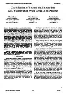
accuracy more significantly than mother wavelets. For all wavelets, decomposition beyond level 7 improves accuracy limitedly and even
but increase the computational cost, sometimes exponential (e.g.,
decreases accuracy. We further study the most effective bands and
RBF kernel SVM [16]), on the other hand.
features for seizure detection. An interpretation to our results is
For space sake, we pick seizure detection, the most commonly
that seizure and non-seizure EEGs differ across all conventional
EEG classification problem [4], as an example to study. This
frequency bands of human EEG rhythms. The best accuracy of seizure detection achieved in this research is 92.30% using coif3
paper aims at: 1. finding the best mother wavelet for seizure
from levels 2 to 7.
detection; 2. finding the trade-off between decomposition level and seizure detection accuracy. The machine learning approach
Keywords-Seizure detection, EEG, wavelet, decomposition level
to seizure detection is to classify seizure and non-seizure EEGs I.
INTRODUCTION
recorded simultaneously from multiple channels [3] , [17]. worldwide,
Seven families of 54 total mother wavelets are used in this
epilepsy is the second most COlmnon neurological disorder.
research. For each mother wavelet, we decompose the signal to
Epilepsy is characterized by recurring seizures caused by ab
the maximum allowed levels, the full-level decomposition. Not
Affecting approximately
60
million
people
normal discharges in the brain [1]. Directly recording the neuro
only do we study the relationship between decomposition level
electric activities, electroencephalogram (EEG) is a gold standard
and accuracy, we also perform feature selection [3] and wavelet
in epilepsy diagnosis. Diagnostic tasks for epilepsy, such as
band selection. Choosing suitable features that can best represent
seizure detection [2]-[4], spike detection [4] , [5] and focus
the characteristics of the EEG signals is important for EEG
localization [6] , [7] , usually require long-term EEG recording
classification [10] . Features used in this research are those well
up to a few days. Therefore, many computer-aided solutions
known in wavelet-based EEG signal classification [13] , [18] .
have been developed to assist neurologists. Combining signal
Results show that given deep enough decomposition, all
processing and machine learning, most of those approaches
mother wavelets deliver similar results. Furthermore, consistently
model the problem as classification of signals, such as epileptic
across all mother wavelets, decomposition beyond certain level
vs. healthy for epilepsy diagnosis [8] , [9] , ictal (on seizure) vs.
provides little accuracy improvement and even sometimes de
interictal for seizure onset detection [10], [11], etc. The most
creases performance instead. Our explanation to the results is
commonly classification problem is seizure detection [4] where
that too many decomposition levels will cause feature vector
patients' seizure and non-seizure EEG need to be identified [12]. Applying Discrete Wavelet Transform (DWT) on epilepsy
redundancy. Seizure and non-seizure EEG differs across all
related EEG signal classification is gaining ground in recent
accuracy of 92.30% is achieved by RBF-kernel SVM [19] when
978-1-4673-6799-8115/$31.00 ©2015
IEEE
conventional frequency bands of human EEG rhythms. The best
1596
B. Wavelet Families
using coif3 as mother wavelet and its 7 features from levels 2-7.
In this paper, we test 7 most commonly used wavelet fam II.
PROBLEM FORMULATION AND DATASET
ilies' performance on epileptic focus localization using EEG
We formulate the problem of seizure as classifying multi
signal [4], [14]. The 7 wavelet families are: BiorSplines, Coiflets,
channel EEG recordings (seizure and non-seizure). The dataset
Daubechies, DMeyer, Haar, ReverserBior and Symlets. They
used in this work was collected at the Children's Hospital Boston
include 54 family members (mother wavelets) in total as shown
(MIT, in short), consists of EEG recordings from pediatric
in Table I.
subjects with intractable seizures. Subjects were monitored for up
C.
to several days following withdrawal of anti-seizure medication
Decomposition bands In this research, each wavelet will be tested through full-level
in order to characterize their seizures and assess their candidacy
decomposition. The maximum level L of decomposition is jointly
for surgical intervention [20]. Recordings, grouped into 23 cases,
determined by the signal and the mother wavelet to satisfy the
were collected from 22 subjects (5 males, ages 3-22; and
condition:
17 females, ages 1.5-19). The International 10-20 system of
L
-� 9085 � �80 LI X.
Figure 5: Regression Curve of Decomposition Level and Accu racy using best member in each wavelet family
combinations of bands and features (e.g., for bior1. 1, j
13,
it has
8191
x
511
=
4185601
=
combinations of bands and
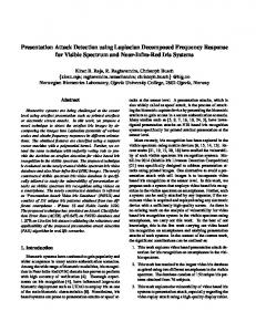
scale function
1.5
wavelet function
1.5
features). For each of the combinations, a cross validation is 0.5
performed. Because of the high time complexity of band and
0.5
\ 1/
0
feature selections, we only perform them on mother wavelets that exhibit the best performance in their respective families.
-0.5
-0.5
-1
-1
The steps to find the best wavelet, decomposition level,
a
15
10
Lowpass Analysis Filter
frequency bands and features are abstracted into Fig 3.
0.5
.�(!)
20
Jt
(')�
-0.5
0
0
10
15
20
Lowpass Synthesis Filter
-1
0
10
15
20
Highpass Synthesis Filter
0.5
0.5
'(!)
Jt
(!)�
-0.5
0 -0.5
15
Highpass Analysis Filter
0.5
-0.5
10
a
0
10
15
20
-1
0
10
15
20
N
...------