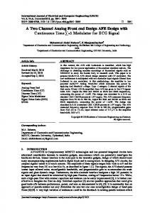
Abnormality classification of ECG signal using DSP processor Dr. Rahul Kher, Shivang Gohel Associate Professor EC department, G.H. Patel College of Engineering and Technology Vallabh Vidhyanagar, India Student of (M.E) Embedded System, G.H. Patel College of Engineering and Technology Vallabh Vidhyanagar, India
[email protected],
[email protected]
Abstract— Heart ailments have replaced communicable diseases as the biggest killer in rural & urban India. So diagnosis of heart diseases is a crucial part in preventing the fatalities occurring because of these diseases. These diseases are commonly known as arrhythmias of electrocardiogram. An electrocardiogram is a significant recording for measuring the electrical activity of the heart. It provides us with a complete picture of the condition of heart. The main purpose of this study is to develop an efficient arrhythmia detection algorithm based on the morphological characteristics of arrhythmias in ECG signal. Each arrhythmia of ECG has different morphology; we exploit this fact to minimize the computation required in classical methods. This system is ideal for use in real time applications.
Index Terms— ECG; Signal Analysis; Abnormality; Classification
I.
INTRODUCTION
In India, chronic diseases are projected to account for 53% of all deaths [1]. WHO has projected that over the next 10 years in India, over 60 million people will die form a chronic disease. Deaths from chronic diseases will increase by 18%. So diagnosis of these diseases is crucial if we want to reduce the number of deaths by these diseases. The electrocardiogram (ECG) is one of the simplest and oldest cardiac investigations available. And it provides a wealth of information about the heart of the patient. An ECG is simply a representation of the electrical activity of the heart muscle as it changes with time, usually printed on paper for easier analysis. The majority of the clinically useful information in the ECG is originated in the intervals and amplitudes defined by its features. The improvement of precise and rapid methods for automatic ECG feature extraction is of chief importance, particularly for the examination of long recordings [2]. The ECG feature extraction system provides fundamental features (amplitudes and intervals) to be used in subsequent automatic analysis. In recent times, a number of techniques have been proposed to detect these features [3]
[4] [5]. The ECG features can be extracted in time domain [6] or in frequency domain [7]. Our aim is to reduce the extensive analysis performed by the physicians thereby saving their time as well as efforts. This analysis requires a lot of medical knowledge to identify the type of abnormality since there exists around 35-40 types of abnormalities of ECG signal for human heart. So manual recognition of all these types has been proved to be a time consuming and tedious task, making the results subjective to the examiner. So this gave rise to a need for an automation to perform this classification. A lot of work has been done to make this process automated, but very less research has been done using Digital Signal Processing. So our aim is to make a standalone system that classifies ECG abnormalities using Digital Signal Processor. The system proposed here exploits the morphological characteristics of the ECG signal to accurately classify the abnormalities. This approach yields accurate results while reducing computational efforts required by the classical methods. The morphological characteristics that can be used to extract information from the ECG signal are listed below.
Time Intervals Voltage extremes(amplitude) Duration Location
Rest of the paper is organized as follows. Section II provides an overview of previous work done by researchers in the field of ECG abnormality classification. Section III describes the results of an algorithm implementation. Section IV describes the system design and section V concludes this contribution.
II.
RELATED WORK
The method in this research paper processes a noisy ECG signal obtained from recordings memorized in a database and to obtain scalograms of some forms of the ECG. From the database we extract the noisy ECG, which is digitally filtered and displayed. For ECG processing authors have used continuous wavelet transform to detect different pathologies [8]. In this paper the authors have proposed an algorithm that uses independent component analysis (ICA) to improve the performance of ECG pattern recognition [9]. In this paper, Harr Wavelet Transform (HWT) is used in order to extract features from the ECG signal. Pre-processing and the classification of ECG signals is done using Forward Feed Neural Network. And the MIT-BIH database is used to evaluate the proposed algorithm [10].
In this paper a system for collecting cardiac signals with minimal equipment is proposed. This system reduces the size of the circuitry by using a processor specializing in audio processing which is the Texas Instruments TMS320C6713 DSP development board. This system focuses on the extraction and processing of the cardiac signal in real time [11]. In this paper the authors have used the distribution of the voltage extreme values of the signal and the time distribution of proper extreme values to extract heartbeats [12]. In this paper, ECG classification is carried out based on the correlation coefficient approach. A window is positioned at the QRS complex for the matching of the constituent elements. The window only spans the duration of the QRS complex rather the whole cardiac Cycle. The beneficial property helps to minimize the computation in order to achieve classification quickly [13].
Figure 1 Record 105[14]
Figure 2 Record 119[14]
The flowchart below explains the method used to classify the PVC abnormality from ECG.
III. RESULTS A little example of how to use the morphological characteristic of the signal is shown here. Here abnormal ECG waveforms with PVC (Premature Ventricular contraction) are shown. Now the main characteristic of PVC is that it appears as an intermediate QRS wave between two regular QRS waves, and its amplitude is significantly higher than a regular QRS wave. So, we have exploited this fact to classify the abnormal signals having PVC. In this classification, whichever beat has the amplitude greater that the PVC threshold, is classified as a PVC beat. This simple yet elegant solution is quite accurate. The flowchart below explains the method used to classify the PVC abnormality from ECG. The table below shows its accuracy. It is evident that using these morphological characteristics can yield accurate results with very less computation. Figure 3 Flowchart of the algorithm
The table below shows its accuracy. It is evident that using these morphological characteristics can yield accurate results with very less computation.
Table 1 Results
Sample file No.
No. of PVC beats
No. of detected PVC beats
Accuracy A. HARDWARE
119
23
23
100%
106
30
29
97%
105
07
07
100%
112
00
00
100%
122
19
18
96%
Total beats=79
IV.
Total detected beats=77
PVC Atrial Flutter Atrial Fibrillation P-Wave missing T-wave alternans
Overall Accuracy=97.46%
SYSTEM DESIGN
First the ECG signal having abnormality is pre-processed. In this usually de-noising is done to remove artifacts like power line interference, base line wander etc. The abnormal ECG is downloaded from databases available online. After this stage, the de-noised ECG and a template ECG signal are applied to a correlation algorithm to classify the type of abnormality in the abnormal signal.
We are using TI’s TMS320C6713 DSP kit for implementing the algorithm. The features are listed below.
Features of TMS320C6713 DSP kit:
32bit floating point processor operating frequency of 225MHz Embedded JTAG support via USB High-quality 24-bit stereo codec Four 3.5mm audio jacks for microphone, line in, speaker and line out 512K words of Flash and 16 MB SDRAM Expansion port connector for plug-in modules On-board standard IEEE JTAG interface +5V universal power supply
B.
SOFTWARE
As IDE we are using Code Composer Studio. Its features are listed below.
Features of CCS:
Code Composer Studio (CCStudio or CCS) is an integrated development environment (IDE) to develop applications for Texas Instruments (TI) embedded processors. It includes an optimizing C/C++ compiler, source code editor, project build environment, debugger, profiler, and many other features.
V.
CONCLUSION
Figure 4 Block diagram of proposed system
Every abnormality of ECG has a different morphology. Taking advantage of the fact, we can reduce the computing necessary in popular approaches like, wavelet transform and component analysis. Thus, making this system ideal to implement on a processor which has memory and timing constraints. The major abnormalities which we will be addressing are listed below.
We intend to develop a Digital Signal Processor based system for classification of arrhythmia like Ventricular Premature Contraction (VPB), atrial flutter, atrial fibrillation, P-wave missing and T-wave alternans (TWA) in ECG signals. In this direction, a threshold-based algorithm to detect the Premature Ventricular contraction (PVC) has been implemented using MATLAB. The experimental results show that the PVC detection accuracy for five subjects is 97.46%. In
future, the similar algorithm will be implemented on DSP processor for detection and classification of other ECG arrhythmia mentioned above. REFERENCES [1] www.who.int/chp/chronic_disease_report/media/ind ia.pdf [2] S. Z. Mahmoodabadi, A. Ahmadian, and M. D. Abolhasani, “ECG Feature Extraction using Daubechies Wavelets,” Proceedings of the fifth IASTED International conference on Visualization, Imaging and Image Processing, pp. 343-348, 2005. [3] Juan Pablo Martínez, Rute Almeida, Salvador Olmos, Ana Paula Rocha, and Pablo Laguna, “A WaveletBased ECG Delineator: Evaluation on Standard Databases,” IEEE Transactions on Biomedical Engineering Vol. 51, No. 4, pp. 570-581, 2004. [4] Krishna Prasad and J. S. Sahambi, “Classification of ECG Arrhythmias using Multi-Resolution Analysis and Neural Networks,” IEEE Transactions on Biomedical Engineering, vol. 1, pp. 227-231, 2003. [5] Cuiwei Li, Chongxun Zheng, and Changfeng Tai, “Detection of ECG Characteristic Points using Wavelet Transforms,” IEEE Transactions on Biomedical Engineering, Vol. 42, No. 1, pp. 21-28, 1995. [6] Y. H. Hu, S. Palreddy, and W. Tompkins, “A Patient Adaptable ECG Beat Classifier Using A Mixture Of Experts Approach”, IEEE Transactions on Biomedical Engineering vol. 44, pp. 891–900, 1997. [7] Costas Papaloukas, Dimitrios I. Fotiadis, Aristidis Likas, Lampros K. Michalis, “Automated Methods for Ischemia Detection in Long Duration ECGs”, 2003. [8] Adriana Maria Ciupe and Nicolae Marius Roman, “Study of ECG signal processing using wavelet transform”, the 9th international symposium on advanced topics in electrical engineering, may 7-9, Bucharest, Romania, 2015. [9] Mohammad Sarfraz, Ateeq Ahmed Khan and Francis F. Li, “Using Independent Component Analysis to Obtain Feature Space for Reliable ECG Arrhythmia Classification”, IEEE International Conference on Bioinformatics and Biomedicine, Belfast, UK, November 2-5, 2014. [10] K.Muthuvel, L.Padma Suresh, S.H.Krishna Veni ,K.Bharathi Kannan, “ECG Signal Feature Extraction and Classification using Harr Wavelet Transform and Neural Network”, International Conference on Circuit, Power and Computing Technologies [ICCPCT], Nagercoil, India, march 21-22, pp. 13961399, 2014.
[11] Meddour Cherif, Malika-Djahida Kedir-Talha, Malika Tighidet, “Acquisition and processing on DSP of a cardiac signal”, 5th International Conference on Information and Communication Systems (ICICS), Karachi, Pakistan, December 4-15, pp. 4-8, 2013. [12] Adam Szczepa´nski, Khalid Saied, and Alois Ferscha, “A New Method for ECG Signal Feature Extraction”, ICCVG, Part II, LNCS 6375, pp. 334–341, 2010, Springer-Verlag Berlin Heidelberg. [13] Chuang-Chien Chiu Tong-Hong Lin And Ben-Yi Liau, “Using correlation coefficient in ecg waveform for arrhythmia detection”, biomedical engineering applications, basis & communications, vol. 17, no. 03, pp. 147-152, 2005. [14] www.physionet.org/physiobank/database/mitdb