2013 IEEE MTT-S International Microwave Workshop Series on RF and Wireless Technologies for Biomedical and Healthcare Applications (IMWS-BIO)
Detection and Localization of Brain Strokes in Realistic 3-D Human Head Phantom A.T. Mobashsher, P.T. Nguyen, A. Abbosh School of ITEE, The University of Queensland, St Lucia, QLD4072, Australia. Email:
[email protected] Abstract — Stroke is the second leading cause of death worldwide. The treatment depends on the location and extent of stroke and it should be started within 4.5 hours of the attack. Thus rapid detection is a must. Existing imaging systems do not provide a solution to monitor the patient in real time and not portable, thus requires patient’s movement. Addressing these limitations, a portable diagnostic system is proposed in this paper employing monostatic radar approach using only one antenna covering 73% fractional bandwidth centered at 1.85 GHz. A realistic three-dimensional (3-D) head phantom is generated to simulate different stroke conditions. The phantom anatomically holds the structure of a real human head with frequency dispersive electrical properties of respective biological tissues of head that are rigorously fitted with fourth-order Debye model for the simulation environment. A confocal imaging technique is used to map the abnormalities inside the head. The obtained images indicate the potential of the presented technique to detect and locate the position of both hemorrhagic and ischemic strokes. Index Terms — Stroke diagnostic system, ischemic stroke, hemorrhagic stroke, confocal imaging, monostatic radar.
I. INTRODUCTION Stroke occurs when a part of brain is damaged due to the lack of the blood supply as a blood vessel bursts (hemorrhagic stroke), spilling blood into the surrounding brain cells or is blocked by a clot (ischemic stroke), interrupting the circulation of necessary oxygen and nutrients. Stroke is a critical and quite common disease claiming a life in every six seconds worldwide, which makes it the second leading cause of death. In Australia, stroke is the second biggest killer after coronary heart disease and a leading cause of disability. Wrong medical analysis will be fatal for the stroke affected patient. Moreover, the patient must receive drug treatment within 4.5 hours of the onset symptoms for a full recovery. Hence, the treatment of a stroke affected patient mostly depends on the fast detection and localization of the stroke. The existing imaging techniques fail to provide a real time, low cost, fast and portable system that is accessible at rural medical clinics or can be carried by a paramedical team. To overcome these limitations, a new compact stroke detection system has to be developed that monitors the patient continuously in real time either at the bedside or in emergency room. Microwave imaging (MI) is a good candidate in this regard because of its non-invasive, non-ionizing and costeffective features. However, due to the fact that MI technology extends to a diverse range of fields including microwaves and antennas, signal and image processing as well as software engineering, the challenge of doing research in
978-1-4673-6096-8/13/$31.00 ©2013 IEEE
this field is also diverse. The band of operation for stroke detection is proposed to be in low microwave frequencies [1]. This raises an issue to the total size of the imaging system as the size of the microwave and antenna hardware components mostly depend on the frequency operation. Antennas with directive radiation are also needed to increase the transceiving power. Moreover, realistic 3-D head phantoms are also rare in software simulation environment to test the imaging and detection techniques. Mainly simplified head phantoms are used in the reported literature [1, 2]. Some of them used 2-D MRI slice to investigate the stroke scenario in mathematical model [3]. However, the construction of a realistic head phantom is one of the serious issues that the diagnostic system faces to get initial detection results using designed antennas and image algorithms in simulation environment. This paper describes an MI system that is portable and of low cost. A realistic 3-D human head phantom is generated to accurately simulate the stroke circumstances. Unlike most of the reported manuscripts [1-4], both hemorrhagic and ischemic strokes are considered. The system effectively identifies and locates the position of the strokes. Top GML 1032 substrate
Feeding Point
Semi-circular slot
70 mm
60 mm
Z
θ
Y
φ X Bow-tie flare 15 mm Shorting wall
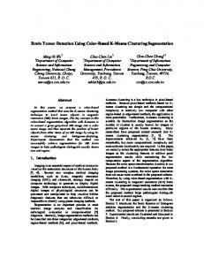
Fig. 1.
Circular sector Circular slot
Isometric view of the designed wideband antenna.
II. COMPACT WIDEBAND ANTENNA DESIGN A wideband antenna is designed for the stroke diagnostic system. An initial version of the antenna is described in [5]. The antenna is engineered to provide directional radiation with a low profile and compact size in order to meet the requirements of a portable detection system. The overall shape of the antenna is 70 × 60 × 15 mm3. The antenna is constructed with two GML 1032 substrates with dielectric constant of 3.32, loss tangent of 0.0023 and thickness of 1.52 mm. The top substrate consists of two bow-tie flare shaped
2013 IEEE MTT-S International Microwave Workshop Series on RF and Wireless Technologies for Biomedical and Healthcare Applications (IMWS-BIO)
120 Measured real-part of rel. permittivity Debye model real-part of rel. permittivity Measured imaginary-part of rel. permittivity Debye model imaginary-part of rel. permittivity
100
Relative Permittivity
elements which are fed from the center of the antenna. Edge length of each flare is increased by adding two circular sectors. Two circular slots are separated 0.5 mm apart from each other and enforced with two semi-circular ones on each bow-tie flare to achieve lower operating frequencies. The bow-tie elements are connected to the rectangular patch printed on the lower substrate slab from two opposite sides with two shorting walls. The walls and reflector creates a folded shape and reflects the backward radiation created by the bow-tie flares. The antenna supports a wide bandwidth of 1.35 GHz covering from 1.17 to 2.52 GHz, which is equivalent to 73% fractional bandwidth with 1.85 GHz center frequency. A stable gain of around 5 dBi is attained with a front to back ratio of 10 dB along the direction of radiation (+ve Z-axis).
80
60
40
20
0
0
0.5
1
1.5
2
Frequency (GHz)
Fig. 2. Comparison between permittivities of white matter.
measured
2.5
and
3
3.5
Debye
model
Direction of Rotation
III. GENERATION OF REALISTIC HUMAN HEAD PHANTOM Anatomically realistic numerical brain phantom is built using MRI images. This phantom considers the frequency dispersive properties of the biological tissues of a real human head. To that end, a fourth-order Debye model is derived and included in the numerical model over the band from 0.5 to 2.5 GHz, which is widely used in microwave-based head imaging as a reasonable compromise between penetration and resolution [6], [7]. The head model is created using a combination of Matlab and CST Microwave Studio from MRI slices of a real patient. The head phantom is comprised of a three-dimensional grid of 256 × 256 × 128 cubic voxels where the size of each voxel is 1.1 mm × 1.1 mm × 1.1 mm. In order to obtain the best estimation of the Debye parameters compared with measured data from [8], the measured properties of the head tissues at the available few discrete frequencies over the spectrum of 0.5 - 2.5 GHz are interpolated to get a larger number of discrete values and more accurate representation of the dispersive characteristics. The new data set are fitted to a fourth-order Debye model represented by the following formula. 4
ε r′ ( w) = ε ∞ + ∑ i =1
Δε 1 + jw τ
+
i
i
σ
(a)
0
The complex relative permittivity ε '( w) as a function of angular frequency is composed of frequency dependent permittivity ε (w) and conductivity σ (w ) . It is expressed in terms of Debye model of five terms in addition to a conductivity term in which σs is the static value; ε0 denotes the permittivity of free space; ε∞ is the permittivity at infinite frequency; εsi is the static permittivity, Δεi= εsi- ε∞. As an example of the fitted model, Fig. 2 depicts the electrical properties of the one of the main head tissues (white matter) based on the derived fourth-order Debye. A comparison in the same figure indicates that the derived model accurately emulates the measured properties across the band of interest. The data set generated by Debye model is then imported into CST microwave studio. Thus, the CST model not only demonstrates physical structure of realistic human model but also its electromagnetic properties.
978-1-4673-6096-8/13/$31.00 ©2013 IEEE
0.51 0.5 0.49 0.48 0.47 0.46 0.45 0.44 0.43
(1)
s
jwε
Bleeding
0.42
(b)
0.41
Fig. 3. (a) Simulation setup of the diagnostic system with cubical hemorrhagic stroke and first antenna position, (b) reconstructed image of the human head model; while the white dashed square represents the bleeding position.
IV. INVESTIGATION OF HEMORRHAGIC STROKE To simulate the hemorrhagic stroke, a cubic object with 20 mm arm length is inserted inside the realistic head phantom (shown in Fig. 3 (a)). Since this type of stroke is caused by bleeding, the electrical properties of blood are assumed for the target. In order to investigate the hemorrhagic stroke, the wideband antenna is placed at 0° angle in front of the head model. A Gaussian pulse covering a bandwidth from 1.1 to 2.6 GHz is emitted from the antenna and the scattered time domain signals are received by the same antenna. The process is repeated in 16 different positions with an angle of 360°/16°
2013 IEEE MTT-S International Microwave Workshop Series on RF and Wireless Technologies for Biomedical and Healthcare Applications (IMWS-BIO)
= 22.5° apart from each other. The direction of antenna rotation is shown in Fig. 3 (a). Fermat’s principle is used to determine the path of travel of the emitted signal from the antenna. As there is no matching liquid used in this experiment, high reflected signal is presumed from the exterior of the head phantom. The common background signals are then cancelled out from the received signal. Finally the image is constructed using the monostatic confocal imaging approach adopted from [4], and modified for the current technique. Figure 3 (b) exhibits the reconstructed image for hemorrhagic stroke. The white square marked on the image represents the position of the bleeding. It is seen that the image can locate the approximate position of the stroke affected cells. Direction of Rotation
Clot
be attributed to the basic assumption made by the algorithm that the propagating medium is homogeneous, which lead to some error in deciding the correct path of the travelling wave. Moreover, the electrical properties of the clot are closer to the white matter, which makes the detection more challenging. The future plan for this project includes using some classification techniques [9] to differentiate between the two types of strokes. Those techniques will be tested on an experimental system that uses a realistic head phantom prototype [10]. VI. CONCLUSION A compact microwave imaging system employing only one antenna that rotates around the head has been described. A realistic 3-D human head model is developed to simulate accurately the conditions of both hemorrhagic and ischemic strokes. Monostatic radar approach is utilized to receive the backscattered signals at 16 equiangular positions around the head model. Finally, the confocal imaging technique accumulates the information obtained from the antenna and used to reveal the position of the bleeding and clot inside the head phantom. The future work will involve using classification techniques to differentiate between the stroke types and improving the detection accuracy. REFERENCES
(a) 0.19
0.18
0.17
0.16
0.15
(b)
0.14
Fig. 4. (a) Test setup of the diagnostic system with cubical ischemic stroke and the first antenna position, (b) reconstructed image of the human head model; while the white dashed square represents the clot position.
V. INVESTIGATION OF ISCHEMIC STROKE In case of ischemic stroke simulation, the target is placed in the same spot and filled with the properties of clot (permittivity and conductivity of 30 and 0.5 S/m respectively [1]). As shown in Fig. 4 (a), same experimental setup discussed in the previous section is followed here. The back scattered signal is received in 16 points using a single antenna. The constructed image is shown in Figure 4 (b). It is seen that the algorithm effectively detects the location of the stroke, although it assumes a bit larger size than the original. This can
978-1-4673-6096-8/13/$31.00 ©2013 IEEE
[1] B. Mohammed, A. Abbosh, and D. Ireland, “Stroke detection based on variations in reflection coefficients of wideband antennas,” IEEE Int. Symp. Antennas Propag., pp. 1-2, Chicago, July 2012. [2] B. Mohammed, A. Abbosh, D. Ireland, and M. Bialkowski, “Compact wideband antenna for microwave imaging of brain,” Prog. Electromag. Research C, vol. 27, pp. 27-39, 2012. [3] R. Scapaticci, L. Di Donato, I. Catapano, and L. Crocco, “A feasibility study on microwave imaging for brain stroke monitoring,” Prog. Electromag. Research B, vol. 40, pp. 305324, 2012. [4] S. Mustafa, B. Mohammed, A. Abbosh, “Novel preprocessing techniques for accurate microwave imaging of human brain,” IEEE Antennas Wireless Prop. Lett., vol. 12, pp. 460-463, 2013. [5] A.T. Mobashsher, and A. Abbosh, “Wideband unidirectional antenna for head imaging system,” IEEE Int. Symp. Antennas Propag., pp. 674-675, Orlando, July 2013. [6] D. Ireland, and A. Abbosh, “Modeling human head at microwave frequencies using optimized Debye models and FDTD method,” IEEE Trans. Antennas Prop., vol.61, no.4, pp.2352-2355, 2013. [7] D. Ireland, K. Bialkowski, and A. Abbosh, “Microwave imaging for brain stroke detection using Born iterative method,” IET Microw Antennas Prop, vol.7, no.11, pp.909-915, August 2013. [8] S. Gabriel, R. Lau, and C. Gabriel, “The dielectric properties of biological tissues,” Physics Med Biology, vol. 41, no. 11, pp. 2271-2293, 1996. [9] S. Mustafa, A. Abbosh, B. Henin, and D. Ireland, “Brain stroke detection using continuous wavelets transform matching filters,” Int. Biomed. Eng. Conf, pp.194-197, Cairo, December 2012. [10] A.T. Mobashsher, B.J. Mohammed, S. Mustafa, and A. Abbosh, “Ultra wideband antenna for portable brain stroke diagnostic system,” IEEE MTT-S Int. Microwave Workshop Series RF Wireless Tech. Biomed. Healthcare App., Singapore, December 2013.