Cumhuriyet Üniversitesi Fen Fakültesi
Cumhuriyet University Faculty of Science
Fen Bilimleri Dergisi (CFD), Cilt:36, No: 3 Özel Sayı (2015)
Science Journal (CSJ), Vol. 36, No: 3 Special Issue (2015)
ISSN: 1300-1949
ISSN: 1300-1949
An Efficient Method for Detection of Masses in Mammogram Images Javad HADDADNIA1,*, Omid RAHMANI-SERYASAT2, Hossein GHAYOUMI-ZADEH2, Hamidreza RABIEE2 1.
Associate Professor, Electrical and Computer Engineering Department, Hakim Sabzevari University, Sabzevar, Iran 2.
Department of Electrical and Computer Engineering, Hakim Sabzevari University, Sabzevar, Iran
Received: 01.02.2015; Accepted: 05.05.2015 ______________________________________________________________________________________________ Abstract. Breast cancer is one of the most common cancers among women. Mammography is currently the most effective method for early detection of breast cancer. In this paper, a method is proposed for detecting masses in mammogram images. First, based on a specific algorithm, image is segmented and a number of the suspicious regions are obtained. Then, many features are extracted from these regions. To reduce the features, a supervised feature selection method is used. In the final step, a cost-sensitive classifier has been used for classification of the samples. This approach was tested on all images having mass from mini-MIAS data set. Based on the classification results, the percentage of true positive detection rate was 91% false-positive detection was 14% and the area under ROC curve was achieved 96%. Keywords: Mammogram images, Ranklet features, Co-occurrence matrix, composite classifier, unbalanced data sets, fractal dimension.
_____________________________________________________________________________ 1. INTRODUCTION Breast cancer is considered the most important factor of deaths related to cancers among women. As we know the best prevention method is early detection that leads to improved treatment and reduced loss of human lives. According to national cancer institute of America, in every 3 minutes it is diagnosed one woman is affected with breast cancer and every 13 minutes one woman is died as a result of this disease. Also, it is estimated that 1/8 % of women are affected with breast cancer, and 1/30 % of them die of it. Mammography is currently the most effective method for early and reliable detection of breast cancer [1]. Micro-calcifications and masses are the main symptoms of breast cancer. Mass detection is more difficult than Micro-calcifications because the characteristics of masses may be ambiguous or similar to main tissue of breasts. Masses are often located in areas with dense breast tissue and have smooth borders than Micro-calcifications and on the other hand have many forms. These factors cause mass detection to be considered as a challenging problem. Various methods have been proposed for detecting masses in mammogram images. For example, Dominguez and colleagues [2] have improved clearness of mammography based on statistical criteria. Then suspicious areas are segmented using multi-level thresh holding. Some features are extracted from each region and at the end; a grading system based on suspicious areas is used. Ozex and colleagues [3] used a template to identify masses. In that system, initially suspicious areas are identified using different thresh holdings, then a template for classification of suspicious area to mass or non-mass is used. Zheng and colleagues [4] combined several artificial intelligence techniques with Discrete Wavelet Transform. Artificial intelligence techniques used include the analysis of fractal dimension, MMRF (Multi-resolution Markov Random Field) and algorithm of Dogs & Rabbits for clustering. At the end, a classifier _____________
*Corresponding author. Email address:
[email protected] Special Issue: The Second National Conference on Applied Research in Science and Technology http://dergi.cumhuriyet.edu.tr/cumuscij ©2015 Faculty of Science, Cumhuriyet University
HADDADNIA, RAHMANI-SERYASAT, GHAYOUMI-ZADEH, RABIEE based on decision trees is used to classify areas. Dizung and colleagues [5] using Iso-intensity contours gained a number of suspected areas and then extracted co occurrence matrix characteristics from each area and used neural network as a classifier. Bovis and colleagues [6] built 4 co occurrence matrices for each area, and from these 4 co occurrence matrices extracted 70 features, and for classification of areas used RBF and MLP. Most of the existing techniques in the field of mass detection are combination of image processing techniques and algorithm identification templates, but often they are unaware of the fact that the issue of mass detection is inherently sensitive to the costs. This means that if a mass is wrongly classified as a normal tissue, its risk is much greater than when normal tissue is classified as a mass. Also, another case that negatively affects the classification performance is imbalanced data sets. Since the number of normal regions is much more than abnormal regions, the classification during the training phase will bias to the normal samples. One of the things that is often overlooked, is feature selection phase. Especially when the number of extracted features is high, the selection process is necessary because presence of irrelevant features causes declines in performance of classification. Another limitation of the proposed methods in articles is that most of them are used for detection of just one special group of masses (like Speculated mass or Circumscribed mass) and cannot be generalized to different masses in terms of type and size. In this paper, first each mammogram is segmented using special techniques and some preliminary suspicious area is achieved. This method is effective in detecting all kinds of masses and allows the radiologist to determine in what range (in respect of size) the existing are identified. Then, to reduce suspicious areas that cannot have mass, a lot of features were extracted from each region. Using a supervised feature selection method, a proper subset of the extracted features was chose. In the following, sampling method was used to eliminate bias created on normal samples. At the end a combined classifier method was used that is sensitive to cost and uses a cost matrix for applying the considered cost. In the following of Section 2 a method is proposed for finding suspicious areas. In section 3, the features extracted from the suspicious area are introduced and their selection method is examined. Section 4 will describe the classification system designed in this paper. In Section 5, testing methods and the results obtained are summarized and conclusions are presented in Section 6. 2. FINDING SUSPICIOUS AREAS First, to reduce the volume of computations, the image size is reduced from 1024 x 1024 to 512 x 512, then to remove impulsive noises on the image of the mammogram that may drastically affect the next steps, the median filter of size 3*3 is used. After this pre-processing, mammograms should be segmented. The purpose of this image segmenting is to obtain some suspicious areas or ROI (Region of interest) within which a mass may be hidden. In general, some ROI may be detected on image, all of which are indicative of a mass. In other words, the ROI mapping - to -mass is not a one - to - one mapping. At this point, method of splitting into half was used to find the roots of inspiration, and a number of suspicious areas are provided to mass. The algorithm steps are as follows: 1-The image is divided by a number of overlay cells with same sizes. (Grading image into cells with dimensions S*S). 2-In each cell, the pixel with the highest gray level is found. Its Coordination is shown with the index and its value with m. 3-F indicates the beginning of the range in which the answer is sought and R is also the end of range. Initially, F is assumed 0, and R is assumed as m.
2270
An Efficient Method for Detection of Masses in Mammogram Images
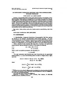
4-Th = (F+R)/2 is considered and an iso-intensity contour with value of Th is applied to the image. 5-The region within contour that includes index is considered. If the surface area exceeds a limit (Area_Max) or circularity of the region is greater than maximum (Circ_Max), then F=Th and we go back to step 4. Otherwise, if the surface area is less than a minimum (Area_Min), then put R=Th and we return to step 4. If the two above conditions are not maintained then this area is determined as a primary suspicious areas, and we go to next cell and Step 2 is followed. Another condition for stop of process on a cell is that the difference between two successive Th is less than a limit (Δ). In this study, Area_Max is considered as equal 8000 and Area_Min equal to 155 because there is no mass whose area is greater than 8000 (ie. a mass with a radius of 50) and smaller than 155 (a mass with a radius of 7). It should be noted that these values are obtained about images of 512 x 512. One of advantages of this method is that it provides degree of freedom for radiologist, so that radiologist himself can determine value of these parameters and specify dimensions in which he is going to discover system of existing masses. Circ_Max is determined, practically and through successive experiments, as 7. Also, in this paper Δ is considered as 4 gray levels and s as 32. The higher the S, the number of suspicious areas is reduced and the number of false positive samples (FP; the samples that were incorrectly identified by the system as mass) is reduced, but also the rate of (TP) (really true examples) and is decreased and vice versa. So, there should be balance between S, FP and TP. Through Consecutive tests it is found that the best value for S is equal to 32. Figure 1 shows an example of the result of the implementing the mentioned segmentation algorithm. Now it's time to give label to the regions obtained from the segmentation stage (TP or FP). The Label related to each area is determined according to the following procedure: First, a Bounding Box is drawn in a way that the considered area is completely included. Suppose that (LX, LY) indicates the length and width of the rectangle, and (Xcog, Ycog) represents the center of gravity of the considered area, and {(Xb, Yb), Rb} represents the center and radius of the mass after biopsied. If │Xb-Xcog│