JOURNAL OF CLINICAL MICROBIOLOGY, May 1997, p. 1108–1111 0095-1137/97/$04.0010 Copyright q 1997, American Society for Microbiology
Vol. 35, No. 5
An Improved Colorimetric PCR-Based Method for Detection and Differentiation of Entamoeba histolytica and Entamoeba dispar in Feces DAWN BRITTEN,1 STUART M. WILSON,1† RUTH MCNERNEY,2 ANTHONY H. MOODY,3 PETER L. CHIODINI,3 AND JOHN P. ACKERS1* Department of Medical Parasitology1 and Department of Clinical Sciences,2 London School of Hygiene and Tropical Medicine, London WC1E 7HT, and Hospital for Tropical Diseases, London NW1 0PE,3 United Kingdom Received 30 September 1996/Returned for modification 9 December 1996/Accepted 11 February 1997
The epidemiological implications of the recent separation of “Entamoeba histolytica” into two separate species, pathogenic E. histolytica sensu stricto and commensal E. dispar, will not become apparent without methods of distinguishing between them which are applicable to large numbers of specimens. We have modified a PCR-based method to produce such a technique which may be completed in 1 day while still identifying 1021 E. histolytica and 1 to 10 E. dispar trophozoites per g of feces when present separately and 10 E. histolytica and 100 E. dispar trophozoites per g in the presence of 106 trophozoites per g of the other species. Applied to fecal specimens from 18 patients from which E. histolytica or E. dispar had been grown and identified to the species level by hexokinase isoenzyme analysis, the method in every case yielded the correct result. Positive and negative results are easily distinguished by eye, and we are now applying this technique to a large-scale epidemiological study of amebiasis in the eastern Mediterranean region. PCR-based methods (2, 3, 11, 13, 15, 19) have shown the most promise; we have chosen to concentrate on the latter. The PCRsolution hybridization enzyme-linked immunoassay (SHELA) technique (3), in which specific colorimetric product detection replaces conventional gel electrophoresis and Southern blotting, provided an excellent starting point; our aim in this work was to speed up and simplify the whole process (including DNA extraction), so that multiple samples could be analyzed within 1 working day while maintaining sensitivity and specificity.
By conventional light microscopic diagnosis, some 500 million people meet the World Health Organization’s criteria (23) for amebiasis (21). However, it has long been known that 10% at most will suffer from invasive disease, with the remainder being asymptomatic cyst passers. Over the last 20 years it has become increasingly clear that these two conditions are caused by infection with two different but morphologically identical species of parasite, which have now been formally named Entamoeba histolytica (which is pathogenic, causes invasive amebiasis, and is still frequently referred to as the pathogenic strain of E. histolytica) and E. dispar (which is probably truly nonpathogenic and which is still found to be described as the nonpathogenic strain of E. histolytica) (7). The epidemiological implications of this new insight are still largely unknown (for example, it is not clear what proportion of patients infected with E. histolytica sensu stricto are asymptomatic), and studies to fill this gap will depend on the development of methods for differentiating between E. histolytica and E. dispar in large numbers of samples. The many biochemical, immunological, and genetic differences between the two species (7) provide plenty of scope for developing such tests, but the majority rely on establishing the organisms in culture. This process is not only time-consuming, but even with two different media, the process also fails with at least 30% of cystpositive fecal samples (17); on the other hand, culture followed by isoenzyme analysis is the only technique which has so far been validated with several thousand samples from many parts of the world (16). Of the available methods which do not require culture, detection of E. histolytica-specific antigen in feces (1, 10) and
MATERIALS AND METHODS Reagents. All reagents were obtained from Sigma (Poole, United Kingdom) unless otherwise stated and were of the highest available purity. Double-distilled water (DDW), which was sterilized by autoclaving, was used to make all solutions. Cultures. E. histolytica HM-1:IMSS was grown axenically in TYI-S-33 medium (8). E. dispar 53 clone 1 (isolated from a patient at the Hospital for Tropical Diseases, cloned by micromanipulation, and identified as E. dispar by hexokinase mobility and reaction with monoclonal antibodies [17]) was grown in the medium described by Robinson (14). Cells were harvested after 48 h of growth by centrifugation at 48C (900 3 g for 15 min), washed twice with chilled phosphatebuffered isotonic saline (PBS; pH 7.2), resuspended in PBS, and counted. Samples. Fecal samples obtained from patients attending the Hospital for Tropical Diseases were examined for ova and parasites directly and after concentration by the formol-ether method (4); those positive for cysts of E. histolytica or E. dispar or for hematophagous trophozoites were immediately inoculated into Robinson’s medium (14). Two fecal specimens (cyst negative) from a patient with a clinically diagnosed amebic liver abscess were also included. The remainder of the fecal sample was stored at 2208C until DNA was extracted (preliminary indications are that long-term storage at 48C is not satisfactory). Solutions. Apart from those solutions mentioned elsewhere in the text, the following solutions were prepared. The PCR mixture was prepared by following the manufacturer’s recommendations, in which 200 ml was prepared by combining 110 ml of DDW, 40 ml of 103 buffer A (0.5 M KCl, 0.1 M Tris-HCl [pH 9.0 at 258C], 1% Triton X-100), 32 ml of MgCl2 (final concentration, 2 mM) (both buffer A and MgCl2 were from Promega Kit M1861), 16 ml of a stock solution containing 20 mmol of each deoxynucleoside triphosphate (27-2035-01; ultrapure; Pharmacia), and, at the last minute, Taq polymerase (2 ml; 10 U; M1861; Promega). The optimum Mg21 concentration was determined by preliminary experiments. TBS contained 2.54 g of Tris-HCl, 0.47 g of Tris base, and 8.76 g of sodium chloride per liter; TBST (22) was TBS plus 5 ml of Tween 20 per liter (we have found that it is satisfactory and economical to reduce the Tris concentration considerably from the recommended concentration of 0.1 M); and TBSTM (12) (always freshly prepared) was TBST plus 30 g of dried skim milk powder (Tesco or Marvel; [Cadbury]) per liter. Peroxidase substrate consisted of 240 mg of
* Corresponding author. Mailing address: Department of Medical Parasitology, London School of Hygiene and Tropical Medicine, Keppel Street, London WC1E 7HT, United Kingdom. Phone: 44 171 927 2346. Fax: 44 171 636 8739. E-mail:
[email protected]. † Present address: Mycobacterium Reference Laboratory, Public Health Laboratory, Dulwich Hospital, London SE22 8QF, United Kingdom. 1108
PCR-BASED DIAGNOSIS OF E. HISTOLYTICA AND E. DISPAR
VOL. 35, 1997
TABLE 1. Primers and probes used for PCR amplification and product detection Primer or probe
Sequence
Primers Eh1 ................59-biotin-TCA AAA TGG TCG TCG TCT AGG C-39 Eh2 ................59-CAG TTA GAA ATT ATT GTA CTT TGT A-39 Ed1 ................59-TCG GAT CCT CCA AAA AAT AAA GTT T-39 Ed2 ................59-biotin-ACA GAA CGA TAT TGG ATA CCT AGT A-39 Probes EH .................59-digoxigenin-CCC GAG GTT CTT AGG AAA TGG A-39 ED .................59-fluorescein-AAT GAG GTT GTA GCA GAG CCC C-39
3,39,5,59-tetramethylbenzidine per ml and 0.0078% hydrogen peroxide in 0.2 M citrate buffer (pH 3.95). Differentiation of E. histolytica and E. dispar. (i) Isoenzymes. Organisms from positive cultures were harvested and lysed, and the mobilities of the two characteristic hexokinase bands were determined after electrophoresis in agarose as described previously (18); comparison with the mobilities of the bands from standard isolates enabled the two species to be clearly distinguished in all cases. (ii) PCR. To minimize the risk of contamination, three rooms were used for the PCR analysis, with each room designated for a specific function, as follows: white room, storage of PCR reagents and preparation and aliquoting of reaction mixtures; gray room, storage of samples, extraction of DNA, and addition of DNA to the reaction mixture; and red room, amplification, analysis, and storage of PCR product. Separate sets of equipment, glassware, etc., were kept in each room and were never moved. Materials traveled only in the direction white room3gray room3red room. (iii) DNA extraction from feces (gray room). Feces were thawed, and about 1 g was mixed with PBS (if necessary) to give a semifluid consistency, frozen-thawed (with solid carbon dioxide in ethanol and then water at room temperature) three times to lyse any cysts present, and then centrifuged at 900 3 g for 5 min. A total of 250 ml of supernatant was withdrawn, and DNA was purified by the method of Boom et al. (5) (except that 120 ml of lysis buffer contained only 1.3 g of Triton X-100), in which the material is first absorbed to 8 mg of diatomaceous earth (D5384; Sigma) in the presence of the chaotrope guanidinium thiocyanate and then eluted with 50 ml of TE buffer. The method can be completed in 2 h and effectively prevents the action of parasite nucleases which frequently degrade Entamoeba DNA. (iv) PCR mixtures (white room). For the PCR all four primers (Table 1; 18 ml containing 25 pmol of each primer) were added to one tube, and a pellet of wax (Lambwax; melting temperature, 57 to 588C; Raymond A. Lamb, London NW10, United Kingdom) was added. Primer sequences are homologous to the published sequences of repetitive elements in ribosomal DNA episomes of E. histolytica and E. dispar (9); specifically, they are based on those of Aguirre et al. (3), slightly modified to bring the melting temperatures closer. We also transferred the biotin label from the probes to one member of each pair of primers. The tubes were spun in a microcentrifuge (5 s), and the wax was overlaid with 20 ml of the PCR mixture. (v) Sample addition (gray room). The tubes were moved to the gray room, where 2 ml of fecal DNA in TE buffer was added. No more than 24 tubes (one for each sample, standard, or control) can be included in any one run; at least one tube with authentic E. histolytica at least one tube with authentic E. dispar, and at least two negative control tubes containing 2 ml of sterile DDW instead of DNA were always included in each run. The tubes were capped and moved to the red room. (vi) Amplification and detection (red room). Amplification conditions were optimized by using DNA extracted from E. histolytica and E. dispar trophozoites and were as follows: 2 min at 948C and then 35 cycles of 948C for 30 s, 588C for 1 min, and 728C for 2 min, followed by 10 min at 728C. If at this point a delay was unavoidable, the tubes were held at 808C. At the end of the amplification step 2 ml of probe mixture containing 10 pmol of probe EH and 10 pmol of probe ED (Table 1) was added to all tubes, which were held at 998C for 10 min (denaturation) and then at 378C for at least 15 min (hybridization). The probes were designed to match specific sequences expected in the two different PCR products; the EH probe is similar to that used by Aguirre et al. (3) but is shorter, while the ED probe is both shorter and homologous to a different region. A nonmatching tail was added to the ends of the probes; this prevents them from acting as primers at any point during the PCR. Each probe was labeled with a different, antibody-detectable tag. During hybridization, 450 ml of TBSTM, prewarmed to 378C, was added to each tube, where it rested on the solidified wax. Specific detection of amplified DNA was achieved colorimetrically rather than by gel electrophoresis. During the amplification, microtiter wells (four per sample, two-by-eight-well strips; catalog number 756061; Greiner, Frickenhausen, Germany) were coated with avidin (10 mg/ml in 40 mM carbonate buffer [pH 9.8]) at 378C for 1 h and were washed three times with TBST (22). (Alternatively,
1109
plates may be coated overnight at 48C and stored at this temperature for several days.) One-half of the plate was labeled “Eh” and the other half was labeled “Ed.” At the end of the hybridization stage each tube was opened, the wax plug was pierced with a tip, the contents were mixed with a pipette and fresh tip, and 100 ml was transferred to each of four wells in the coated plate: one pair in the Eh half and one pair in the Ed half. The plate was incubated at 378C for 10 min and was washed three times with TBST; to each well in the Eh side, 100 ml of peroxidase-labeled antidigoxigenin (Anti-digoxigenin–POD Fab fragments; 1207 733; Boehringer, Mannheim, Germany) was added, and to each well in the Ed side, 100 ml of similarly labeled antifluorescein (1426 346; Boehringer) was added. Both antibodies were diluted 1:5,000 in TBSTM. After 30 min at room temperature the plate was washed three times with TBST and once with TBS before 100 ml of peroxidase substrate was added; the color was allowed to develop over 10 min at room temperature, and negative and positive wells could be easily distinguished by eye. Initially, however, 50 ml of 2 M hydrochloric acid was added to each well, and the optical density (OD) at 450 nm was read. The ODs of the negative control wells were measured and were averaged for each half of the plate; these values were then subtracted from the readings from the other wells in the same half. The ODs of the negative control wells were always close to zero with either probe (mean OD, 0.002; highest OD, 0.006).
RESULTS The ability of the procedure described here to specifically identify one species, if necessary in the presence of a large excess of the other, was tested by adding known numbers of E. histolytica or E. dispar trophozoites to separate portions of a microscopically negative fecal sample, extracting the DNA, and carrying out the assay. The results are presented in Table 2, which indicates that 1021 E. histolytica and 1 to 10 E. dispar trophozoites per g of feces may be easily detected. Samples with mixtures of the organisms were also examined, and 10 E. histolytica and 100 E. dispar trophozoites per g could easily be identified in the presence of 106 trophozoites per g of the other species. The generally lower sensitivity for E. dispar may be due to the sequence of its repetitive element, which does not allow for optimal primer design. Fecal samples from 18 patients (specimen numbers commencing with H) from which E. histolytica or E. dispar had been grown and identified by isoenzyme analysis were then TABLE 2. Sensitivity and specificity of the methoda No. of trophozoites added/g E. histolytica HM-1:IMSS
E. dispar 53 clone 1
106 104 102 101 100 1021 None None None None None None 106 106 102 101 100 None
None None None None None None 106 104 102 101 100 1021 102 101 106 106 106 None
OD of antidigoxigenin (E. histolytica)
OD of antifluorescein (E. dispar)
1.96 .2 .2 .2 1.76 0.42 20.02 0 20.02 20.01 20.01 0 1.74 1.86 .2 1.38 0 20.01
20.02 0 0 0 0 0 1.78 1.65 1.67 0.55 0.41 0.04 0.54 0.12 1.92 1.87 .2 0
a Cultured and washed trophozoites were added to portions of the same normal fecal sample to give the final concentrations indicated here. The samples were then processed together. OD values (at 450 nm) are the means of two assays, to two significant figures. Antidigoxigenin detects the EH probe; antifluorescein detects the ED probe (see Materials and Methods and Table 1).
1110
BRITTEN ET AL.
J. CLIN. MICROBIOL.
TABLE 3. Results of assaying specimens from 19 patients from which amebas were grown and identified to the species level by isoenzyme typinga Specimen
H97 H106 H91 H4 H53 H61 H9 H27 H90 H95 H70 H83 H86 H29 H49 H16 H62 H92 6500 (feces) 6500 (second feces) 6500 (liver pus)
Diagnosis by isoenzyme analysis
Symptoms and clinical diagnosisb
Soft, poorly formed feces; IFAT, ,1/80; treated for AD NA NA ACP ACP AD; IFAT, .1/80 NA Microscopically confirmed AD; IFAT, ,1/80 NA Plasmodium falciparum malaria and strongyloidiasis; IFAT, ,1/80 ACP Loose stools; IFAT, ,1/80 NA Abdominal cramps, loose stools, weight loss; diloxanide furoate prescription resulted in no benefit ACP; cutaneous larva migrans NA ACP; “viral illness.” NA Amebic liver abscess Amebic liver abscess Amebic liver abscess
OD of antidigoxigenin (E. histolytica)
OD of antifluorescein (E. dispar)
E. E. E. E. E. E. E. E. E. E.
histolytica dispar dispar dispar dispar histolytica dispar histolytica dispar dispar
1.77 0 20.01 20.01 0.01 0.43 0.02 1.52 0 0
0.09 1.1 0.4 1.3 0.85 0 1.42 0.01 0.18 0.88
E. E. E. E.
dispar dispar dispar dispar
20.02 0 20.01 0
1.56 1.77 1.6 1.78
E. dispar E. dispar E. dispar E. dispar No growth No growth Not done
20.02 0 0 0 2.0 1.0 .2
1.54 1.83 1.44 0.19 0.19 0 0.04
a OD values (at 450 nm) are the means of two assays, to two significant figures. Antidigoxigenin detects the EH probe; antifluorescein detects the ED probe (see Materials and Methods and Table 1). Where available, brief clinical details are given. b AD, amebic dysentery; IFAT, immunofluorescence titer obtained by using whole trophozoites of E. histolytica 200:NIH as antigen; ACP, asymptomatic cyst passer; NA, data not available.
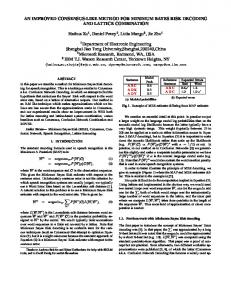
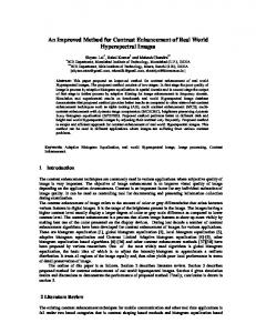
assayed in a blinded manner; the results (Table 3) indicate that for samples from 16 patients a strong signal (OD, $0.4) clearly identified the species present. For two patients (specimens H90 and H92), both infected with E. dispar, the initial OD was low, although it was clearly distinguishable from the EH probe signal, but retesting yielded positive results (ODs, 0.47 and 0.49, respectively). Specimen 6500 came from a patient presenting with an amebic liver abscess without dysentery; cysts were not found in his feces, and no ameba grew in culture. Nevertheless, E. histolytica was easily detectable in his feces. The first result with the ED probe gave a signal which, while well below our cutoff value of 0.4, was nevertheless similar to that given by specimens H90 and H92. We wondered if this was a genuine mixed infection, but retesting of the original sample and testing of a second sample gave negative results and no support to this idea. We were also able to obtain a specimen of the liver abscess contents (pus), prepare DNA, and assay it as described above; a very clear E. histolytica signal was obtained (Table 3). Although amebic liver abscesses are seldom aspirated, it is clear that E. histolytica may be identified there by this method if one so desires. Examination of a typical plate (Fig. 1) showed that positive wells could easily be picked out without recourse to OD measurement, and another 18 samples (2 infected with E. histolytica and 16 infected with E. dispar) were examined as described above, but the plates were simply scored visually. In every case the result was unambiguously correct. DISCUSSION We have developed a sensitive, accurate, and rapid method of identifying E. histolytica and E. dispar in human feces suitable for use with large numbers of specimens. The method is
FIG. 1. Microtiter plate at the end of the assay when no hydrochloric acid has been added and the results are assessed visually. Peroxidase-labeled antidigoxigenin was added to all the wells in the left-hand Eh column and similarly labeled antifluorescein was added to the left-hand Ed column (see Materials and Methods). The wells contained DNA extracted from the following samples: a normal fecal sample to which 104 E. histolytica trophozoites/g had been added (A and B), the same fecal sample in A and B to which 104 E. dispar trophozoites/g had been added (C and D), and E uninoculated feces (E and F); rows G and H were blanks containing distilled water.
PCR-BASED DIAGNOSIS OF E. HISTOLYTICA AND E. DISPAR
VOL. 35, 1997
based on the PCR-SHELA technique (3), and our improvements have involved a simplification of the DNA extraction step and small changes in the primer and probe sequences. We have also transferred the biotin label to the primers and used two different antibodies to detect probe bound to the PCR product; in our hands these changes have resulted in improved sensitivity and a much lower background signal. The very high sensitivity achieved (less than 1 E. histolytica organism and 1 to 10 E. dispar organisms) is due to the choice of PCR primers, which are similar to those of Romero et al. (15) and which are based on the sequences of repetitive elements in the abundant (several hundred per trophozoite) 25-kb ribosomal DNA episomes, and the sensitivity of the colorimetric detection of product. Extremely limited information is available about the number of cysts excreted by infected humans; one study (20) of a carrier (i.e., someone who was probably infected with E. dispar) suggested that the maximum output was about 1.25 3 106 cysts/g of feces. The rate varied from day to day, and on some days cysts were not detectable; it is, however, impossible to estimate the minimum number which could have been counted and thus whether the method described here is potentially capable of detecting all infected individuals with a single fecal specimen from each patient. Using similar primers but a more complex DNA extraction process and detecting the PCR product by Southern blotting, Rivera et al. (13) achieved a rather lower sensitivity. So far, unlike Romero et al. (15), we have not detected any mixed infections; however, some of our specimens came from travelers, while the specimens of Romero et al. were from long-term residents of an area where the infection is endemic. We are now examining large numbers of specimens from subjects in Egypt, Turkey, and Bethlehem to determine the true incidence of infection with these species in the eastern Mediterranean region. The major potential application of this method is in the investigation of the complex ecology of infections with E. histolytica and E. dispar, particularly the fact that the latter is common in populations in which the former is rare, even though both organisms presumably have identical modes of transmission. In this application large numbers of specimens will need to be processed, and the simplification of previous colorimetric procedures which we describe here should make this more practicable. The design of the primers and probes should ultimately make possible the development of a one-tube assay with a further increase in convenience. The obvious alternatives to PCR-based methods are antigen detection procedures; the latter are available in kit form and are more suited to field use, whereas PCR-based methods (with a number of different primers) are potentially capable of extending species identification to isolate identification (6). At present, however, none of the available methods for distinguishing between E. histolytica and E. dispar are inexpensive enough to find any application in routine clinical practice in areas where the infection is endemic; they may be of real use in managing cases of imported or sexually transmitted intestinal protozoan infections in people in areas where more resources are available. ACKNOWLEDGMENTS This research was supported by the Commission of the European Community’s Avicenne Programme (Contract AVI*-CT93-0008). We are extremely grateful to Aura Aguirre for all her help and
1111
advice during this project, to the staff of the Hospital for Tropical Diseases for the extra effort that they put into obtaining specimens for us, and to C. Graham Clark for reading the manuscript. REFERENCES 1. Abd Alla, M. D., T. F. H. G. Jackson, V. Gathiram, A. M. el Hawey, and J. I. Ravdin. 1993. Differentiation of pathogenic Entamoeba histolytica infections from nonpathogenic infections by detection of galactose-inhibitable adherence protein antigen in sera and feces. J. Clin. Microbiol. 31:2845–2850. 2. Acuna-Soto, R., J. Samuelson, P. De Girolami, L. Zarate, F. Millan Velasco, G. Schoolnick, and D. Wirth. 1993. Application of the polymerase chain reaction to the epidemiology of pathogenic and nonpathogenic Entamoeba histolytica. Am. J. Trop. Med. Hyg. 48:58–70. 3. Aguirre, A., D. C. Warhurst, F. Guhl, and I. A. Frame. 1995. Polymerase chain reaction-solution hybridization enzyme-linked immunoassay (PCRSHELA) for the differential diagnosis of pathogenic and non-pathogenic Entamoeba histolytica. Trans. R. Soc. Trop. Med. Hyg. 89:187–188. 4. Allen, A. H. V., and D. S. Ridley. 1970. Further observations on the formolether concentration technique for faecal parasites. J. Clin. Pathol. 23:545– 546. 5. Boom, R., C. J. A. Sol, M. M. M. Salimans, C. L. Jansen, P. M. E. Wertheim-van Dillen, and J. Van Der Noordaa. 1990. Rapid and simple method for purification of nucleic acids. J. Clin. Microbiol. 28:495–503. 6. Clark, C. G., and L. S. Diamond. 1993. Entamoeba histolytica: a method for isolate identification. Exp. Parasitol. 77:450–455. 7. Diamond, L. S., and C. G. Clark. 1993. A redescription of Entamoeba histolytica Schaudinn, 1903 (Emended Walker, 1911) separating it from Entamoeba dispar Brumpt, 1925. J. Eukaryot. Microbiol. 40:340–344. 8. Diamond, L. S., D. R. Harlow, and C. C. Cunnick. 1978. A new medium for the axenic cultivation of Entamoeba histolytica and other Entamoeba. Trans. R. Soc. Trop. Med. Hyg. 72:431–432. 9. Garfinkel, L. I., M. Giladi, M. Huber, C. Gitler, D. Mirelman, M. Revel, and S. Rozenblatt. 1989. DNA probes specific for Entamoeba histolytica possessing pathogenic and nonpathogenic zymodemes. Infect. Immun. 57:926–931. 10. Haque, R., L. M. Neville, S. Wood, and W. A. Petri, Jr. 1994. Detection of Entamoeba histolytica and E. dispar directly in stool. Am. J. Trop. Med. Hyg. 50:595–596. 11. Katzwinkel-Wladarsch, S., T. Loscher, and H. Rinder. 1994. Direct amplification and differentiation of pathogenic and nonpathogenic Entamoeba histolytica DNA from stool specimens. Am. J. Trop. Med. Hyg. 51:115–118. 12. McNerney, R., I. A. Frame, J. A. Vexenat, J. A. Fonseca de Castro, M. K. Howard, R. Dillon, S. Wilson, and M. A. Miles. 1993. Visceral leishmaniasis in Teresina, N. E. Brazil: towards a DNA probe kit and its adaptation to processing blood-contaminated samples. Arch. Inst. Pasteur Tunis 70:405– 418. 13. Rivera, W. L., H. Tachibana, M. R. A. Silva-Tahat, H. Uemura, and H. Kanbara. 1996. Differentiation of Entamoeba histolytica and E. dispar DNA from cysts present in stool specimens by polymerase chain reaction: its field application in the Philippines. Parasitol. Res. 82:585–589. 14. Robinson, G. L. 1968. The laboratory diagnosis of human parasitic amoebae. Trans. R. Soc. Trop. Med. Hyg 62:285–294. 15. Romero, J. L., S. Descoteaux, S. Reed, E. Orozco, J. Santos, and J. Samuelson. 1992. Use of polymerase chain reaction and nonradioactive DNA probes to diagnose Entamoeba histolytica in clinical samples. Arch. Med. Res. 23:277–279. 16. Sargeaunt, P. G. 1988. Zymodemes of Entamoeba histolytica, p. 370–387. In J. I. Ravdin (ed.), Amebiasis: human infection by Entamoeba histolytica. John Wiley & Sons, Inc., New York, N.Y. 17. Sehgal, R., M. Abd-Alla, A. H. Moody, P. L. Chiodini, and J. P. Ackers. 1995. Comparison of two media for the isolation and short-term culture of Entamoeba histolytica and E. dispar. Trans. R. Soc. Trop. Med. Hyg. 89:394. 18. Strachan, W. D., P. L. Chiodini, W. M. Spice, A. H. Moody, and J. P. Ackers. 1988. Immunological differentiation of pathogenic and non-pathogenic isolates of Entamoeba histolytica. Lancet i:561–563. 19. Tachibana, H., S. Kobayashi, E. Okuzawa, and G. Masuda. 1992. Detection of pathogenic Entamoeba histolytica DNA in liver abscess fluid by polymerase chain reaction. Int. J. Parasitol 22:1193–1196. 20. Tsuchiya, H. 1932. Observations on “encystment cycle” of Entamoeba histolytica in a carrier. Proc. Soc. Exp. Biol. Med. 29:230–232. 21. Walsh, J. A. 1986. Problems in recognition and diagnosis of amebiasis: estimation of the global magnitude of morbidity and mortality. Rev. Infect. Dis. 8:228–238. 22. Wilson, S. M., R. McNerney, P. M. Nye, P. D. Godfrey Faussett, N. G. Stoker, and A. Voller. 1993. Progress toward a simplified polymerase chain reaction and its application to diagnosis of tuberculosis. J Clin. Microbiol 31:776–782. 23. World Health Organization. 1969. Amoebiasis. World Health Organization Technical Report Series No. 421. World Health Organization, Geneva, Switzerland.