Current Bioinformatics
277
Send Orders for Reprints to
[email protected] Current Bioinformatics, 2016, 11, 277-290
ISSN: 1574-8936 eISSN: 2212-392X
Understanding Effects of Psychological Stress on Physiology and Disease Through Human Stressome - An Integral Algorithm
Impact Factor: 0.921
Sushri Priyadarshini and Palok Aich* BENTHAM SCIENCE
School of Biological Sciences, National Institute of Science Education and Research (NISER), Institute of Physics Campus, Sachivalaya Marg, Bhubaneswar 751 005, Odisha, India Abstract: Psychological stress perturbs normal physiological function or homeostasis. Restoration of normalcy demands more supply of energy. A physiological mechanism via activated stress response system is aimed at providing quick energy to deal with such emergency situations. If stress response system remains activated for longer period, maintaining physiological homeostasis becomes difficult because of higher demand for energy which eventually leads to increased susceptibility to infection or Palok Aich disease. Although there are reports, associating psychological stress with physiological functions and diseases, a clear understanding of mechanism of stress manifestation is yet to be established. In order to facilitate extensive exploration and prediction of possible mechanisms, integration of molecular (gene-level) data pertaining to psychological stress, physiological processes and stress-associated diseases is needed. We report power of text-mining in combination with our data-integration methods and mathematical formulation to develop integrated geneassociation networks. These networks can be analyzed to gain holistic insights into the relationship between psychological stress-associated genes (stressome) and related physiological functions and diseases. We built the human psychostressome networks to understand and predict pathways and candidate genes responsible for perturbing balance among various physiological functions and disease manifestation. Using the current methodology, we were able to predict involvement of serotonin receptors and uridine 5'-diphospho-glucuronosyltransferases in mediating effects of psychological stress.
Keywords: Disease, gene networking, meta-analysis, physiology, psychological stress. 1. INTRODUCTION Organisms continually interact with the environment leading to situations affecting their physiological homeostasis [1-3]. Stress is anything that causes perturbation to homeostasis; so dependence on the organism’s stress response system (SRS) is critical for its survival. SRS is complex and highly conserved [4, 5]. Complexity of SRS regulation has intensified with development of autonomic nervous system (ANS) in higher life forms such as humans [6-8]. SRS activation triggers cascades of physiological events, such as elevated blood pressure and impaired heart rate affecting blood circulation, slower digestion, impeded metabolism and increased blood sugar level. This series of events are designed to give targeted focus along with very fast supply of energy to withstand the emergency situation [9]. This mode of high alert or hyper-vigilance is triggered by the sympathetic nervous system (SNS) [10] and is meant to be extended during emergencies for brief periods. A hyperactive or chronically active sympathetic nervous system can disturb the energy homeostasis of the body and affect several other vital physiological functions in order to conserve energy. If body remains under the action of SNS for an extended period of time, its energy reserves start depleting. Neurotransmitters (like endorphins, serotonin and dopamine) are needed to *Address correspondence to this author at the School of Biological Sciences, National Institute of Science Education and Research (NISER), Institute of Physics Campus, Sachivalaya Marg, Bhubaneswar 751 005, Odisha, India; Tel: +916742304062; Fax: +916742304070; E-mail:
[email protected] 2212-392X/16 $58.00+.00
modulate the ANS and restore parasympathetic state or the regenerative phase. High sympathetic neurological activity leads to overstimulation of these neurotransmitters which eventually leads to their exhaustion [11]. Hyperactivity of sympathetic neurological responses may also lead to higher blood insulin level [12], disruption of hormonal regulation [13] and impairment of detoxification process. For example, adrenalin, as a sympathetic response mediating hormone, triggers liver to dump its glucose reserve into blood. High levels of glucose in the blood alert pancreas to release high amounts of insulin [14] which may lead to various deleterious health conditions [15, 16] like insulin resistance [17], obesity [18], type-2 diabetes [19], heart diseases [20, 21] and heightened inflammatory state [22, 23]. The body naturally prefers to be in the parasympathetic or regenerative state, where most of the physiological processes function with an optimal activity. Physiology should, therefore, have robust feed-back signaling mechanisms to allow its transition from sympathetic to parasympathetic mode after a state of heightened sympathetic SRS activity. Although these mechanisms are not well understood, there are bits and pieces of evidence that support the above assumption. One such mechanism is via production of cortisol during stress response. Cortisol is extremely important because it helps to counteract negative effects of stress and tries to maintain homeostasis. Stress induced sympathetic activity increases blood glucose content through catecholamine action [24]. High glucose content in blood stream stimulates production of pro-inflammatory cytokines [25]. IL1b has been shown to © 2016 Bentham Science Publishers
278
Current Bioinformatics, 2016, Vol. 11, No. 2
activate the HPA and cause release of CRH from PVN in the brain, leading to increase in circulating GCs [26, 27]. GCs play important roles in glucose management and immune function such as suppression of pro-inflammatory response. This mechanism leads to a feedback signaling from hyperactive SNS to activation of para-sympathetic signaling and restoration of homeostasis. A very high level of cortisol is required under intense influence of stress, which results in disruption of the hormonal system. High and persistent cortisol demands lead to adrenal fatigue, a situation where adrenal glands can no longer produce enough cortisol [28]. In order to synthesize more cortisol, physiological system uses up precursors needed for synthesizing other hormones like aldosterone, estrogen, progesterone, testosterone and DHEA. This phenomenon, known as “cortisol steal”, creates deficiency of these hormones and increases the allostatic load on physiology by disturbing nervous, endocrinal, digestive, immune and cardiovascular systems. These systems are under autonomic nervous control. Perturbation of various physiological processes has cascading consequential effects on several organs and tissue systems to cause dysautonomia. As a result, SRS negatively affects ANS and might lead to a plethora of disorders such as chronic fatigue, high blood pressure, circulation disorders, gastrointestinal disorders, heart diseases, ulcers, autoimmune disorders, anxiety disorders, depression, addiction, and panic attacks. The neuro-endocrine signaling conveying the status quo at gene and molecular level during stress response thus involves a large number of organ systems, signaling cascades and neuro-endocrinal circuitry which are largely unexplored. In order to explore such complex issues with statistical confidence, one of the methods generally used is metaanalysis, where comparable physiological parameters across various studies are taken into account to establish the significance of causative molecular or signaling events. Studies reported so far, on psychological stress, have only been able to establish a few signaling events at neuroendocrine level to understand psychological stress response and associated changes in human physiology [29, 30]. However, meta-analysis of these reports could not be attempted as effects of stress are not reported to be associated with stress physiology and stress-associated disorders in the same studies. Another major approach is gene-networking which allows segregation of multiple parameters across various dispersed studies. We took lead from our previous reports [31, 32], to derive predictive association of genes with physiology and diseases through analysis of the genenetwork topologies, using the power of text-mining. The advantage of gene-networking analysis is that large dimension data across various studies can be compressed into a single or a few reducible networks. Such output makes comparative analysis feasible at molecular (gene) level with the potential to develop critical tools with predictive power in areas where studies are limited in numbers, diverse or unrelated and scattered. We report here effects of psychological stress on various physiological functions and diseases using analysis of text-mining based geneassociation networks with the goal to understand and predict role of psychological stress on human physiology and diseases.
Priyadarshini and Aich
2. METHODS 2.1. Development of Gene Association Network for Psychological Stress (Human Stressome) Text-mining based gene-association network for psychological stress of humans (human stressome) was generated by scanning literature using genes of entire human genome as gene-based keyword search. Cytoscape plug-in for Agilent Literature Search was used for keyword based text-mining and network generation. For text-mining, this plugin uses each gene as a keyword along with the provided context to search against pubmed and OMIM database, and creates a node (gene) for each match it finds in the relevant context search (the details of the search methods can be found in our earlier work. Once, the text-mining based gene association network for human stressome was created, MCODE plugin was used to identify putative biological complexes/gene clusters followed by in-built network analysis plugin of Cytoscape. MCODE allows retention of an edge in the network only if the nodes it connects is over a stringent threshold. Here we used MCODE score 2.5 and a minimum of 4 nodes as threshold for a MCODE clustering. MCODE clustering thus eliminates unconnected or sparsely connected nodes that might lead to many false positive predictions. This method as already described in our earlier work [31] also removes publication bias. It also provided the node degree and clustering co-efficient for each gene in the network after clustering, which were used in GO enrichment of human stressome using GOriLLA server [33]. To test the robustness of the human stressome genenetwork thus generated, genes of this network were compared with those pulled out by the web-based textmining server, GeneClip [34]. The nature of distribution of literature evidences for each gene from both the text-mining tools was tested by pearson correlation analysis. 2.2. Effects of Stress on Diseases Human stress-disease network was further developed for analyzing the association between stress genes (obtained from human stressome network) and disease susceptibility. DO enrichment of genes from human stressome was done using DOSE package in R (Bio Conductor) [35]. The DO enrichment profile of human stressome (supplementary file SF1) showed that all the diseases enriched by these genes could be broadly classified into five major categories, each of which were then comprehensively populated with disease names (Table 1). Gene-association networks were generated for each of these diseases using Agilent Literature Search plug-in. The keyword used for text-mining was the disease name for each disease network. Effects of psychological stress on disease susceptibility was probed by studying gene associations among each of these disease networks as well as with human stressome by integrating association data of all these networks into a single network that retains the biological significance. Gene-associations of each disease category were used to create disease-gene associations and the strength of such associations was calculated as described below. Algorithm, as shown in Fig. (1), was hence designed for network condensation leading to a single network
Developing Human Psychostressome
Table 1.
Current Bioinformatics, 2016, Vol. 11, No. 2
279
List of the disease categories and diseases used to populate each category based on DO enrichment of human stressome.
Disease Category (DCi)
Disease Name (Dij) Encephalomyelitis, Inflammatory bowel disease, Monoclonal gamopathy, Multiple sclerosis, Graft-versus-host disease,
Autoimmune disorders (# 36)
Primary Biliary Cirrhosis, Celiac disease, Antiphospholipid syndrome, Wegener's granulomatosis, Systemic necrotizing vascolitides, Kawasaki disease, Sarcoidosis, Autoimmune lymphoproliferative Syndrome, Chronic inlammatory demyelinating, Poltradiculoneuropathy, Chronic active hepatitis, Primary Sclerosing cholangitis, cryoglobulinemia, Raynaud's phenomenon, Myasthenia gravis, polymyalgia rhematica, Temporal arteritis, Lambert-Eaton myasthenic syndrome, Throboangitisobliterans, Neuromyotonia /Issacs' syndrome, Pernicious Anemia, Takayasu's Arteritis, Stiff man syndrome, Opsoclonus-myoclonus sysndrome, Acute inflammatory demyelinating Polyneuropathy polyarteritisnodosa, Hypersensitivity vasculitis, Meniere's disease CNS Vasculitis, paraneoplastic neurological disorders, Cogan's syndrome, Autoimmune inner ear disease Anthrax, Avian Influenza, Babesiosis, Boils and skin infection, Brucellosis, Camplylobacteriosis, Chrancroid, Chickenpox, Chikungunya, Chlamydia, Cholera, Creutzfeldt-Jacob disease, Cryptosporidiosis, Dengue, Diphtheria, Epidemic Keratoconjunctivitis, Fifth Disease, Gastroenteritis, Giardiasis
Infectious diseases (# 70)
Metabolic Syndrome (# 15) Neuro-degenerative disdorders (# 8) Neurophysiological disorders (# 11)
Gonorrhoea, Haemolytic Uraemic Syndrome, Haemophilus Influenza Type B, Hand foot and mouth disease, Hepatitis A, Hepatitis B, Hepatitis D, Hepatitis E, AIDS, Infectious Mononucleosis, Influenza, Japanese Encephalitis, Keratoconjunctivitis, Kunjin Virus Disease, Legionnaires Disease, Leprosy, Leptospirosis, Listeriosis, Lyme disease, Lymphogranulomavenereum, Malaria, Measles, Meningococcal disease, Mumps, Murray Valley Encephalitis, Pandemic Influenza, Pertusis, Plague, Pneumococcal Disease, Poliomylitis, Psittacosis, Q Fever, Rickettsia, Rotavirus Infection, Rubella, Salmonellosis, SARS, Shigellosis, Shingles, Smallpox, Syphilis, Tetanus, Tuberculosis, Tularemia, Typhoid, Whooping Cough, Yellow Fever, German Measles, Hepatitis C, Botulism Metabolic Syndrome, Obesity, Hyperthyroidism, Hypothyroidism, Diabetes, Dyslipidemia, Hypolipidemia, Galactosemia, Phenylketonuria, Inborn errors of metabolism, Metabolic Acidosis Tay-Sachs disease, Addison's disease, Gangliosidosis, G-6PD deficiency Alzheimer's disease, Amyotrophic lateral disease, Parkinson’s disease, Primary progressive aphasia, progressive supranuclear palsy, Creutzfeldt_Jacob disease, Huntington's disease, Amylotrophic lateral sclerosis
Attention deficit hyperactivity disorder, Autism, Delayed onset of speech, Anxiety, Bipolar disorder, Depression, Dyslexia, Epilepsy, Obsessive-Compulsive Disorder, Schizophrenia, Social phobia
(Human Stress-Disease Network) that could easily be analyzed for association with stress.
Connectivity (C) between diseases Di and Dj (DiDj) in a disease category is true,
DCi = (Di1, Di2, Di3, ...., Din) or Dij ∈DCi (example of select
if
diseases are shown in Table 1) where, DCi denotes disease category of i-th type consisting of j-number of Dij elements. Similarly,
diDC ∩ d jDC = C for C ≠ 0,
Strength (Sij) of edge or connectivity between two can be calculated by diseases (DiDj),
(
)
SijD = ⎡⎣ diDC + d jDC × C ⎤⎦ / tin
Dij = (gij1, gij2, gij3, ....., gijn) or gijk ∈Dij , where genes
Strength ( SiD ) of disease node Dij can be denoted as ratio
gijk(s) are elements of a disease Dij. Diseases Di1 to Din are in unions as Di1 ∪ Di 2 ∪ Di 3 ......∪ Din leading to disease category DCi.
between number of edges ( di i ij ) connecting its nearest neighbors (diseases) and number of edges connecting its g ↔DCij di nearest neighbors ( diDC ), therefore, SiD = i DC di
We also define that if ni is the number of nodes in Dij
g ↔D
n
where tin = ∑ ni is the total number of nodes in Dij and i
n
diDC = ∑ tin representing total number of nodes in DCi. i
Genes were associated with each other as nodes for each disease, but for comprehensive representation of human disease network, the association data was integrated into a single network, where each disease was represented as a mega node and edges (connections) were formed between any two diseases Di and Dj, following the rules described below.
In the composite disease network, genes were represented as offshoot nodes of disease nodes, and edges were formed between any gene gi and disease Di, based on the following rules: Edges between gi and Dij will form, if, gi ∈Dij ⊆ DCi Strength ( SiDC ) of offshoot gene node is given by n
SiDC = ∑ SijD j=1
In order to study effects of stress on physiological homeostasis, similar text-mining based gene-association
280
Current Bioinformatics, 2016, Vol. 11, No. 2
Priyadarshini and Aich
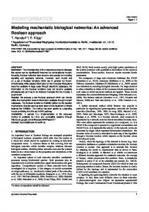
Fig. (1). Schema to build disease-gene network. Individual gene networks of diseases in various disease categories are to build final diseasegene network. Mathematical formulation is given in Methods section.
networks were generated for select stress-affected physiological processes viz., ageing, digestion, growth and development, immunity, metabolism respiration, reproduction. Genes which are already reported to affect each physiological process were manually identified, and expanded to candidate-set genes using gene prioritization algorithm (through Endeavour) as described in [31]. Top 2% of the prioritized candidate genes were used as keywords to construct query for literature search using Agilent Literature Search plugin for each gene-association network. Significant gene-association clusters were then identified using MCODE plug-in as described by the current group previously [31, 32]. Using the formulations described in the preceding sections, various associations between genes and diseases as well as genes and these physiological functions were generated. Similar mathematical basis was also used to build the composite disease, gene and physiology network, details of which can be seen in cytoscape readable supplementary file SF2. Genes conserved for human stress-disease network and human stressome were used for GO Enrichment Analysis using GOriLLA server. Genes from Human stress-disease network were extended to second order neighbors as background set for GO enrichment. 3. RESULTS Gene association network for psychological stress was created using entire human genome (hence forth referred as “human stressome”) as keyword (see methods) by Agilent Literature Search plugin (Fig. 2). Out of all the known human genes, 477 genes were found to be associated with psychological stress. GeneClip, another software which employs algorithm for text-mining based keyword search on pubmed abstracts, yielded 177 genes, out of which 100 genes were shared by both methodologies. A statistical comparison by t-test revealed that both text-mining methods pulled out
similarly distributed gene sets based on frequency of occurrence. A correlation analysis of the genes obtained from two methodologies, Geneclip and Genome search, further revealed formation of tight clusters (Fig. 3). 3.1. Functional Enrichment of Stress Genes in Human Stress-Network To understand the biological implication of the human stressome (Fig. 2a), Gene GO enrichment of the genes from this network was carried out by two methods, viz. GO enrichment based on 1) clustering coefficient (degree involvement of a node in the participating cluster) 2) node degree (the number of connections of a node). Clusteringcoefficient based GO enrichment showed enrichment of Gprotein coupled receptor signaling pathway and cellular glucuronidation to be most significantly enriched. It was seen that genes from these enrichment terms actually formed two sub-clusters: serotonin receptors sub-cluster (seen as highly enriched HTRs, –viz. HTR4, HTR3A, HTR2C, HTR7, HTR6, HTR5A, HTR1B, HTR1A, HTR2A in Fig. 2c) and glucuronidation enzymes sub-cluster (seen as enriched UGTs, –viz. UGT1A3, UGT2B7, UGT1A1, UGT2B15, UGT1A6, UGT1A9 in Fig. 2d) in human stressome. Genes from these two sub-clusters contributed to all of the GO-terms enriched by clustering-coefficient based GO enrichment. The HTRs were implicated in GO terms of synaptic transmission, control of appetite, blood vessel size regulation, as well as catecholamine and dopamine secretion. Node-degree based GO enrichment, revealed that along with genes responsible for serotonin receptor signaling and glucuronidation, other genes associated with GO terms of oxidation-reduction processes, mRNA splicing, isoprenoid biosynthesis (and MVK), and steroid biosynthesis were also significantly enriched and clustered together (Fig. 2b). There was also significant enrichment of genes responsible for regulation of NO biosynthesis process such as TNF, IL10,
Developing Human Psychostressome
Current Bioinformatics, 2016, Vol. 11, No. 2
281
Fig. (2). Gene association network of psychological stress for entire human genome. Networks shown are (a) all gene association, (b) master regulators of integrated stress response, (c) serotonin-receptor mediated signaling pathway and (d) cellular glucuroidination. Nodes sizes are directly proportional to their degree, and color is directly proportional to clustering coefficient (green for lower and red for higher clustering coefficient).
IL4, JAK2, IL1B, IL6 and IFNγ along with other cytokines and apoptotic genes such as BCL2. 3.2. Integrated Human Stress-Disease Network In order to understand how psychological stress affects disease susceptibility, we did a DO enrichment of human stressome and grouped the diseases into five major disease categories as described in methods section. Gene-association networks were developed for multiple diseases based on the DO enrichment profile of human stressome and all the associations from each of the disease networks and human stressome was condensed into a single network (the stressdisease network) through the network condensation algorithm as described in methods (Fig. 1). The human stress-disease network contained all the association information among the disease and stress genes merged in a single network which represented individual contribution of each gene to the diseases they are associated with at multiple dimensions. The same network also showed how many of these disease-associated genes were from human stressome network and how the diseases were related among themselves based on conserved genes between them (Fig. 4). Since all genes in this network were associated with one or
the other disease, 3 types of GO enrichment was done to understand how (1) disease-genes associated with stress contributed to overall disease profile (through enrichment of only stress genes against a background of all genes of stressdisease network), (2) disease-genes not associated with stress contributed to overall disease profile (through enrichment of only those genes which contribute to diseases but are nonstress genes against a background of all genes of stressdisease network), (3) disease genes, independent of their nature with respect to stress, contributed to normal physiology (through enrichment of all disease genes against a background of all genes of stress-disease network). Detailed enrichment results are listed in supplementary file SF3. It was seen that out of 612 disease-genes in human stress-disease network, 168 genes were associated with stress and 444 genes were associated with diseases but not with stress. The list shows that genes associated with psychological stress are capable of affecting large number of genes associated with diseases in humans. We found that stress-associated disease genes indicated an enrichment of positive regulation of icosanoids, prostaglandins secretion and calidiol 1-monooxygenase activity. Positive regulation of peptidyl-serine phosphorylation and negative regulation of catabolic processes were seen which was absent in human
282
Current Bioinformatics, 2016, Vol. 11, No. 2
Priyadarshini and Aich
stressome GO enrichment profile. It was also observed that there were positive regulation of B-type cell activation, isotype switching and activation induced cell death of Tcells. However, GO enrichment of disease genes not associated with stress showed that amino-acid transport (Lglutamate) was positively regulated, along with positive regulation of glutametargic synaptic transmission, which involved and OXTR genes among others. IFNβ and TNFα production were positively regulated, along with production of cytokines. Analysis of gene network revealed that among other diseases, Hepatitis-C was the one most affected by stress and contribution of PGDH gene(stress associated) to the human stress-disease network was highest. A positive correlation (0.82 with p ≤ 0.003) between genes of stressdisease network (obtained through text-mining) and genes obtained through disease ontology enrichment analysis of human stressome for the same disease terms was obtained. Such correlation suggested robustness of the methodologies used for generation of stress-disease network. 3.3. Involvement of Stress Genes at Physiological Regulatory Levels In order to study effects of psychological stress on physiological homeostasis and hence disease susceptibility, we generated a gene-association network for each physiological process that is known to be affected by stress viz. ageing, digestion, reproduction, respiration, growth and development, immunity, and metabolism using Agilent Literature Search plugin as described in methods section. Each of these gene-association networks, representing a physiological process, was analyzed in the light of "disease" genes and “stress” genes. Stress genes were colored red and disease genes were colored cyan and the node size showed its contribution to the stress-disease network (Fig. 5). The nodes colored blue represented the genes associated with the physiological process, but no associations with stress or disease have been reported in literature so far. The biological implication of these networks was done through GO enrichment of their genes. 3.3.1. Ageing
Fig. (3). Classification analysis of Psychological stress associated genes clustered on the basis of frequency of occurrence as obtained through text-mining by two different methods (GeneClip and Agilent Literature Search). Clustering of associated genes classified based on correlated frequency of occurrence is shown.
GO-biological processes enrichment profile of ageing network showed that response to steroid hormones, and stress induced premature senescence were among the highly enriched terms; thereby implying that genes responsible for ageing are also responsive to stress hormones and stress might be capable of affecting genes responsible for acceleration of senescence or ageing. It was also seen that the stress genes TP53, MAPK14 and CDKN1a contributed to other significantly enriched terms like positive regulation of ROS metabolic process, cellular response to hypoxia, stress-induced premature and cell-cycle check-point regulation. Regulation of lipid homeostasis and fat cell differentiation were also enriched which implied involvement of lipid synthesis and metabolism in stressassociated ageing process. Among the genes from disease network contributing to ageing, the most abundantly present genes were PTGS2, SRC, non-receptor tyrosine kinase and APC. ATM serine/threonine kinase gene, known to contribute to cell cycle control, was also among the significantly enriched genes and was associated with
Developing Human Psychostressome
Current Bioinformatics, 2016, Vol. 11, No. 2
283
Fig. (4). Comprehensive network of genes associated with psychological stress and diseases (stress-disease network). Relative contribution of each gene (circular nodes) to its associated diseases is represented by node size. Size of each disease (rectangular node) represents its relative abundance of contributing genes.
negative regulation of serine/threonine kinase (highly enriched). 3.3.2. Development GO enrichment of the genes from development network displayed overall negative regulation of lymphocyte as well as leukocyte proliferation and activation. While serotonin receptor signaling was enriched the most, regulation of insulin secretion was also enriched in this network. Response to growth factor binding was also enriched and was contributed to by IL2Ra, IL2Rb, and ERBB2. However, when genes contributing to both stress and diseases were enriched against all genes present in the gene network for development, activation of immune system, particularly T-
cell activation was seen. It was also seen that negative regulation of developmental process was enriched and all the contributing genes viz. CCL2 (also known as monocyte chemo-attractant protein 1, MCP1), IRF1, CXCR3, IFNγ, TLR4, ZHX2, F2 (coagulation factor II), EPHB2, TP53, SERPINE1, IL6, SIRT1, BDNF, BCL2, IL4 belonged to stress and disease category implying that these stressassociated genes might impair developmental process. 3.3.3. Digestion Enrichment of hexose and glucose metabolism, T-cell differentiation, granulocyte chemotaxis, inflammatory and myeloid cell apoptotic process were seen as a result of GO biological process enrichment of digestion network. Positive
284
Current Bioinformatics, 2016, Vol. 11, No. 2
regulation of JAK-STAT cascade was enriched along with positive regulation of innate immune responses by TLR signaling through TRIF-dependent signaling (Myd88independent signaling), and vitamin metabolism. Enrichment of stress and disease attributed genes against digestion gene background however didn’t show very different enrichment scores except for glucose and hexose metabolism. The enrichment also showed that PDX1 and TRH contributed to the enriched corticosteroid response term. 3.3.4. Immunity Enrichment of immunity network showed that immunityassociated genes enriched fatty-acid metabolism (PRKAG2, PRKAA1, SYK, FABP3, UCP3, STAT5A, LPL, LIPE, DECR1, PTGS2, CYP1A1,A2,B1, FASN and PPARγ, suggesting the role of these fatty-acid metabolism genes in immune regulation. Two of the immune genes (STAT1 and IFNG) were seen to be associated with negative regulation of epithelial cell differentiation involved in kidney development. Results further revealed that TLRs 3, 6, 7 and NOD2 significantly enriched PAMP-dependent induction by symbiotic relation of host innate immune response (implying that symbiotic bacteria could have profound effect on innate immunity). Along the line TGFβ1, CCL5 (also known as RANTES), CCL3 (also known as macrophage inflammatory protein 1 alpha, MIP1a) and CD4 were similarly enriched. These genes are known to be involved in modification of physiology of organisms involved in symbiotic interaction with host. These genes are also known to have profound activating effect on brain microglial cells [36]. However, when genes associated with stress and disease from the network were enriched against immunity network, it was observed that trans-membrane receptor protein serine/threonine kinase signaling pathway (comprising of TP53, EGR1, SP1, NCOR2, RHOA, TGFB1, SERPINE1, MYC and MAP3K7) was highly enriched showing that serine threonine kinase phosphorylation events are necessary for mediating stress response/effects. By analyzing gene network, it was seen that MYC and RHOA were tightly associated with a cluster of ageing genes. This cluster is composed of receptors for tyrosine kinase along with WNTsignaling receptors and fibroblast growth factor. Results revealed that cellular response to growth factor stimulus was also significantly enriched (initially absent in the enrichment of immunity network), hinting that fibroblast growth factor production might be high during stress and compromised immune conditions. 3.3.5. Metabolism Enrichment map of metabolism network showed that positive regulation of neutrophil migration and chemotaxis, positive regulation of IL23 production, positive regulation of IL12 production, positive regulation of acute inflammatory process, and negative regulation of IL17 were significantly enriched. It also showed enrichment of COX activity and vitamin metabolic process enrichment along with positive regulation of monoxygenase activity and negative regulation of glucose transport. Enrichment map obtained by comparison of genes contributing to stress and diseases against metabolism showed enrichment of positive regulation of IL8 production, regulation of fibroblast growth factor signaling, regulation of fatty acid transport, response
Priyadarshini and Aich
to hypoxia, TGFβ signaling and regulation of NO biosynthetic process. 3.3.6. Reproduction The enrichment map of reproduction network showed that gene involved in regulation of stress response, response to cytokine signaling and enhancement/activation of immune system were enriched (esp. TLR signaling through LPS). This result hints at regulations of psycho–neuro immunity on reproduction process. The genes ATF4 and ATM serine/threonine kinase were associated with MAPK, JUN, CREB1, CREBBP and HSPA4 and 5. It was also seen than IL6 was the highest contributing node in reproduction network. Type 2 immunity cytokines viz. IL6, IL2, IL4, IL27, TNF and STAT3 were also among the significant contributors. 3.3.7. Respiration GO enrichment map of respiration network showed enrichment of NO biosynthesis, stress activated MAP kinases, and positive regulation of phosphatidyl serine and tyrosine residues. However, when stress and disease genes were enriched against respiration background, positive regulation of lipid synthesis was enriched, along with glucose and hexose metabolism. 3.4. Physiological processes and stress-mediated disease susceptibility network Finally, to do an integrated analysis of how stress affects physiology to increase disease susceptibility, a combined network of the genes affecting several physiological processes (reported in preceding section) along with genes associated with diseases and stress was generated (Fig. 6). The physiological processes are represented as square nodes and the genes associated with stress, physiology and diseases are represented as circular nodes with different colors indicative of association. An analysis of this network not only reveals how each of the physiological process are related to established stress and disease genes, but also lends predictive function assignment to genes that do not have direct established associations with the physiological processes, but might have indirect implications on them as revealed by network topology. As can be seen from the integrated gene network of physiological processes, there are genes that are not directly associated with these physiological processes, however, they are associated with their first order (nearest) neighbors. This analysis of integrated gene network revealed that genes, which are not directly associated, might even have associations (direct/indirect) with physiological processes that are yet to be reported. Therefore, the current method allows predictive assignment of these genes in the associated physiological processes. 4. DISCUSSION Stress is already established to have immune modulatory effects. Several studies as well as meta-analysis have shown how stress can affect immune system and increase susceptibility to immune and inflammatory disorders [31, 37, 38]. Enrichment analysis of stressome and stress-disease
Developing Human Psychostressome
Current Bioinformatics, 2016, Vol. 11, No. 2
285
Fig. (5). Gene association network of physiological processes where genes associated with diseases (from stress- disease network) are represented with cyan color and genes associated with stress are in red color. Nodes sizes are directly proportional to their relative contribution to stress-disease network.
network also showed involvement of immune functions like leucocyte activation, migration, TLR signaling, MYD88 and NFκB mediated inflammatory pathways for which sufficient biological explanation exists. Since, immunity has been well explored for associations with stress, in this study we have focused additionally on avenues studied less intensively with respect to stress. 4.1. Low Energy State Induced Chronic Compensatory Mechanism During Stress Response Adipose tissue is one of the most important endocrine organs, which also acts as a reservoir of energy and might be one the immediate targets of sympathetic nervous stimulation. Studies show that epinephrine causes a significant rise in adipose tissue blood flow and net efflux of NEFA under the action of both HSL and LPL [39]. The same is also hinted by GO enrichment of phospholipase Cactivation term in human stressome. This analysis suggests involvement of adipose tissue in providing instantaneous
energy during sympathetic response to stress. This response might drain vital reserves of the system; so the physiology would try to replenish the energy storage by activating compensatory mechanisms during an extended stress exposure. Various isotypes of HTRs are implicated in diverse mechanisms of stress response. Neuronal signaling through HTR1c and HTR2a is known to increase appetite and body weight. In rodent models of hypertension, neuronal signaling through HTR1c and HTR2a has been shown to have stimulatory effect on plasma levels of ACTH and epinephrine. Enriched HTRs (esp. HTR2c and HTR2a) are implicated in phospholipid biosynthesis; suggesting signaling through these receptors might contribute to lipid accumulation during stress. HTR2c and HTR2a are already established in promoting adipocyte differentiation and fat cell development, thereby contributing to obesity [40]. This adipogenic signaling through HTRs in adipose tissue is boosted by corticosteroids [41], implying that cortisol aids in fat storage to compensate action of epinephrine.
286
Current Bioinformatics, 2016, Vol. 11, No. 2
Priyadarshini and Aich
Fig. (6). Combined network of genes affecting the physiological processes, stress and diseases. Genes, associated with stress, diseases and both stress and diseases are colored in red, green and yellow, respectively. Edges showing associations between physiological processes (a) Development, (b) Ageing. (c) Immunity, (d) Metabolism and (e) Digestion] and genes, are colored as described in the figure sidebar with acronyms where AA-ageing associations, DiA- Digestion associations, DvA- Development associations, IA- Immunity associations, MAMetabolism associations, RpA- Reproduction associations, RsA- Respiration associations, pp - not directly associated with any physiological process.
ANS, through epinephrine, initiates stress response system (coping mechanism) by targeting physiological energy reservoirs and mobilizing energy. The oxidative metabolic mechanisms triggered in the process drain the system of vital energy and also lead to accumulation of oxidative toxic by-products like ECs and PGs. Low energy state of physiology is detected and relayed across the system to activate various compensatory mechanisms to replenish energy shortage. Compensatory mechanism to replenish energy is achieved by slowing down catabolism and by increasing anabolic processes. An interesting hint that emerged from the current work is a very intriguing question as to how low energy state of a physiology is detected and
conveyed to appropriate sites. One of the known mechanisms is by glucocorticoid negative feed-back on the HPA axis. It has long been demonstrated that cortisol negates epinephrine-induced limbic arousal during acute stress response. However, during sustained or chronic stress, where cortisol effect dominates, there should be regulatory mechanisms to normalize its deleterious effects. GO enrichment profile of stressome network revealed that stress is associated with steroid hormone biosynthesis along with increased redox reactions, isoprenoid biosynthesis and NO biosynthesis process. Important to note here is that enrichment of stress as well as disease genes from the
Developing Human Psychostressome
physiological processes network too showed similar enrichment profile with more specific child terms such as enrichment of response to corticosteroids, hypoxia and serine tyrosine kinase signaling. NO synthesis was also enriched by stress genes in most of the physiological processes. These profiles clearly show that stress response system works in a manner that creates mobilization of energy by increasing catabolic oxidative metabolism. This goes with the earlier established notion of high energy demand of stress, which is adequately met by stress response mechanism of the physiology by mobilizing energy produced through oxidative metabolic processes. It is also known that chronic stress increases susceptibility to a wide range of metabolic and inflammatory disorders. Enrichment profile of stress-disease network showed enrichment of calidiol 1-monooxygenase activity and Ecs, PGs secretion. It was seen that negative regulation of catabolism too was enriched. This observation hints that stress mediated diseases might result due to accumulation of Ecs and PGs in the physiological system. Increasing amounts of these endotoxic oxidation endproducts might trigger suspension of catabolism through negative feed-back signaling. High levels of cortisol have been reported to increase PGs level in vitro [42]. Stress has been shown to increase lipid peroxidation and protein oxidation in liver, brain and plasma [43]. It is also known that organs/sites of high oxygen consumption, and/or high lipid content are highly susceptible to redox reactions. PTGS2 which is highly represented among the enriched terms oxidizes arachidonic acid derived from lipids to produce PGs, Ecs, prostacyclins, isotrenes, etc. These oxidation derived metabolites during stress response accumulate and exert deleterious effects on the concerned organs. 4.2. Detoxification Systems During Stress Response and Associated Disorders UGTs are major phase 2 drug metabolizing enzymes and are highly expressed in liver and GI-tract (also present in kidney, brain skin, etc in lesser extents). It has been shown by Krishnaswamy et al. [44-46] that serotonin is a highly specific substrate for human intestinal UGTs, and serotonin UGT activity is induced by oxidative stress. This observation suggests that apart from detoxification, intestinal UGTs might contribute to intestinal homeostasis of 5-HT. Liver is one of the most important organs in terms of detoxification or getting rid of foreign substances or toxins (including micro-organisms), especially from the gut, with major percentage of blood being filtered from the portal vein, which carries blood from intestines. Liver is also the major site for breakdown of fats. It has been reported that UGTs are responsible for cholesterol homeostasis in liver. During sustained stress response, when there is continuous release of esterified fatty acids from fat deposits of body, liver is under high pressure and might become susceptible to malfunction due to over-activity. A functional analysis of the GO enrichment profile of human stressome thus aptly prioritizes liver as one of the major organs to be affected during stress response. Moreover, efferent sympathetic system has been implicated in stress-induced exacerbation of liver diseases, while, the efferent parasympathetic nervous system elicits an inhibitory effect on the development of hepatic inflammation
Current Bioinformatics, 2016, Vol. 11, No. 2
287
[47]. The stress-disease network also consequently showed that liver associated disease (Hepatitis C) to be the most over-represented disorder. Signaling through HTR1c and HTR2a have also been reported to increase mean arterial blood pressure and renin activity [48] as is also reflected by the stressome enrichment profile. NO is known to control blood pressure. NO is produced by iNOS and eNOS from L-arginine and known to counteract angiotensin II and inhibit glomerulosclerosis, interstitial fibrosis, microvascular lesions and podocyte stress [49] in hyper-cholesterolomic rodents. Adrenergic innervations have been identified in the renal vasculature and renal sympathetic activity tends to be increased during depleted circulating volume [50-52], thereby implying renalcontrol on blood pressure as a stress responsive measure. This shows that kidneys on the other hand could be another major organs highly affected by stress. As was seen from the stress-disease network, PTGS2 was also associated with L-glutamate transport and glutaminergic nerve transmission. It is already known that glucocorticoids mediate stress-induced accumulation of extracellular glutamate. This creates exito-toxicity in the nervous system and might be one of the leading causes of neuro-degenerative disorders [53]. PGDH is a stress gene which is highest represented in stress-diseases network. This again emphasizes the relevance of lipid metabolism/turnover and resulting accumulation of oxidized products as one of the major causes of stress-associated diseases. Previous gene-networking based studies for two metabolic disorders (T2D, hypertension) have also pointed out oxidative stress as one of the underlying mechanisms [54]. This could also be due to heavy bias of studies related to oxidative stress as is also seen from reviews [55]. However, the algorithm used in generation of stress-disease network normalizes such bias. Hence, we could see enrichment of HTRs and UGTs as the most enriched clusters, along with enrichment of oxidative process genes. 4.3. Predictive Assignment of Function Apart from identifying the underlying clues through over-representation/enrichment analysis, our methods of gene-networking also allow the assignment of function to genes based on prediction. As can be seen from Fig. (6a-e), the physiological processes ageing, digestion, development, immunity, respiration, reproduction, metabolism are directly associated only with those genes in literature which are connected through colored edges. However, some of these directly associated genes (direct hubs) act as hubs for tightly clustered genes which are not directly associated with the physiological processes. These genes, owing to their tight associations with direct hubs, might also indirectly/directly regulate the physiological processes. Lack of their association to the concerned physiological processes might be an indicator of lack of studies for these associations. Furthermore, this network also shows which physiological processes are closely associated with each other owing to the number of conserved genes among them; e.g. respiration and metabolism appear to be closely regulated, while digestion and reproduction show close regulation. Current report thereby produces an idea which reveals genes that could be
288
Current Bioinformatics, 2016, Vol. 11, No. 2
responsible for possible cross-talks among various physiological processes and their association with psychological stress. CONCLUSION The gene-networking method reported here, helps solve the issue of handling and representing large multidimensional data. Gene networking also makes enrichment or over-representation analysis easier compared to genenetworking methods developed so far. The methodology reported here, normalizes publication bias and reduces the risk of over-representation of existing reports. Using the current methodology, we get an idea of the holistic biology represented by stress-associated genes in literature and how this information can be used in understanding stressmediated disease susceptibility. LIST OF ABBREVIATIONS ANS APC ATF4 BCL2 BDNF CCL CD4 CDKN1a CREB1 CREBBP CXCR3 CYP1A1 DECR1 DHEA DO EGR1 EPHB2 FABP3 FASN FDFT1 GCs GO HPA HSL HSPA4, 5 HTRs ICs IFN IL6 ILR IRF1 JAK2 JUN
= = = = = = = = = = = = = = = = = = = = = = = = = = = = = = = = =
Autonomic nervous system Adenomatous polyposis coli Activating transcription factor 4 B cell CLL/lymphoma 2 Brain derived neurotrophic factor Chemokine (C-C motif) ligand Cluster of differentiation 4 Cyclin dependent kinase inhibitor 1A cAMP responsive element binding protein 1 CREB binding protein Chemokine (C-X-C motif) receptor 3 Cytochrome P450, family 1 member A1 2,4 Dienoyl-CoA reductase 1 Dehydroepiandrosterone Disease ontology Early growth response protein 1 EPH receptor B2 FABP3 fatty acid binding protein 3 Fatty acid synthase Farnesyl-diphosphate farnesyl transferase 1 Glucocorticoids Gene ontology Hypothalamic pituitary axis Hormone sensitive lipase Heat shock protein 4 and 5 5-hydroxy tryptamine receptors Icosanoids Interferon Interleukin Interleukin receptor Interferon regulatory factor Janus kinase 2 Jun proto-oncogene
Priyadarshini and Aich
LIPE LPL LPL MAP3K7
= = = =
MCODE MVK NCOR2 NEFA NFκB NO NOD2
= = = = = = =
NOS OMIM OXTR PAMP PDX1 PGDH PGs PPARγ
= = = = = = = =
PRKAA1
=
PRKAG2
=
PTGS2 PVN RHOA ROS SERPINE1 SIRT1 SNS SRS STAT
= = = = = = = = =
SYK TGF TLR TNF TP53 TRH UCP3 UGTs ZHX2
= = = = = = = = =
Hormone sensitive lipase Lipoprotein lipase Lipoprotein lipase Mitogen activated protein kinase kinase kinase 7 Molecular complex detection Mevalonate kinase Nuclear receptor co-repressor 2 Non-esterified fatty acids Nuclear factor kappa B Nitric oxide Nucleotide-binding oligomerization domaincontaining protein 2 Nitric oxide synthetase Online mendelian inheritance in men Oxytocin receptor Pathogen associated molecular pattern Pyridoxal phosphate synthase protein Phosphoglycerate dehydrogenase Prostaglandins Peroxisome proliferator activated receptor gamma 5'-AMP-activated protein kinase subunit gamma-2-activated protein kinase catalytic subunit alpha-1 5'-AMP-activated protein kinase subunit gamma-2 Prostaglandin endoperoxide synthase 2 Para ventricular nucleus Ras homolog family member A Reactive oxygen species Serpin peptidase inhibitor, clade E member 1 Sirtuin 1 Sympathetic nervous system Stress response system Signal transducers and activators of transcription Spleen tyrosine kinase Tumor growth factor Toll like receptor Tumor necrosis factor Tumor protein p53 Thyrotropin releasing hormone Uncoupling protein 3 UDP-glucuronosyltransferases Zinc fingers and homeoboxes 2
CONFLICT OF INTEREST There is no financial or non-financial competing interest for this report.
Developing Human Psychostressome
ACKNOWLEDGEMENTS Authors would like to thank NISER for providing infrastructure. SP as a PhD candidate executed most of the work while PA conceptualized the work as supervisor and also developed theoretical basis of the work.
Current Bioinformatics, 2016, Vol. 11, No. 2 [20]
[21] [22]
SUPPLEMENTARY MATERIAL
[23]
Supplementary material is available on the publisher’s web site along with the published article.
[24]
REFERENCES
[25]
[1]
[2]
[3]
[4]
[5] [6] [7]
[8]
[9] [10] [11]
[12]
[13]
[14] [15] [16]
[17] [18]
[19]
Elmquist JK, Coppari R, Balthasar N, Ichinose M, Lowell BB. Identifying hypothalamic pathways controlling food intake, body weight, and glucose homeostasis. J Comp Neurol 2005; 493(1): 6371. Romano GH, Harari Y, Yehuda T, et al. Environmental stresses disrupt telomere length homeostasis. PLoS Genet 2013; 9(9): e1003721. Glazachev OS, Sudakov KV. [The interactions of functional systems at the homeostatic level in normal children and adolescents and in a radioecologically unfavorable environment]. Usp Fiziol Nauk 1999; 30(3): 73-92. Slavov N, Airoldi EM, van Oudenaarden A, Botstein D. A conserved cell growth cycle can account for the environmental stress responses of divergent eukaryotes. Mol Biol Cell 2012; 23(10): 1986-97. Wenk M, Ba Q, Erichsen V, et al. A universally conserved ATPase regulates the oxidative stress response in Escherichia coli. J Biol Chem 2012; 287(52): 43585-98. Joels M, Baram TZ. The neuro-symphony of stress. Nat Rev Neurosci 2009; 10(6): 459-66. Morris KE, St Laurent CD, Hoeve RS, et al. Autonomic nervous system regulates secretion of anti-inflammatory prohormone SMR1 from rat salivary glands. Am J Physiol Cell Physiol 2009; 296(3): C514-24. Ulrich-Lai YM, Herman JP. Neural regulation of endocrine and autonomic stress responses. Nat Rev Neurosci 2009; 10(6): 397409. Nicolaides NC, Kyratzi E, Lamprokostopoulou A, Chrousos GP, Charmandari E. Stress, the stress system and the role of glucocorticoids. Neuroimmunomodulation 2014; 22(1-2): 6-19. Mayer EA, Fanselow MS. Dissecting the components of the central response to stress. Nat Neurosci 2003; 6(10): 1011-2. Rea WJ, Patel K. Reversibility of Chronic Disease and Hypersensitivity: Clinical Environmental Manifestations of the Neurocardiovascular Systems. CRC press: Taylor & Francis group; 2014. Masuo K, Mikami H, Ogihara T, Tuck ML. Sympathetic nerve hyperactivity precedes hyperinsulinemia and blood pressure elevation in a young, nonobese Japanese population. Am J Hypertens 1997; 10(1): 77-83. Cortelli P, Gambetti P, Montagna P, Lugaresi E. Fatal familial insomnia: clinical features and molecular genetics. J Sleep Res 1999; 8 Suppl 1: 23-9. Jarhult J, Falck B, Ingemansson S, Nobin A. The functional importance of sympathetic nerves to the liver and endocrine pancreas. Ann Surg 1979; 189(1): 96-100. de Kloet ER, Joels M, Holsboer F. Stress and the brain: from adaptation to disease. Nat Rev Neurosci 2005; 6(6): 463-75. Vink EE, Blankestijn PJ. Evidence and consequences of the central role of the kidneys in the pathophysiology of sympathetic hyperactivity. Front Physiol 2012; 3: 29. Jamerson KA, Julius S, Gudbrandsson T, Andersson O, Brant DO. Reflex sympathetic activation induces acute insulin resistance in the human forearm. Hypertension 1993; 21(5): 618-23. Tentolouris N, Liatis S, Katsilambros N. Sympathetic system activity in obesity and metabolic syndrome. Ann N Y Acad Sci 2006; 1083: 129-52. Seals DR, Bell C. Chronic sympathetic activation: consequence and cause of age-associated obesity? Diabetes 2004; 53(2): 276-84.
[26]
[27]
[28] [29] [30]
[31] [32]
[33] [34]
[35]
[36]
[37]
[38] [39] [40]
[41]
[42]
289
Schultz HD, Li YL, Ding Y. Arterial chemoreceptors and sympathetic nerve activity: implications for hypertension and heart failure. Hypertension 2007; 50(1): 6-13. Barretto AC, Santos AC, Munhoz R, et al. Increased muscle sympathetic nerve activity predicts mortality in heart failure patients. Int J Cardiol 2009; 135(3): 302-7. Straub RH, Wiest R, Strauch UG, Harle P, Scholmerich J. The role of the sympathetic nervous system in intestinal inflammation. Gut 2006; 55(11): 1640-9. Levick SP, Murray DB, Janicki JS, Brower GL. Sympathetic nervous system modulation of inflammation and remodeling in the hypertensive heart. Hypertension 2010; 55(2): 270-6. Sherwin RS, Shamoon H, Hendler R, et al. Epinephrine and the regulation of glucose metabolism: effect of diabetes and hormonal interactions. Metabolism 1980; 29(11 Suppl 1): 1146-54. Shanmugam N, Reddy MA, Guha M, Natarajan R. High glucoseinduced expression of proinflammatory cytokine and chemokine genes in monocytic cells. Diabetes 2003; 52(5): 1256-64. Berkenbosch F, van Oers J, del Rey A, Tilders F, Besedovsky H. Corticotropin-releasing factor-producing neurons in the rat activated by interleukin-1. Science 1987; 238(4826): 524-6. Schotanus K, Makara GB, Tilders FJ, Berkenbosch F. ACTH response to a low dose but not a high dose of bacterial endotoxin in rats is completely mediated by corticotropin-releasing hormone. Neuroimmunomodulation 1994; 1(5): 300-7. Raison CL, Miller AH. When not enough is too much: the role of insufficient glucocorticoid signaling in the pathophysiology of stress-related disorders. Am J Psychiatry 2003; 160(9): 1554-65. Fisher JP, Young CN, Fadel PJ. Central sympathetic overactivity: maladies and mechanisms. Auton Neurosci 2009; 148(1-2): 5-15. Silverman MN, Heim CM, Nater UM, Marques AH, Sternberg EM. Neuroendocrine and immune contributors to fatigue. PM R 2010; 2(5): 338-46. Priyadarshini S, Aich P. Effects of psychological stress on innate immunity and metabolism in humans: a systematic analysis. PLoS One 2012; 7(9): e43232. Sood P, Priyadarshini S, Aich P. Estimation of psychological stress in humans: a combination of theory and practice. PLoS One 2013; 8(5): e63044. Eden E, Navon R, Steinfeld I, Lipson D, Yakhini Z. GOrilla: a tool for discovery and visualization of enriched GO terms in ranked gene lists. BMC Bioinformatics 2009; 10: 48. Wang JH, Zhao LF, Lin P, et al. GenCLiP 2.0: a web server for functional clustering of genes and construction of molecular networks based on free terms. Bioinformatics 2014; 30(17): 25346. Yu G, Wang LG, Yan GR, He QY. DOSE: an R/Bioconductor package for disease ontology semantic and enrichment analysis. Bioinformatics 2015; 31(4): 608-9. Rock RB, Gekker G, Hu S, et al. Role of microglia in central nervous system infections. Clin Microbiol Rev 2004; 17(4): 94264. Segerstrom SC, Miller GE. Psychological stress and the human immune system: a meta-analytic study of 30 years of inquiry. Psychol Bull 2004; 130(4): 601-30. Glaser R, Kiecolt-Glaser JK. Stress-induced immune dysfunction: implications for health. Nat Rev Immunol 2005; 5(3): 243-51. Samra JS, Simpson EJ, Clark ML, et al. Effects of epinephrine infusion on adipose tissue: interactions between blood flow and lipid metabolism. Am J Physiol 1996; 271(5 Pt 1): E834-9. Kinoshita M, Ono K, Horie T, et al. Regulation of adipocyte differentiation by activation of serotonin (5-HT) receptors 5HT2AR and 5-HT2CR and involvement of microRNA-448mediated repression of KLF5. Mol Endocrinol 2010; 24(10): 197887. Brummett BH, Kuhn CM, Boyle SH, et al. Cortisol responses to emotional stress in men: association with a functional polymorphism in the 5HTR2C gene. Biol Psychol 2005; 89(1): 948. Neulen J, Zahradnik HP, Flecken U, Breckwoldt M. The effect of cortisol on the synthesis of prostaglandins (PGF2 alpha, PGE2) by human endometrial fibroblasts in vitro with and without addition of estradiol-17 beta or progesterone. Prostaglandins 1989; 37(5): 58795.
290 [43]
[44]
[45]
[46]
[47] [48]
Current Bioinformatics, 2016, Vol. 11, No. 2
Priyadarshini and Aich
Liu J, Wang X, Shigenaga MK, et al. Immobilization stress causes oxidative damage to lipid, protein, and DNA in the brain of rats. FASEB J 1996; 10(13): 1532-8. Kohle C, Badary OA, Nill K, Bock-Hennig BS, Bock KW. Serotonin glucuronidation by Ah receptor- and oxidative stressinducible human UDP-glucuronosyltransferase (UGT) 1A6 in Caco-2 cells. Biochem Pharmacol 2005; 69(9): 1397-402. Krishnaswamy S, Duan SX, Von Moltke LL, Greenblatt DJ, Court MH. Validation of serotonin (5-hydroxtryptamine) as an in vitro substrate probe for human UDP-glucuronosyltransferase (UGT) 1A6. Drug Metab Dispos 2003; 31(1): 133-9. Krishnaswamy S, Hao Q, Von Moltke LL, Greenblatt DJ, Court MH. Evaluation of 5-hydroxytryptophol and other endogenous serotonin (5-hydroxytryptamine) analogs as substrates for UDPglucuronosyltransferase 1A6. Drug Metab Dispos 2004; 32(8): 862-9. Chida Y, Sudo N, Kubo C. Does stress exacerbate liver diseases? J Gastroenterol Hepatol 2006; 21(1 Pt 2): 202-8. Pinilla L, Gonzalez LC, Tena-Sempere M, Aguilar E. 5-HT1 and 5HT2 receptor agonists blunt +/- -alpha-amino-3-hydroxy-5methylisoxazole-4-propionic acid (AMPA)-stimulated GH secretion in prepubertal male rats. Eur J Endocrinol 2001; 144(5): 535-41.
Received: March 9, 2015
[49]
[50] [51] [52]
[53]
[54]
[55]
Attia DM, Feron O, Goldschmeding R, et al. Hypercholesterolemia in rats induces podocyte stress and decreases renal cortical nitric oxide synthesis via an angiotensin II type 1 receptor-sensitive mechanism. J Am Soc Nephrol 2004; 15(4): 949-57. Moss NG. Renal function and renal afferent and efferent nerve activity. Am J Physiol 1982; 243(5): F425-33. DiBona GF. Neural control of renal function: cardiovascular implications. Hypertension 1989; 13(6 Pt 1): 539-48. Osborn JL, Holdaas H, Thames MD, DiBona GF. Renal adrenoceptor mediation of antinatriuretic and renin secretion responses to low frequency renal nerve stimulation in the dog. Circ Res 1983; 53(3): 298-305. Meldrum B. Amino acids as dietary excitotoxins: a contribution to understanding neurodegenerative disorders. Brain Res Brain Res Rev 1993; 18(3): 293-314. Jesmin J, Rashid MS, Jamil H, Hontecillas R, Bassaganya-Riera J. Gene regulatory network reveals oxidative stress as the underlying molecular mechanism of type 2 diabetes and hypertension. BMC Med Genomics 2010; 3: 45. Gadek-Michalska A, Tadeusz J, Rachwalska P, Bugajski J. Cytokines, prostaglandins and nitric oxide in the regulation of stress-response systems. Pharmacol Rep 2013; 65(6): 1655-62.
Revised: August 1, 2015
Accepted: August 26, 2015