Feature Extraction in Retinal Images using Machine Learning D.Nagarajan Department of Mathematics Hindustan Institute of technology & Science, Padur ,Chennai,India 603103.
[email protected] Abstarct- Retinal image processing has turn one of the drift in the area of image processing. The retinal vasculature being singular for each single serves to be a origin of eye disease. This paper presents a method of serving the image and extracting image using machine learning and utilizing it to execute the eye dieses examining. The image is pre-processed and allow the k value with respect to the KNN classifier is decided after iterative assuring and sorting is did to categorize the arteries and veins from the overall vasculature. Finally the image is compared with the test image to check the authenticity.
Joyce John Department of Computer Science and Engineering. .Hindustan Institute of technology & Science, Padur ,Chennai, India 603103.
[email protected]
pattern from the exploiter that could be not favorable. Retina iris images could disclose diseases such as blood pressure; this may be bitter. It could be bruising for popularity of retina-based recognition arrangements. Database This litigate has been carried out employing the globally available DRIVE database.
Keywords: KNN classifier, image pre-processing, wavelet transform, vasculature, graph-based approach. I.
INTRODUCTION
The retina is the alone emplacement where blood vessels can be right away projected non-invasively. Now a days determinations propose that the images furnish a beginning for the diagnosis of some retinal diseases more with the pointss of a iris image.
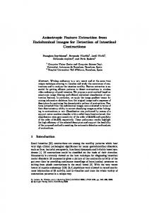
Fig. 1 Illustration of Robert Hill system for retinal scanning Blood vessel forms in the retina, is unequaled to each and every iris image. Retina is one of the reliable biometric characteristic because of its feature. The retinal points have not been lots used because of the technical restriction and expensive devices [1-4]. Some recognition studies based on retina iris images. It have been executed till Pattern of retina's blood vessels seldom converts throughout lifespan. Retina has not adjoin with surround dissimilar the former biometrics such as finger print; therefore, it is saved from outside issues. Moreover, people have not approach to the retina and hence could not delude recognition arrangements. Retinal details being little and exact is advantage when compared to other iris eye diseases; this property leads to quick recognition and manifest than other. Retina has more or less limitations [5]. People may endure from eye diseases such as cataract or glaucoma, these diseases makes recognition chore much complicated to a dandy level. The scanning process demands a lot of
978-1-5386-4304-4/18/$31.00 ©2018 IEEE
II.
RELATED WORK
2.1 METHODS FOR A/V CLASSIFICATION There are optic and geometric characteristics that alter favoritism between veins and arteries, So many methods have been searched these places for A/V sorting. Arteries are bright red while veins are dimmer, and in general artery caliber is little and vein caliber is bigger. Arteries also have dimmer and so it ponders light as a bright reflex. Another features of the retinal vessels are that, at least in the region almost optic disc, veins rarely baffle veins and arteries rarely baffle arteries. But both can bifurcate to narrower vessels, and veins and arteries can baffle each other [6]. Behdad Dashtbozorg et.al. suggested method classifies the entire vascular settling on the type of each point and assigning one of two labels to every vessel segment. Lastly the classification of vessel segment as Artery and Vein is carried out by combining results with a set of features [7].Gavin Robertson et.al. suggested method in which retinal vasculature is raised using the morphological.Further the thresholding technique in vessel map which is further used in the identification process of the retinal vessels. In regard to this process corroding and dilation process are repeated times so as to make the image reduced and authorize for fit to be processed. Then the reconstruction is performed to raise the image. Gaussian and Laplacian-of-Gaussian filter is in the enhancement process. Enhancement process is by hysteresis thresholding and background correction step [8]. Roscher R et.al. suggested a method investigating two of the machine learning algorithms: KNN and SMO.Whereas SMO can be used in classification of images which vary widely [9].Sekar C. C et.al. said the issues in planning dynamic kernel based SVMS for assortment and note of images. This approach used techniques for scheming matching kernel (IMK) and Gaussian mixture model (GMM).IMK used for matching a