Figure S1: Multiple sequence alignment of the 3' half of snR80. Conservation of snR80 across diverse yeast, showing conserved sequence and structural ...
IL HS FATHL LENGV DIRA I OQLLG SNL ST TO I. IL HS Y ATHL F E O G V N I K I I 99 L LG SNL STT I ... Ape-A L P PDSRI I E. AV Y KRL KSL A KRAGL. Tho-A I ...
Figure S1. Multiple sequence alignment of membrane-bound carbonic anhydrase proteins: pufferfish CA. XII, human CA XII, human CA VI, and human CA IV.
Figure S1. Sequence alignment of a hemoglobin alpha subunit. Tyrosine C7 (Tyr 42, human) is highly conserved. P69905.
Figure S1 Sequence alignment of TAG from different organisms (S. aureus MSSA476; S. aureus MRSA252; S. typhi; E. coli; gi|152977981, Actinobacillus ...
266, KPA171202, PMH5 and the âhly mutant were harvested from cultures grown to different time points. (24 h, 48 h, 72 h, 144 h) and incubated with HA for 10 ...
Free for academic and non- commercial use. http://www.soe.ucsc .edu/~kent/ ... et/apps/mediawiki/c loudburst- .... Includes adaptor trimming, SNP calling and.
Figure S1. DNA sequence chromatograms of wt or mutant viruses. To verify the mutations introduced in to the genomes as well as to detect possible revertants ...
The alignment consists of transferrin sequences from organisms of various taxa. The disulphide bridges (Cys11-Cys49, Cys21-Cys45, Cys271-. Cys370 ...
LKELEK QE QINSKASRRQET TKI RAELKEI ETRKTLQKI NESRS WE FEKI NKI DRPLARLI KKKREKNQI DTI KNDK GDI 400. OR F2 PA8. ORF2 PA (corrected).
SLCO4A1. PRDM16. ADRB2. ELOVL3. DIO2. PGC1ACPT1B. 0. 5. 10. 15. Re la tive m. RNA. E xp re ssio n vehicle. Rosi. GQ-16 average ± SEM; *p
Md Shakir Hussain Haider, Wajihul Hasan Khan, Farah Deeba, Sher Ali, Anwar Ahmed, Irshad H Naqvi, Ravins Dohare,. Hytham A. Alsenaidy, Abdulrahman M.
K3,8,10,14R, the four lysines at K3, 8, 10, and 14 of endogenous Htz1 were mutated to ... Data are the average of at least three independent experiments ± SD.
Supplementary Figure 3 Sequence alignment of the DnaJ-like zinc finger protein from 10 CsVYL's homologous. The DnaJ-like CR domain are highlighted in ...
H Z ITT TIL TILL ATT TILLITI TILTI. C I I II || K || 1 || 1 | | | | | | LA ALIA A. LA O 9 b A A A A LA I. H I L M N M M. å³ AID1. Z LI. I I O. TI. |. II I I ALI I I II I M N M L H | I |.
Pop500m-1km Population between 500m and 1km from HCF 5 ... 3 Population Services International, Scaling Up Point-of-Use Water Treatment in Rwanda.
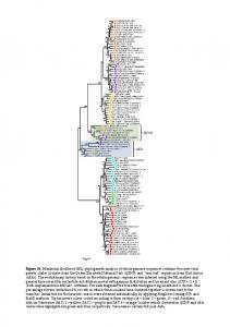
genetic clades (isolates from the Queen Elizabeth National Park (QENP) and âunusualâ sequences from East Africa. (uEA)). The evolutionary history based on ...
XIII I II I II I III TILL LITT TIL A LI TIITLITI TIL LITT TIL TILT I. O Z ITT TILL LITT TIL TIL TIL TIL TITTI. > ALLT TIL TIL TILL MITT TIL TILL TILL. A AI I I I II I TIL I I I I II II.
injection in Freund's adjuvant, with a total of three times of immunization with a time interval of two weeks. Upon collecting the anti-X protein polyclonal serum,.
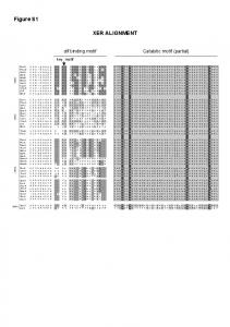
Figure S1. Sequence alignment between the four PIIIA binding ... - MDPI
Sequence alignment between the four PIIIA binding domains of the varied NaV subtypes. Loops with low sequence identify as well as the voltage sensing ...
Figure S1. Sequence alignment between the four PIIIA binding domains of the varied NaV subtypes. Loops with low sequence identify as well as the voltage sensing domain (VSD) were truncated from the sequence alignment. The P loop from the four domains were labelled as PI to PIV.
Figure S2. The top 10 conformations of PIIIA at the binding site of NaV1.4 from docking in AutoDock. A) and B) show the top 10 conformations of PIIIA together at the binding site of NaV1.4. Obviously, the top 10 conformations of PIIIA at the binding site are in varied orientations. C)–K) show the orientation of the top 10 conformations of PIIIA at the binding site of NaV1.4, respectively.
Figure S3. Binding modes of the top 10 conformations refined using MD. A)–J), Comparison of the MD refined 10 conformations of PIIIA bound with NaV1.4 (in varied colors)
with
conformations prior to MD (in gray). MD refinement could result in significant conformation deviation from that determined using docking.
Figure S4. Surface electrostatic potential of PIIIA and NaV channels. A) shows the structure of PIIIA and five key residues on its surface; B) shows the electrostatic potential of PIIIA;C)-K), show the surface electrostatic potential of the pore domain/ or PIIIA binding site of NaV subtypes.
Figure S5. Conformation of the NaV1.4 embedded in membrane in MD simulations. Root mean square deviation (RMSD) for the backbone of NaV1.4 embedded in membrane in 100 ns nonrestraint MD simulations. A),B) show the backbone RMSD of NaV1.4 in apo and bound with PIIIA, respectively. C,D) show the binding mode of PIIIA to NaV1.4. Residues at the PIIIA were labelled in italic.
Table S1. Energetic contribution for the side chain of the key residues at the binding site of NaV subtypes.