ARTICLE
Multiple linear regression model of Shiga toxin inhibitory activity of dihydroquinazolinone derivatives of Retro-2cycl Liza T. Billones1 and Junie B. Billones2* Department of Epidemiology and Biostatistics, College of Public Health Institute of Pharmaceutical Sciences, National Institutes of Health and Department of Physical Sciences and Mathematics, College of Arts and Sciences University of the Philippines Manila Taft Avenue, Ermita, Manila, Philippines 1000 1 2
Q
uantitative Structure-Activity Relationship (QSAR) study was carried out on dihydroquinazolinone derivatives of Retro-2cycl using multiple linear regression methodology. The model shows that the activities of these compounds against Shiga toxin (Stx) are dependent on the partial charges on N10 and C3, absolute hardness (η), and a lipophilicity parameter milogP. Stx tend to be inhibited by a lipophilic and electronically soft compound with electron-rich N10 and electron deficient C3 atoms. Furthermore, a retro-2cycl derivative is likely to be active against Stx if its AM1-level HOMO energy is above -8.74 kcal/ mol and its topologic polar surface area (tPSA) does not exceed 35.0 Å2. These data are crucial in structure optimization of Retro2cycl-based Shiga toxin inhibitors.
INTRODUCTION
KEYWORDS
The deleterious effects of Shiga toxin infection arise from Stx’s ability to inhibit the protein synthesis in target cells (Thompson et al. 1976) by catalytically inactivating the 60S ribosomal subunit (Reisbig et al. 1981). In particular, Shiga toxins bind to glycolipid Gb3 receptor in the extracellular surface of cell plasma membranes through the carbohydrate moiety (Jacewicz et al. 1986). After binding to the cell surface through its B-subunit (STxB), Stx is efficiently internalized from clathrin-coated pits (Sandvig & van Deurs, 1996) by endocytosis and is transported retrogradely to the endoplasmic reticulum (ER) via the endosomes, TGN protein, and Golgi complex (Ghosh et al. 1998, Sandvig et al. 2004, Johannes et al. 2008). In the ER, the enzymatically active
QSAR, dihydroquinazolinone, Retro-2, multiple linear regression, Shiga toxin, leave-one-out (LOO), leave-group-out (LGO)
*Corresponding author Email Address:
[email protected] Submitted: July 25, 2013 Revised: September 16, 2013 Accepted: September 26, 2013 Published: November 29, 2013 Editor-in-charge: Gisela P. Padilla-Concepcion Vol. 6 | No. 2 | 2013
The bacteria Shigella dysenteriae and some Escherichia coli strains are toxic to humans because they produce several structurally and functionally related protein toxins called Shiga toxins (Stx) (O’Brien & Holmes 1987), which cause common diseases such as diarrhea, hemorrhagic colitis, and a deadly complication known as hemolytic uremic syndrome (HUS) (Tarr et al. 2005). Shiga toxins have caused widespread disease and numerous deaths worldwide (Sandvig 2001). Furthermore, the ubiquity of bacteria secreting Shiga-like toxins in different types of food, including milk, apple juice and vegetables make them a serious threat to human health (Kaper 1998, Uchida et al. 1999, Bower 1999).
Philippine Science Letters
231
A1 fragment (STxA) is translocated to the cytosol and inactivates the ribosomes through inhibition of the binding of amino-acyltRNA to 60S ribosomal unit (Paton & Paton 1998), leading to a general inhibition of protein synthesis. To date, there are no known effective drugs against Stxproducing bacterial infections. Although the monoclonal antibody eculizumab has been reported to be effective in three-year old patients with severe HUS caused by Stx-producing E. coli (STEC) (Lapeyraque et al. 2011), it has not been shown to be beneficial to adults with HUS (Menne et al. 2012). Patients afflicted with the disease were just given general supportive care and those who suffered acute renal failure were subjected to dialysis. In addition, the efficacy and benefits of plasma exchange were not established and its applicability to HUS is still dubious (Tarr et al. 2012). The present strategies in toxin inhibition have been focused on the blockade of intracellular retrograde trafficking using small organic molecules (Wahome et al. 2010, Barbier et al. 2012) and inorganic ion (Mukhopadhyay & Linstedt 2012). Not long ago, a high-throughput screening study identified a lead compound called Retro-2cycl as a selective Stx retrograde trafficking blocking agent that neither mar the cellular morphology nor affect the other trafficking pathways (Stechmann et al. 2010). Very recently, the same group reported the elaboration of a cyclic variant of the lead, which showed similar mode of action but with better potency against Stx (Noel et al. 2013). In this account, the relationship between the experimental Stx inhibitory activity (EC50) (Noel et al. 2013) and the computed whole-molecule/structure-based properties of dihydroquinazolinone derivatives of Retro-2cycl were defined using multilinear regression (MLR) modeling technique. Additionally, the crucial properties (i.e. chemical markers) that discriminate the group of inactive compounds from the actives were deciphered as well. The knowledge of the discriminating factors that confer activity and the key bioactivity-determining parameters unveiled in the QSAR model provide invaluable insights for further structural refinement of Retro-2cycl-like Shiga toxin inhibitors. METHODOLOGY The structure of dihydroquinazolinones and their Shiga toxin cytotoxicity data (EC50) were obtained from the literature (Noel et al. 2013). The common QSAR descriptors such as the logarithm of octanol/water partition ratio (logP), total polar surface area (tPSA), number of atoms (Natoms), molecular weight (MW), number of O and N atoms (NO,N) (i.e. the H-bond acceptors), number of OH and NH groups (NOH,NH) (i.e. the H-bond donors), number of violations (Nviol), number of rotatable bonds (Nrotb), and molecular volume (miV) were determined using Molinspiration (www. molinspiration.com). The Molinspiration protocol for logP prediction involves adding different fragment-based contributions and correction 232
factors and fitting calculated logP with experimental logP for a training set of at least twelve thousand, mostly drug-like molecules. The topologic polar surface area (tPSA) is calculated as a sum of fragment contributions such as O- and N- centered polar fragments (Ertl et al. 2000). The molecular volume (miV) is calculated as sum of fragment contributions to 3D volume optimized by the semi-empirical AM1 method. The number of rotatable bonds (Nrotb) is defined as any single non-ring bond, bonded to nonterminal heavy (i.e. non-hydrogen) atom. The other molecular properties such as total energy (Etot), hydration energy (Ehyd), refractivity (Ref), polarizability (Pol), atomic charges (Cn), dipole moment (ρ), and the energy of the highest occupied molecular orbital (HOMO) and the lowest unoccupied molecular orbital (LUMO) included in QSAR analysis were calculated after optimizing the geometry of the molecules at the semi-empirical Austin-Model 1 (AM1) level of theory (Dewar et al. 1985) using Hyperchem 08 (Hypercube, Inc.). The molecular electrostatic potential map was generated using Spartan 08 (Wavefunction, Inc.). The multiple linear regression equation was generated using SPSS® version 20 running on Mac OS 10.8 system, employing the stepwise protocol (Efroymson 1960). In multiple linear regression, the mean function relates the k independent variables or descriptors (Xi) to the response variable or regressand E(Y|X) in the form: Equation 1 𝐸𝐸(𝑌𝑌|𝑋𝑋) = α + 𝛽𝛽1 𝑋𝑋1 + ⋯ + 𝛽𝛽𝑘𝑘 𝑋𝑋𝑘𝑘 ,
a linear function of the parameters α and βj. The α (intercept) and βj (slopes or coefficients) are estimated using the least squares 2 𝑛𝑛 𝑦𝑦𝑝𝑝𝑝𝑝𝑝𝑝𝑝𝑝also ) examined 𝑃𝑃𝑃𝑃𝑃𝑃𝑃𝑃𝑃𝑃 of the∑predictors method. The collinearity 𝑖𝑖=1(𝑦𝑦𝑒𝑒𝑒𝑒𝑒𝑒 −was 2 = 1 − protocol in SPSS®2. Finally, the 𝐿𝐿𝐿𝐿𝐿𝐿 = using the 𝑞𝑞bivariate correlation 𝑆𝑆𝑆𝑆𝑆𝑆 ∑𝑛𝑛𝑖𝑖=1(𝑦𝑦𝑒𝑒𝑒𝑒𝑒𝑒 − 𝑦𝑦̅) multiple linear regression model was validated using LeaveOne-Out (LOO) and Leave-Group-Out (LGO) techniques (vide infra). The statistical soundness of the model was evaluated on = α + 𝛽𝛽1 𝑋𝑋1 + squared ⋯ + 𝛽𝛽𝑘𝑘 𝑋𝑋correlation (𝑌𝑌|𝑋𝑋) 𝑘𝑘 , 2 the basis of 𝐸𝐸the cross-validated 𝑡𝑡𝑡𝑡𝑡𝑡𝑡𝑡 ∑ (𝑦𝑦 − 𝑦𝑦𝑝𝑝𝑝𝑝𝑝𝑝𝑝𝑝 ) coefficient, 𝑃𝑃𝑃𝑃𝑃𝑃𝑃𝑃𝑃𝑃 𝑒𝑒𝑒𝑒𝑒𝑒 𝑖𝑖=1 2 2 q , which𝑞𝑞is𝐿𝐿𝐿𝐿𝐿𝐿 determined from the ratio of prediction error sum of = =1− 2 𝑡𝑡𝑡𝑡𝑡𝑡𝑡𝑡 𝑆𝑆𝑆𝑆𝑌𝑌 αto+ the 𝛽𝛽1 𝑋𝑋sum ⋯ + 𝛽𝛽𝑒𝑒𝑒𝑒𝑒𝑒 𝑋𝑋𝑘𝑘− , 𝑦𝑦̅𝑡𝑡𝑡𝑡𝑡𝑡𝑡𝑡𝑡𝑡 (𝑌𝑌|𝑋𝑋) = 1 +∑ 𝑘𝑘 (𝑦𝑦 ) deviations the squares 𝐸𝐸 (PRESS) of𝑖𝑖=1 the squares of the 2 according to of the experimental values from their mean (SSY) ∑𝑛𝑛𝑖𝑖=1(𝑦𝑦𝑒𝑒𝑒𝑒𝑒𝑒 − 𝑦𝑦𝑝𝑝𝑝𝑝𝑝𝑝𝑝𝑝 ) 𝑃𝑃𝑃𝑃𝑃𝑃𝑃𝑃𝑃𝑃 2 Equations and 3. 𝑞𝑞𝐿𝐿𝐿𝐿𝐿𝐿2 = =1− 2 𝑛𝑛 𝑆𝑆𝑆𝑆𝑆𝑆 − 𝑦𝑦̅) 2 𝑛𝑛∑𝑖𝑖=1(𝑦𝑦𝑒𝑒𝑒𝑒𝑒𝑒 (𝐶𝐶3) 𝐸𝐸𝐸𝐸502 = 219.574 117.092 − 𝑦𝑦−𝑝𝑝𝑝𝑝𝑝𝑝𝑝𝑝 ) 𝑃𝑃𝑃𝑃𝑃𝑃𝑃𝑃𝑃𝑃(𝑁𝑁10) +∑17.930 𝑖𝑖=1(𝑦𝑦𝑒𝑒𝑒𝑒𝑒𝑒(𝜂𝜂) Equation 2 𝑞𝑞𝐿𝐿𝐿𝐿𝐿𝐿 = =1− 2 𝑛𝑛 𝑆𝑆𝑆𝑆𝑆𝑆 ∑𝑖𝑖=1+ (𝑦𝑦23.967 ̅) 𝑒𝑒𝑒𝑒𝑒𝑒 − 𝑦𝑦 −0.778 𝑚𝑚𝑖𝑖𝑙𝑙𝑙𝑙𝑙𝑙𝑙𝑙 2
∑𝑡𝑡𝑡𝑡𝑡𝑡𝑡𝑡 𝑃𝑃𝑃𝑃𝑃𝑃𝑃𝑃𝑃𝑃 𝑖𝑖=1 (𝑦𝑦𝑒𝑒𝑒𝑒𝑒𝑒 − 𝑦𝑦𝑝𝑝𝑝𝑝𝑝𝑝𝑝𝑝 ) Equation 3 =1− 2 𝑡𝑡𝑡𝑡𝑡𝑡𝑡𝑡 𝑆𝑆𝑆𝑆𝑌𝑌 ∑𝑡𝑡𝑡𝑡𝑡𝑡𝑡𝑡 (𝑦𝑦𝑒𝑒𝑒𝑒𝑒𝑒 − 𝑦𝑦̅𝑡𝑡𝑡𝑡𝑡𝑡𝑡𝑡𝑡𝑡 )2 𝑖𝑖=1 ∑𝑖𝑖=1 (𝑦𝑦𝑒𝑒𝑒𝑒𝑒𝑒 − 𝑦𝑦𝑝𝑝𝑝𝑝𝑝𝑝𝑝𝑝 ) 𝑃𝑃𝑃𝑃𝑃𝑃𝑃𝑃𝑃𝑃 2 𝑞𝑞𝐿𝐿𝐿𝐿𝐿𝐿 = =1− 2 𝑆𝑆𝑆𝑆𝑌𝑌 ∑𝑡𝑡𝑡𝑡𝑡𝑡𝑡𝑡 ̅𝑡𝑡𝑡𝑡𝑡𝑡𝑡𝑡𝑡𝑡 ) 𝑖𝑖=1 (𝑦𝑦𝑒𝑒𝑒𝑒𝑒𝑒 − 𝑦𝑦 RESULTS AND DISCUSSION 𝐸𝐸𝐸𝐸50 = 219.574 (𝑁𝑁10) + 17.930 (𝜂𝜂) − 117.092 (𝐶𝐶3) It is a fundamental thesis in QSAR study that certain + for 23.967 (𝑁𝑁10)𝑚𝑚𝑖𝑖𝑙𝑙𝑙𝑙𝑙𝑙𝑙𝑙 (𝜂𝜂)their 𝐸𝐸𝐸𝐸50 = 219.574 +account 17.930 − 117.092 3) properties of the−0.778 molecule observed(𝐶𝐶 experimental bioactivity. After all, these molecular properties have distinct −0.778factors 𝑚𝑚𝑖𝑖𝑙𝑙𝑙𝑙𝑙𝑙𝑙𝑙 23.967 influence on crucial for+drug action. For example, the 2 𝑞𝑞𝐿𝐿𝐿𝐿𝐿𝐿 =
Philippine Science Letters
Vol. 6 | No. 2 | 2013
and calculated EC50 values in Stx inhibition.
Table 1. The structures of Retro-2 derivatives and the experimental (Noel et al. 2013) and calculated EC50Calc’d values in Stx inhibition. Calc’d
18
Compound Compound Compound
Compound 18Compound
R1 R1 R1
Compound Compound 19 Compound Compound
R1R1 R1
18 1 1 19 20 11 1 1 1 18 1 1 220 2 19 22 22 2 21 2
321 19 20 33 33
22 18 420 44 44 21 22 23 18 19 55 55 521 22 23 18 24 20 19 66 66 622 23 18 24 19 721 77 20 25 23 7 24 19 20 8 21 888 22 25
3 3 3 4 4 4 5 5 5 6 6 6
R1 R1 R1
R2 R2 R2
R3 R3 R3
R2 R2
R3 R3 R3 R3 R3
R2 R2 R2 R2
R3
H
1
2 2
3 4
H HH HH HH
H H HH HH H H HH H H H HHH HH H HH H H H H H HH HHH H
3
5 H H H HHH
6
nd – not determined.
24 20 9 999 21 25 nd 22 – not determined. 23 10 2110 10 22 25 nd 23 – not determined. 24 Vol. 6 | No. 2 | 2013 11 2211 nd11 – not determined. 23 24
4
HH H
H
HHHH H HHHH H
Calc’d Calc’d Expt’l Calc’d Calc’d Expt’l H 0.3 Calc’d -3.1 (LOO) 2.0 (LGO) Calc’d Calc’d Expt’l Calc’d (LOO) (LGO) R4 Expt’l 50 (LOO) Expt’lEC Calc’d Calc’d (LGO) EC50 (LOO)Calc’d R4 EC (LGO) EC50 50 EC50 Calc’d (LOO) R4 Expt’l Calc’d (LGO) Calc’d Expt’l EC (LOO) (LGO) 50 2.0 EC50 EC EC R4 R4 50 H 0.350(M) -3.1 Expt’l EC50(LGO) EC50 (LOO) EC50 (M) R4 EC EC EC EC 50 50 (LOO) (LGO) 50 50 (M) (M) (M) EC50 EC50 R4H EC 50 8.2 7.3 7.3 (M) EC (M) (M) R4 (M) 50Experimental Calculated (LOO) Calculated EC50 (M)EC50 (M) (LGO) R4
H H H H H H HH HH HH H HH HH HH HHH H H H HHH H HH HHH H HH H HH HH HH H H H H HH HH H HH H H H HH H HH HH HH H H H
HH H HH H H H H HH HH H H H H H HH H H H H
HHHHH
H HH HH H H H H
HH
HH
(M) EC (µM) EC(M) (µM) (M) EC(M) EC50(M) EC (µM) 50 (M) (M) (M) 0.3 -3.1 2.0 (M) (M) (M) 27.3 35.0 33.9 (M)7.3 33.9 27.3 (M) 35.0 33.9 8.2 7.3 27.3 35.0 3.7 8.3 6.1 27.327.3 35.0 35.0 33.9 33.9 27.327.3 35.0 35.0 33.9 33.9 27.30.3 35.0-3.1 33.9 2.0 27.3 35.0 33.9 8.7 10.1 8.2 8.73.7 8.2 6.1 8.7 10.1 8.3 10.1 8.2 8.2 7.3 7.38.2 8.2 8.7 10.1 8.7 10.1 5.4 7.5 7.4 8.7 8.7 10.1 10.1 8.2 8.2 8.7 10.1 8.2 8.7 10.1 8.2 13.7 8.9 8.9 13.7 8.9 7.3 8.9 13.75.4 8.9 7.5 8.9 13.7 8.9 8.9 8.2 7.3 3.7 8.3 6.1 13.7 8.9 13.7 8.9 8.9 8.9 7.4 8.98.9 8.9 13.713.7 8.9 3.8 13.7 8.93.7 8.9 3.5 7.80.3 6.6 -3.1 7.5 2.0 7.8 6.6 7.5 7.8 6.6 6.1 7.5 7.83.7 6.6 7.8 6.6 7.8 6.6 7.5 7.8 6.6 7.57.5 7.5 8.3 7.8 6.6 5.4 7.5 7.5 7.4 7.8 6.6 7.5 3.8 3.7 3.5 5.45.5 6.4 6.8 6.5 6.5 5.40.3 6.4 -3.1 6.5 2.0 5.4 6.4 6.5 5.4 6.4 6.5 5.4 6.4 6.56.5 6.5 5.4 6.5 7.4 8.2 7.3 7.3 5.45.4 6.46.4 5.4 6.47.5 6.4 6.5 5.4 3.8 3.7 3.5 5.15.5 6.5 6.8 6.0 6.5 5.10.3 6.5 -3.1 6.0 2.0 5.18.0 6.5 4.5 6.0 4.7 5.1 5.1 5.1 6.56.5 6.5 6.0 6.0 6.0 50
50
50
3.7 8.3 6.16.0 6.0 8.2 5.1 5.1 7.3 6.5 6.5 7.3 3.8 5.1 3.7 6.5 3.5 6.0 5.5 6.8 6.5 0.3 -3.1 2.0 8.0 4.5 4.7 19 12.0 7.0 8.2 7.3 7.3 3.7 5.4 7.5 7.4 12.0 12.0 8.3 7.0 7.0 7.0 7.07.0 19 6.1 12.0 7.0 197.0 nd nd nd 5.5 6.8 196.5 8.0 4.5 4.7 19 19 7.47.4 8.2 7.3 7.3 4.0 6.5 4.0 6.5 7.4 3.7 8.3 6.1 4.0 6.5 5.4 7.5 7.4 1919 4.0 6.5 7.4 3.8 3.7 3.5 nd nd nd 8.0 4.5 1.4 7.5 1.4 3.7 1.4 7.5 8.3 1.4 5.4 7.5 3.8 3.7 nd nd 5.5 6.8
14.8 14.8 5.4
11.7 11.7 7.5
4.7 8.1 6.1 7.58.17.4 3.5 nd 6.5
7.5
continued on the next page 11.7
11.7 7.4 H 14.8 11.7 H 3.8 3.7 3.5 H nd nd nd H 5.5 6.8 6.5 H 8.0 4.5 4.7 Philippine Science Letters H 4-F 10.6 11.4 11.3 H 4-F 10.6 11.4 11.3 H 3.8 3.7 3.5 H 4-F 5.5 10.6 6.8 11.4 6.5 H H 8.0 4.5 4.7
H
8.1
8.1
11.7
233
11.3
H 3.4 5.2 5.2 15 8 H H 4.0 6.5 7.4 H H 4.0 6.5 7.4 8 H H 4.0 6.5 7.4 8 H 10.2 9.4 7.9 12 H 4-F 10.6 11.4 11 H 4.04.0 14.86.5 11.7 11.7 10 H H 4.0 6.5 7.4 H 6.5 7.4 11.7 11.3 10 88 8888 HHH HHH 7.4 6.5 7.4 H H H H H4.0 4.0 4.2 14.8 6.5 7.4 6.2 6.4 11.7 13 H H 4.0 6.5 7.4 H HH 1.44.8 7.5-0.3 8.1 -2.0 9 14 continuation 3.4 5.2 15 99 1. The structures of Retro-2 derivatives and theH H Table experimental (Noel and calculated7.5 EC 5.2 values in8.1 Stx inhibition. H HHet al. 2013)1.4 1.4 7.5 8.1 9
99 9999
16 Compound 12 13 11 1110 14 1510 10 10 10 1010 10 10 10 10 16 13 14 11 12 12 11 15 1711 11 11 11 1111 11 11 11 16
R1
R2
H HH HH H H
R3 H H H
H H H HH HH HH H H H H HHH HH H H H
H H HHH H H
1.4 1.4 1.4 1.4 1.4 1.4 1.4
50 7.5 8.1 7.5 8.1 7.5 7.5 8.1 7.57.5 8.1 8.1 8.1 7.5 8.1
1.4 Experimental 7.5 Calculated 8.1 (LOO) Calculated (LGO) H 2.1 0.9 0.6 R4 H 10.2 9.4 7.9 4.2(µM) 6.2(µM) 6.4(µM) EC EC EC 4-F 10.6 11.4 11.3 HH 4-F 14.8 11.7 -2.0 11.3 4.8 10.6 11.7-0.3 11.4 3.411.7 H 14.8 11.7 11.7 H 14.8 11.7 11.7 HHH 14.8 14.8 11.75.2 11.7 5.2 14.8 11.7 11.7 HHH 11.7 14.8 11.7 14.811.7 11.7 11.7 H 14.8 11.7 11.7 11.7 H 14.8 11.7 11.7 H 14.8 11.7 H 2.1 0.9 0.6 H 4.2 6.2 6.4 4.8 10.2 9.4 7.9 4-F 10.6 11.4-0.3 11.3 -2.0 H 10.2 9.4 H 3.4 5.2 5.2 7.9 4-F 10.6 11.4 11.3 4-F 10.6 11.4 11.3 4-F 10.6 11.4 11.3 H 1.5 1.2 4-F 10.6 11.4 11.3 4-F 10.6 11.4 11.3 4-F 10.6 11.4 11.3 4-F 10.6 11.411.4 11.3 11.3 11.3 1.6 4-F 10.6 11.4 4-F 10.6 4-F 10.6 11.4 11.3 H 2.1 0.9 0.6 50
HH
50
10.2
50
9.4
7.9 -2.0 5.2 6.4
14 15 1212 13 12 12 17 12 12 12 12 12 13 12 16 12
H H HHH H H H HH
4.8 -0.3 3.49.4 5.2 4.2 6.2 10.2 9.4 7.9 10.2 9.4 10.2 7.97.9 1.5 1.2 10.2 10.2 9.4 10.2 9.4 7.9 10.2 9.4 H 10.2 4.2 9.4 9.47.97.9 6.27.9
13 13 17 13 13 1313 13 13 15 14 13 1613
HH H HHH HH H H
4.2 4.2 6.2 6.21.2 6.4 4.2 6.2 6.4 4.2 6.2 6.4 4.2 6.4 6.2 4.2 6.4 4.2 1.5 6.2 -0.3 6.4 4.2 6.4 3.4 5.2 4.8 6.26.2
17 14 14 14 14 1414 14 14
H H HHH HH H H
1.5 -0.3 1.2 4.8 -0.3 -2.0 4.8 -0.3 -2.0 4.8 -0.3 4.8 -2.0 4.84.8 4.8-0.3 -0.3-0.3 -2.0 -2.0 -2.0 4.8 -0.3 -2.0
H HH
H HH H H
14 13
14 1514 16
14
14 15 15 17 15 15 1515 15 15
17 17
17
18 17
17 1818 18 20 18 18
H
20 20 20
22 20 20 234
2123
21 21 21
21
3.4 3.4 3.42.1
H H H H H H H
HH
5.2 5.2 5.2 0.9
2.1 0.9 2.1 0.9 2.1 0.9 H2.1 2.1 2.1 0.9 2.1 1.5 0.9 2.1 0.9
2.1 0.3 2.1 2.1 2.1
1.5 1.5 1.5 1.5 1.5 1.5
0.9 -3.1 0.9 0.9 0.9
1.5 1.2 1.5 1.2 1.2 1.2
1.2 1.2
H
H
H H HH
H H
5.4
8.28.2 8.2 8.2 3.73.7 3.7 3.8
3.7
Philippine Science Letters
H H H
HH
5.45.4 5.4
H
5.4 5.5
7.5
8.27.37.3
8.2 7.37.3
8.38.3 8.33.7
3.7 8.3 7.57.5 7.5
7.5 5.4 6.8
20 1.6 5.2
-2.0
5.2
5.2 5.2 5.2 0.6 20
0.6 0.6 0.9 0.6 1.2 0.9 0.6 0.6 0.6
0.6 2.0 0.6 0.6 0.6
1.2 1.6 1.2 1.6 1.6 1.6
1.6 1.6
H 8.2 7.3 1.27.3 H H 1.5 1.5 1.2 1.6 H 1.5 1.2 1.6 H H 1.5 0.3 1.2 -3.1 1.6 H 1.5 1.2 -3.1 2.02.0 1.6 HH H 0.3 -3.1 H H 0.30.3 0.3 0.3-3.1-3.1 -3.1 3.7 8.3 2.0 H 2.0 6.1
H
21
19 19 19 19 1919
1.6 20 -2.0 -2.0 5.2 -2.0 0.6
3.4 5.2 5.2 3.4 5.2 5.2 3.4 5.2 5.2 3.4 5.2 3.4 5.2 5.2 3.43.41.5 5.2 5.2 1.2 5.2 5.2 3.4 5.2 5.2
H H HHH H H H
17 17 17 17 1717
4.2 4.22.1 4.8 6.2 6.2 0.9 -0.3 4.2 6.2 6.4
H H HH
HH H H H
1816 16 16 16
1.6 6.4 5.2 -2.0 6.4 0.6 6.4
4.8 -0.3 3.4 5.2 4.82.1 -0.30.9 4.8 3.4 -0.3 5.2-2.0
H HHH H HH H
16 17 1616 16 1616 16 16
1.6
7.9 7.9 0.6 6.4 7.9
9.4 9.4 0.9
H H HH H H
H H HHH HHH H
15 15 15 15 16 15
19 17 17
10.2 10.22.1
7.4 20 20 20 20 7.3 20 7.3 20 7.3 7.3 7.3 7.3
20
0.6 0.6 1.6
20
1.6 1.6
20 1.6
2.0
2.0
20
7.3 20 7.3
continued on the next page 6.1 6.1 3.5 6.1 20 20
8.36.120 20
6.1
Vol. 6 | No. 2 | 2013
7.47.4 7.4
7.4
7.5 6.5
7.4
HH H 0.3 -3.1 2.0 0.30.3 -3.1 2.0 -3.1 2.0 1818 7.3 7.3 19 18 0.3 8.2 -3.1 2.0 18 H H H 0.3 -3.1 2.0 18 H 8.2 7.3 7.3 19 H 8.2 7.3 7.3 19 H 20 H HH 8.2 3.7 19 continuation 8.2 7.3 8.3 7.3 7.3 6.17.3 19 Table 1. The structures of Retro-2 derivatives and the experimental (Noel et al. 2013) and calculated EC values7.3 in Stx inhibition. 7.3 H8.2 8.2 19 8.23.73.7 7.3 7.3 6.1 6.1 192019 H H 8.3 HHH 7.3 7.3 20 8.2 7.3 7.3 H 8.2 7.3 7.3 19 19 H H 8.3 Experimental Calculated (LOO) Calculated (LGO) H H R4 8.28.2 7.37.3 7.37.3 19 19 Compound R1 R2 R3 H H 20 EC3.7 (µM) EC8.3 (µM) EC 6.1 (µM) H HH 3.7 5.4 8.3 7.5 6.1 7.4 20 21 50
50
20 20 20 2020 20 20 2021 21 20 21 21 21 2121 21 2121 21 21 22 212222 22 22 2222 22 22 22 22 22 23 22 222323 2323 23 23 23 23 23 23
H H H HH H H H H
23 24 232424 2424 24 24 24 24 24 24 24 25 2525 25 25 25 25 25 24 25 25 25 25
Vol. 6 | No. 2 | 2013
5.5
H
H H
8.08.0
HH H
5.5
6.8
4.54.5
8.08.0 8.0
H
H HHH
6.5
6.8 6.5 4.7 4.7 6.8 4.7 4.7 4.5 4.7 4.5 4.7 6.5 4.7 4.7 4.54.5 4.7 4.7 4.54.74.5
4.5
8.0
nd ndndnd
4.7
4.5 4.7 not nddeternined nd 4.7 4.5 nd nd nd nd nd nd nd nd
HH H H nd nd ndnd ndnd deternined ndnotnd nd H H H HHnd nd nd 8.0
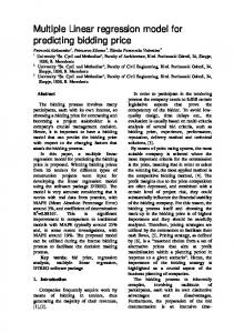
nd determined. nd––nd –not not determined. –– not determined. nd not determined. nd not determined. nd – not determined. of the ratio of octanol-water partition coefficient known nd logarithm – not determined. as–logP been commonly used in QSAR studies to estimate nd25 not has determined. the hydrophobic characteristic of a molecule (Fujita et al. 1964). nd not determined. nd –––not nd notdetermined. determined. Hydrophobicity is an important factor in drug absorption, bioavailability, Caco-2 permeability, hydrophobic drug-receptor interactions, metabolism of molecules, as well as toxicity (Fujikawa et al. 2005, Yang et al. 1996, Kubinyi 1979). The tPSA has been nd –associated not determined. with drug absorption, bioavailability, and blood-brain barrier penetration (Ertl et al. 2000). Moreover, according to nd –thenot determined. Lipinski’s “Rule of Five”, most druggable molecules have logP values not greater than 5, molecular weight not over 500, number of hydrogen bond acceptors is at most 10, and number of hydrogen bond donors is no greater than 5 (Lipinski et al. 2001). Noticeably, the limits are 5 or its multiple, hence the “Rule of 5” description. The common molecular properties of a molecule that are associated with druglikeness were determined using online program Molinspiration. As a representative output, the 2D and 3D structures of compound 1 along with the calculated properties are shown in Figure 1. Additionally, the absolute hardness (η) and absolute electronegativity (χ) values, which affects the donor/
25
H 5.4 7.5 7.4 7.57.4 7.4 HH HH 5.4 5.45.45.45.4 7.5 7.57.5 7.47.4 H HH 7.5 7.5 7.4 HH 7.5 7.4 H 5.45.4 5.4 7.5 7.4 3.7 7.4 7.43.5 5.4 3.8 7.5 HH H 3.83.85.4 3.73.7 7.5 3.5 3.5 7.4 H 3.8 3.83.7 3.7 3.5 HH H 3.5 3.8 3.7 3.5 3.8 3.7 3.5 3.7 3.5 3.5 3.5 H HHH 3.83.83.83.8 3.73.7 3.73.5 H 3.8 3.7 3.5 HH 5.5 6.8 6.5 3.8 3.7 3.5 H 5.5 6.8 6.5 HH 5.53.8 6.8 3.7 6.5 3.5 HH H H 5.5 6.8 6.5 5.5 6.8 6.5 5.5 6.8 6.5 5.5 6.86.8 6.8 6.5 6.5 6.8 6.5 H H H 5.55.5 5.5 6.5 H 5.5 6.8 6.5 HH 8.08.0 HH H H H8.0 4.5 5.5 8.0 4.5 8.0 4.5 8.0 H H 8.0 8.0 4.5
24
50
H 3.7 3.7 8.3 6.1 HHH 8.3 3.73.7 8.38.3 6.16.1 3.7 3.7 8.3 8.3 6.1 6.1 6.1 6.1 H HH H3.7 3.7 8.3 6.1 HH 8.3 6.1 3.7 8.3 H 5.45.4 7.57.5 7.4 7.4
H
23
50
ndndnd nd
ndnd not deternined nd nd
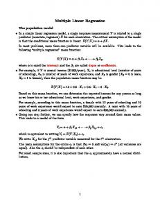
acceptor characteristic of the compound, were computed from the frontier values (Pearson 1988). H orbital energynd nd Figure 2 shows nd the HOMO and LUMO isosurfaces for compound 1. Evidently, both the HOMO and the LUMO in 1 are located within the dihydroquinazolinone core.
H
nd
nd
nd
Over 40 calculated structure-based properties for each molecule were collected and inputted in SPSS spreadsheet as independent variables, while the experimental half-maximal effective concentration (EC50) values served as the response variable. Out of 97 compounds screened experimentally, the 73 compounds, which gave protection factor (PF) values lower than the positive control (Retro-2cycl), were not further subjected for EC50 measurements (Noel et al., 2013). Hence, only 24 Retro2cycl derivatives with measured activity (EC50) against Stx were 21 2121 included in the QSAR study (Table 1). 21 21
Having relatively smaller sample size (n = 24) at hand, a model of Stx inhibition that contains at most four21 predictors, i.e. 1 predictor : 5 samples (Tabachnick 2007), was developed.
Philippine Science Letters
21
21
235 21 21
21
a
a) HOMO of 1
b
The
Figure 1. The SMILES notation, 2D (above) and 3D (below) structures, and molecular properties of compound 1 calculated using an onlinenotation, program Molinspiration. SMILES 2D (above) and 3D (below) structures,
a) HOMO of 1
Figure 2. The frontier orbitals of compound 1 a) HOMO, b) LUMO obtained at semi-empirical AM1 level.
and molecular
b) LUMO of 1
2. The frontier orbitals of compound 1 a) HOMO, b) LUMO obtained s of compound 1 calculated using an online program Figure Molinspiration.
Multiple linear regression analysis on 24-sample, 42-variable to r2 because it was significantly correlated with EC50 (r = -0.44, empirical AM1 dataset yielded a statistically sound four-predictor QSAR model. Plevel. = 0.03) and not with the other predictors. b) LUMO of 1 The model was obtained upon employing the stepwise protocol in regression analysis with modification to address the collinearity Considering that N10 values are negative, the model indicates 2. Thethat frontier compound 1 a) atom HOMO, LUMO obtained issue among the predictors in the model. In particular, Figure the stepwise a moreorbitals negativeofcharge on nitrogen (N10)b)minimizes protocol yielded a five-predictor model comprising of N10, η, C3, EC50 and thus improves the potency. Evidently, the EC50 of AM1logP, and milogP. The rule of thumb on number of predictorsAM1compound empirical level. 1 with H atom on N10 is obviously larger (less potent) requires that one predictor be removed to yield the suitable 4predictor model. Notwithstanding the favorable variance inflation factor (VIF < 5), examination of the correlation matrix revealed that AM1logP is strongly correlated with C3 (r = 0.797**). Ideally, the predictors in the model should be orthogonal, that is, independent of one another. In other words, the information brought by a predictor is unique and not carried by the other predictors in the model. Thus, a four-predictor model with adequate statistics (i.e. n = 24, r = 0.92, r2 = 0.84, s = 2.55, F = 24.80, q2LOO = 0.67, q2LGO = 0.68) was obtained after removing AM1logP (Equation 4). EC50 = 219.574(N10) + 17.930(η) - 117.092(C3) - 0.778 milogP + 23.967 Equation 4 The results showed that the partial atomic charge on N10 alone accounts for 68% in the variability of EC50 values and the combined N10 charge and absolute hardness (η) already explains approximately 80% of the variation in Stx inhibitory activity. With the addition of C3 as one of the predictors, the non-cross validated squared correlation coefficient, r2, improved further by 3%. The milogP was retained despite its minuscule contribution
22
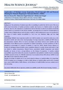
Figure 3. Calculated EC50 based on Leave-One-Out (LOO) method versus experimental EC50 (µM).
Figure 3. Calculated EC50 based on Leave-One-Out (LOO) method versus Exp
236
Philippine Science Letters
EC50 (M).
Vol. 6 | No. 2 | 2013
EC50 (M).
tells that a softer, larger, and more polarizable molecule tend to be more active against Shiga toxin. Lastly, increasing the overall hydrophobicity (milogP) of the compound tend to increase the potency of retro-2cycl derivatives.
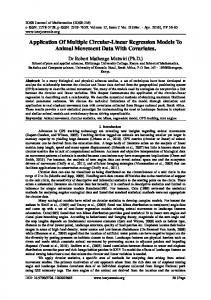
Figure 4. Calculated EC50 based on Leave-Group-Out (LGO) method versus experimental EC50 (µM).
4.
mental
Calculated EC50 based on Leave-Group-Out (LGO)
compared with that of compound 12 whose nitrogen bears a more electron-donating methyl group. In contrast, a more positive ECcharge 50 (M). on C3 improves the activity against STx. Moreover, the model suggests that EC50 decreases as the absolute hardness (η) of the compound decreases. Absolute hardness has been defined as half of the difference between the ionization energy and electron affinity (Pearson 1988). Since the ionization energy and electron affinity are related to HOMO and LUMO energies (Meissler & Tarr 2003), respectively, the η values were calculated from the computed energy of the frontier orbitals. Absolute hardness is closely associated with molecular size and electron density. In general, a molecule is hard if it is small and its electrons are less polarizable. In fact, the absolute hardness displays a strong inverse correlation with molecular volume and polarizability, with r value of -0.57** and -0.64**, respectively. Thus the model
Figure 5. Studentized residual based on Leave-Group-Out method.
The positive coefficient on N10 suggests that the magnitude of the charge must increase to improve the activity. Intuitively, this can be accomplished by installing longer R groups on nitrogen. Surprisingly, only the methylated analogue gave a lower EC50 value and those containing longer alkyl substituents were all inactive (Noel et al., 2013). Examination of the data showed that among analogous compounds (i.e. only R on N10 varying), Me gave the most negative charge on N10, followed by the longer R groups, the unsubstituted (1) being the least negative. Since 1 and the methylated analogue 12 turned out to be both active, the other predictors are worth considering as well. In going from H to Bu the charge on C3 decreases while milogP increases. The trend in C3 is in accord with experiment but that in milogP indicates otherwise. Obviously, C3 outweighs milogP when the contributions of these predictors in the model are considered. The greater potency of 12 over 1 can be explained by the fact that the former is softer than the latter. Moreover, method versus the increase in hardness as alkyl gets longer also accounts for the inactivity of compounds containing Et, Pr, and Bu substituents on N10. The QSAR model was validated using both Leave-One-Out (LOO) and Leave-Group-Out (LGO) methods. In LOO validation (Gong, 1986), one compound was removed from the data set and a multi-linear regression equation was generated based on the n – 1 24 dataset left. The generated model was used to predict the activity of the compound that was taken out of the set. Subsequently, the compound that was pulled out was added back to the data set and another one was omitted and the dataset refitted based on new n – 1 training set of compounds. This procedure was repeated until the biological activity of each and every compound in the dataset has been calculated. In contrast, in the LGO method (Osten, 1988), a group of compounds, typically 20% of sample size, is omitted at each instance. In this work, four compounds (17%) at a time were pulled out to serve as a test set and a model was generated based on n – 4 dataset. After refitting, the generated model was used to calculate the activities of the four compounds that were excluded from the set. The process was repeated in such a way that the new test set did not include those compounds, which were already picked out previously. The first round was completed when the activity of all compounds have been predicted. Three more rounds of calculations were done so that the computed activity of each compound is an average of four values. Figures 3 and 4 were generated using the data in Table 1. The plots of calculated versus experimental EC50 reveals very strong correlation between the predicted and experimental EC50 values (r = 0.87). More importantly, the cross-validated squared correlation coefficients (q2) for both LOO and LGO validation techniques were far greater than the normally recommended cut off of 0.3 for a model to be considered statistically sound (Norinder 2009). It is
Studentized based on Leave-Group-Out method. Philippine Science Letters Vol. 6 Residual | No. 2 | 2013
237
Figure 6. Molecular electrostatic potential (MEP) map for compound 13 (left) and compound 25 (right) calculated at semi-empirical AM1 level. The MEP map locates the negative (red) and the positive (blue) regions in the surface of the molecule.
Figure 6. Molecular electrostatic potential (MEP) map for compound 13 (left) and also better thancompound the common25 cut(right) off of 0.60 (Wold 1991). Thus, value AM1 above level. -8.74 kcal/mol and its topologic calculated at semi-empirical The MEP map locatespolar the surface area the q2 values obtained in this work clearly established the validity does not exceed 35.0 Å2. of the model. negative Furthermore, theand scatter of the(blue) studentized (red) the plot positive regions in the surface of the molecule. residuals (Figure 5) reveals absence of outliers (t < ±3.0) and that CONCLUSION the errors are randomly distributed around zero (t = 0.3) indicating model adequacy. Evidently, the LOO and LGO methods, and the Quantitative structure-activity relationship (QSAR) study residual analysis consistently validate the statistical soundness of has been performed on retro-2cycl derivatives with known the QSAR model. biological action on Shiga toxin. Multiple linear regression analysis was performed on the dataset composed25 of two dozens It is noteworthy that almost half of the retro-2cycl derivatives of retro-2cycl variants with experimental EC50 values which served reported by Noel and coworkers had no activity. It is therefore as the dependent variable, and over four scores of computed equally instructive to find out which structure-based properties structure-based properties which served as the independent of retro-2cycl compounds are critical for biological activity. variables. The LOO- and LGO-validated MLR model unveils Accordingly, the mean values of all the variables for the group that the charges on N10 and C3 atoms, absolute hardness, and of 40 inactive compounds (PF < 1) were compared with those in milogP are essential predictors for STx activity. Specifically, the active group composed of 24 compounds (PF > 99). Student’s the negative charge on N10 and the positive charge on C3 must t-test showed that the two groups show dramatic differences (P increase, the absolute hardness must decrease, and the lipophilic = 0.006) in their HOMO energies, which partly determine the character (milogP) of the compound must increase to improve its absolute hardness. The mean of EHOMO for the actives is -8.68 inhibitory action against Shiga toxins. Furthermore, the inactive kcal/mol and its values spread over the range from -8.61 to -8.74 dihydroquinazolinone derivatives are statistically distinct, their kcal/mol at 95% confidence interval. On the other hand, the EHOMO AM1 HOMO energies are more stabilized (EHOMO < -8.74; EHOMO = values for inactives are remarkably lower (i.e. centered at -8.80 -8.80 kcal/mol) and their topologic polar surface areas are larger kcal/mol and vary from -8.74 to -8.85 kcal/mol). Clearly, the two (tPSA > 35; tPSA = 38.0 Å2) compared with the active variants. groups are remarkably distinct in terms of their HOMO energies Certainly, the information obtained in this work will be useful in (i.e. EHOMO < -8.74 for the inactives and EHOMO > -8.74 for the future efforts to produce second generation of Retro-2cycl-based actives). The other variable that distinguishes the two groups is anti-Stx compounds. In fact, a computer-aided Shiga toxin drug topological polar surface area (tPSA). The average tPSA of active design based on Retro-2cycl structural motif is underway in our 2 2 compounds is 32.1 Å while that of the inactive group is 38.0 Å group. (P = 0.03). Figure 6 displays the molecular electrostatic potential (MEP) map for a representative active compound 13 (tPSA = 23.5 ACKNOWLEDGEMENT Å2) and an inactive compound 25 (tPSA = 51.2). The MEP map locates the negative (red) and the positive (blue) regions in the JBB appreciates the Gawad Sentenaryo (Centennial Award) 2013 surface of the molecule and thus provides a pictorial representation awarded to him by UP Manila. of the polar surface area. Obviously, compound 25 has greater PSA due to the trimethoxy substituent on the thiophenyl moiety. CONFLICTS OF INTEREST In general, a dihydroquinazolinone variant of retro-2cycl tend to be active if its HOMO is relatively destabilized, with AM1 energy None 238
Philippine Science Letters
Vol. 6 | No. 2 | 2013
CONTRIBUTION OF AUTHORS LTB performed the modeling and statistical analyses. JBB performed the calculation of the QSAR predictors. REFERENCES: Barbier J, Bouclier C, Johannes L, Gillet D. Inhibitors of the cellular trafficking of ricin. Toxins 2012; 4: 15−27. Bower JR. Foodborne diseases: Shiga toxin producing E. coli (STEC). Ped Infect Dis J 1999; 18: 909−910. Dewar MJS, Zoebisch EG, Healy EF, Stewart JJP. Development and use of quantum mechanical molecular models. 76. AM1: A new general purpose quantum mechanical molecular model. J Am Chem Soc 1985; 107(13): 3902. Efroymson MA. Multiple regression analysis. In: Ralston A, Wilf HS, Eds. Mathematical Methods for Digital Computers. New York: Wiley, 1960: 191-203. Ertl P, Rohde B, Selzer P. Fast calculation of molecular polar surface area as a sum of fragment based contributions and its application to the prediction of drug transport properties. J Med Chem 2000; 43: 3714-3717. Fujikawa M, Ano R, Nakao K, Shimizu R, Akamatsu M. Relationships between structure and high-throughput screening permeability of diverse drugs with artificial membranes: application to prediction of Caco-2 cell permeability. Bioorg Med Chem 2005 13: 4721–4732. Fujita T, Iwasa J, Hansch C. A New Substituent Constant, �, Derived from Partition Coefficients. J Am Chem Soc 1964; 86: 5175–5180. Ghose AK, Pritchett A, Crippen GM. J Comput
Chem 1988; 9: 80–90. Gong G. Cross-validation, the jackknife, and the bootstrap: excess error estimation in forward logistic regression. J Am Stat Assoc 1986; 81(393): 108–113. Jacewicz M, Clausen H, Nudelman E, Donohue-Rolfe A, Keusch GT. Pathogenesis of Shigella diarrhea. XI. Isolation of a Shigella toxin-binding glycolipid from rabbit jejunum and HeLa cells and its identification as globotriaosylceramide. J Exp Med 1986; 163: 1391–1404. Johannes L, Popoff V. Tracing the retrograde route in protein trafficking. Cell 2008; 135: 1175−1187. Kaper JB. Enterohemorrhagic Escherichia coli. Curr Opin Microbiol 1998; 1: 103−108. Kubinyi H. Nonlinear dependence of biological activity on hydrophobic character: the bilinear model. Farmaco [Sci] 1979; 34(3): 248–76. Lapeyraque AL, Malina M, Fremeaux-Bacchi V, Boppel T, Kirschfink M, Oualha M, Proulx F, Clermont MJ, Le Deist F, Niaudet P, Schaefer F. Eculizumab in severe Shiga-toxinassociated HUS. N Engl J Med 2011; 364: 2561−2563. Lipinski CA, Lombardo F, Dominy BW, Feeney PJ. Experimental and computational approaches to estimate solubility and permeability in drug discovery and development settings. Adv Drug Deliv Rev 2001; 46(1-3): 3–26. Vol. 6 | No. 2 | 2013
Meissler GL; Tarr DA, Acid-base and donor-acceptor chemistry. In: Inorganic Chemistry. 3rd Ed. New Jersey: Pearson Prentice Hall, 2003: 187-188. Menne J, Nitschke M, Stingele R, Abu-Tair M, Beneke J, Bramstedt, J, Bremer JP, Brunkhorst R, Busch V, Dengler R, Deuschl G, Fellermann K, Fickenscher H, Gerigk C, Goettsche A, Greeve J, Hafer C, Hagenmuller F, Haller H, Herget-Rosenthal S, Hertenstein B, Hofmann C, Lang M, Kielstein J T, Klostermeier U C, Knobloch J, Kuehbacher M, Kunzendorf U, Lehnert H, Manns MP, Menne TF, Meyer TN, Michael C, Munte T, Neumann-Grutzeck C, Nuernberger J, Pavenstaedt H, Ramazan L, Renders L, Repenthin J, Ries W, Rohr A, Rump LC, Samuelsson O, Sayk F, Schmidt BM, Schnatter S, Schocklmann H, Schreiber S, von Seydewitz C U, Steinhoff J, Stracke S, Suerbaum S, van de Loo A, Vischedyk M, Weissenborn K, Wellhoner P, Wiesner M, Zeissig S, Buning J, Schiffer M, Kuehbacher T. Validation of treatment strategies for enterohaemorrhagic Escherichia coli O104:H4 induced haemolytic uraemic syndrome: case-control study. BMJ 2012; 345: e4565. Mukhopadhyay S, Linstedt AD. Manganese blocks intracellular trafficking of Shiga toxin and protects against Shiga toxicosis. Science 2012; 335: 332−335. Noel R, Gupta N, Pons V, Goudet A, Garcia-Castillo MD, Michau A, Martinez J, Buisson D, Johannes L, Gillet D, Barbier J, Cintrat J. N‐Methyldihydroquinazolinone derivatives of Retro‐2 with enhanced efficacy against Shiga Toxin. J Med Chem 2013; 56, 3404−3413. Norinder U. Calculated molecular properties and multivariate statistical analysis. In: van de Waterbeemd H, Testa B, Eds. Drug Bioavailability. 2nd Ed. Vol 40. Weinheim: WileyVCH Verlag GmbH, 2009: 399. O’Brien AD, Holmes RK. Shiga and Shiga-like toxins. Microbiol Rev 1987; 51: 206−220. Osten DW. Selection of optimal regression models via crossvalidation. Chemom 1988; 2: 39−48. Paton JC, Paton AW. Pathogenesis and diagnosis of Shiga toxinproducing Escherichia coli infections. Clin Microbiol Rev 1998; 11: 450−479. Pearson, RG. Absolute electronegativity and hardness: application to inorganic chemistry. Inorg Chem 1988; 27: 73. Reisbig R, Olsnes S, Eiklid K. The cytotoxic activity of Shigella toxin. Evidence for catalytic inactivation of the 60S ribosomal subunit. J Biol Chem 1981; 256: 8739–8744. Sandvig K. Shiga Toxins. Toxicon 2001; 39: 1629–1635. Sandvig K, Spilsberg B, Lauvrak SU, Torgersen ML, Iversen TG, van Deurs B. Pathways followed by protein toxins into cells. Int J Med Microbiol 2004; 293: 483−490. Sandvig K, van Deurs B. Endocytosis, intracellular transport, and cytotoxic action of Shiga toxin and ricin. Physiol Rev 1996; 76: 949−966. Stechmann B, Bai SK, Gobbo E, Lopez R, Merer G, Pinchard S, Panigai L, Tenza D, Raposo G, Beaumelle B, Sauvaire D, Gillet D, Johannes L, Barbier J. Inhibition of retrograde
Philippine Science Letters
239
transport protects mice from lethal ricin challenge. Cell 2010; 141: 231−242. Tabachnick BG, Fidell LS. Multiple Regression. In: Using Multivariate Statistics. 5th Ed. London: Pearson/Allyn & Bacon, 2007: 123. Tarr PI, Gordon CA, Chandler WL. Shiga-toxin-producing Escherichia coli and haemolytic uraemic syndrome. Lancet 2005; 365: 1073−1086. Tarr PI, Sadler JE, Chandler WL, George JN, Tsai HM. Should all adult patients with diarrhoea-associated HUS receive plasma exchange? Lancet 2012; 379: 516−517. Thompson MR, Steinberg MS, Gemski P, Formal SB, Doctor BP. Inhibition of in vitro protein synthesis by Shigella dysenteriae 1 toxin. Biochem Biophys Res Commun 1976; 71: 783–788.
240
Uchida H, Kiyokawa N, Horie H, Fujimoto J, Takeda Y. The detection of Shiga toxin in the kidney of a patient with hemolytic uremic syndrome. Ped Res 1999; 45: 133−137. Yang S, Bumgarner JG, Kruk LF, Khaledi MG. Quantitative structure-activity relationships studies with micellar electrokinetic chromatography influence of surfactant type and mixed micelles on estimation of hydrophobicity and bioavailability. J Chromatogr A 1996; 721: 323–335. Wahome PG, Bai Y, Neal LM, Robertus JD, Mantis NJ. Identification of small-molecule inhibitors of ricin and shiga toxin using a cell-based high-throughput screen. Toxicon 2010; 56: 313−323. Wold S. Validation of QSAR’s. Quant Struct Act Relat 1991; 10(3):191-193.
Philippine Science Letters
Vol. 6 | No. 2 | 2013