ORIGINAL PAPER. Numerical simulations of human tibia osteosynthesis using modular plates based on Nitinol staples. DANIELA TARNIÅ¢Ä. 1), D. N. TARNIÅ¢Ä.
Romanian Journal of Morphology and Embryology 2010, 51(1):145–150
ORIGINAL PAPER Numerical simulations of human tibia osteosynthesis using modular plates based on Nitinol staples DANIELA TARNIŢĂ1), D. N. TARNIŢĂ2), D. POPA1), D. GRECU3), ROXANA TARNIŢĂ3), D. NICULESCU4), F. CISMARU1) 1) Department of Applied Mechanics, Faculty of Mechanics, University of Craiova 2)
Department of Anatomy
3)
Department of Surgery
Faculty of Medicine, University of Medicine and Pharmacy of Craiova 4)
Faculty of Arts and Science, Harvard University, Boston
Abstract
The shape memory alloys exhibit a number of remarkable properties, which open new possibilities in engineering and more specifically in biomedical engineering. The most important alloy used in biomedical applications is NiTi. This alloy combines the characteristics of the shape memory effect and superelasticity with excellent corrosion resistance, wear characteristics, mechanical properties and a good biocompatibility. These properties make it an ideal biological engineering material, especially in orthopedic surgery and orthodontics. In this work, modular plates for the osteosynthesis of the long bones fractures are presented. The proposed modular plates are realized from identical modules, completely interchangeable, made of titanium or stainless steel having as connecting elements U-shaped staples made of Nitinol. Using computed tomography (CT) images to provide three-dimensional geometric details and SolidWorks software package, the three dimensional virtual models of the tibia bone and of the modular plates are obtained. The finite element models of the tibia bone and of the modular plate are generated. For numerical simulation, VisualNastran software is used. Finally, displacements diagram, von Misses strain diagram, for the modular plate and for the fractured tibia and modular plate ensemble are obtained. Keywords: modular plates, Nitinol staples, numerical simulation, osteosynthesis.
� Introduction Nitinol, an alloy containing an almost equal mixture of nickel and titanium, was invented in the late 1960s and belongs to a group of materials referred to as “smart materials” because of their unique physical properties that make nitinol so remarkable: shape-memory and superelasticity. Of the SMAs available, NiTi is the only material with an appropriate level of biocompatibility and it became a key component of several revolutionary medical devices. Its properties enable new types of medical devices to be designed and produced in diverse fields of medicine. Applications of Shape Memory Alloys to the biomedical field have been successful because of their advantages over conventional implantable alloys, enhancing both the possibility and the execution of less invasive surgeries. NiTi has been approved for use in orthodontic dental archwires, endovascular stents, vena cava filters, diagnostic and therapeutic catheters, laparoscopic instruments, intracranial aneurisms clips, bone staples, and various orthopedic implants [1]. Several characteristics make NiTi extremely attractive for use in medical devices: the material has good biocompatibility [2], the devices can be pseudo-elastically or thermally deployed,
and the material can apply a constant transformation stress over a wide range of shapes [3]. Biocompatibility studies have shown NiTi to be a safe implant material, which is at least equally good as stainless steel or titanium alloys [4–6]. In orthopedic surgery, NiTi applications currently include compression bone staples used in osteotomy and fracture fixation [1, 7], rods for the correction of scoliosis, [8] shape memory expansion clamps used in cervical surgery [9], clamps in small bone surgery [10], and fixation systems for suturing tissue in minimal access surgery [11]. Other medical applications of NiTi in orthopedic surgery are presented in [12–15]. Typically, a fractured or cut bone is treated using a fixation device, which reinforces the bone and keeps it aligned during healing. Bone plates are surgical internal devices, which are used to assist in the healing of broken or fractured bones. � The AO classification of the tibia/fibula diaphyseal fractures The statistics show that ones of the most frequent fractures of human tibia bone are the diaphyseal fracture, type A (AO Classification [16]). The subgroups of the tibia/fibula diaphyseal fractures [16] are:
146
Daniela Tarniţă et al.
A1 – Simple fracture, spiroid 42–A1.1 Fibula intact; 42–A1.2 Fibula fractured at another level; 42–A1.3 Fibula fractured at the same level. These are simple diaphyseal fractures of the tibia, with a spiroid line of fracture. A2 – Simple fracture, oblique (>300) 42–A2.1 Fibula intact (Figure 1); 42–A2.2 Fibula fractured at another level (Figure 2); 42–A2.3 Fibula fractured at the same level (Figure 3).
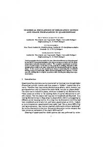
Figure 4 – Fracture 42–A3.1 Fibula intact.
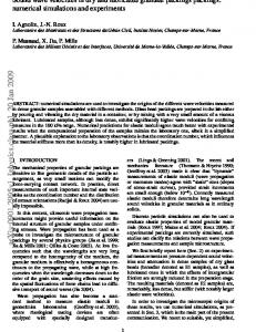
Figure 1 – Fracture 42–A2.1 Fibula intact.
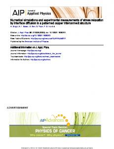
Figure 5 – Fracture 42–A3.2 Fibula fractured at another level.
Figure 2 – Fracture 42–A2.2 Fibula fractured at another level.
Figure 6 – Fracture 42–A3.3 Fibula fractured at the same level.
Figure 3 – Fracture 42–A2.3 Fibula fractured at the same level.
These are simple diaphyseal fractures of the tibia, with an oblique fracture line. An oblique fracture line is defined by its inclination equal to or greater than 300 with respect to the perpendicular to the axis of the tibia. A2 – Simple fracture, transverse (