the National Natural Science Foundation of China under Grant (#51275101), ... Y. Geng, L. Zhang, and G. Li are with the Key Lab of Health Informatics.
35th Annual International Conference of the IEEE EMBS Osaka, Japan, 3 - 7 July, 2013
Pattern Recognition Based Forearm Motion Classification for Patients with Chronic Hemiparesis* Yanjuan Geng, Student Member, IEEE, Liangqing Zhang, Dan Tang, Xiufeng Zhang, and Guanglin Li, Senior Member, IEEE �
Abstract—To make full use of electromyography (EMG) that contains rich information of muscular activities in active rehabilitation for motor hemiparetic patients, a couple of recent studies have explored the feasibility of applying pattern recognition technique to the classification of multiple motion classes for stroke survivors and reported some promising results. However, it still remains unclear if kinematics signals could also bring good motion classification performance, particularly for patients after traumatic brain damage. In this study, the kinematics signals was used for motion classification analysis in three stroke survivors and two patients after traumatic brain injury, and compared with EMG. The results showed that an average classification error of 7.9±6.8% for the affected arm over all subjects could be achieved with a linear classifier when they performed multiple fine movements, 7.9% lower than that when using EMG. With either kind of signals, the motor control ability of the affected arm degraded significantly in comparison to the intact side. The preliminary results suggested the great promise of kinematics information as well as the biological signals in detecting user’s conscious effort for robot-aided active rehabilitation.
I. INTRODUCTION Stroke is the second most common cause of death globally and the major cause of neurological disability [1]. While motor hemiparesis caused by stroke is one of the serious and common disabilities, traumatic brain damage is also an important cause of chronic hemiparesis, especially in the developing countries [1]. Currently, the rehabilitation robots, mostly providing passive physical therapy or resistance exercise, as well as the health care professionals, play an important role in assisting patients in their recovery. A number of stroke survivors and brain injured patients can regain some lost/weakened functions involved in their lower limbs using currently available passive rehabilitation techniques. However, the recovery of upper-limb functions, particularly the fine motor skills, is quite slow and often limited [2]. For the patients with severe impairment, the lack * Manuscript received January 18, 2013. This work was supported in part by the National Natural Science Foundation of China under Grant (#51275101), the National Key Basic Research Program of China (#2013CB329505), and the Guangdong Innovation Research Team Fund for Low-cost Healthcare Technologies. Y. Geng, L. Zhang, and G. Li are with the Key Lab of Health Informatics of Chinese Academy of Sciences (CAS), Shenzhen Institutes of Advanced Technology, CAS, Shenzhen, Guangdong 518055 China (Phone: 86-755-86392219; e-mail: gl.li@)siat.ac.cn.). L. Zhang is also with the Guangzhou Medical College, Guangzhou, 510182, China. D. Tang is with the Guangdong Provincial Industrial Injury Rehabilitation Center, Guangzhou, 510970, China. X. Zhang is with the National Research Center for Rehabilitation Technical Aids, Beijing, 100721, China.
978-1-4577-0216-7/13/$26.00 ©2013 IEEE
of hand dexterity may persist in their remaining years, and impact their daily activities permanently. Therefore, the active rehabilitation techniques that put great emphasis on patients’ conscious effort during physical training have attracted increasing interests of researchers recently. The previous studies have proved that the active rehabilitation methods could enhance the therapeutic effect and have a potential acceleration of brain plasticity [3-7]. For example, Krebs et al. programmed the robot MIN-MANUS to only give assistance as needed when the patients using the system grasp a joystick-like handle to finish the tasks displayed in virtual environment. Their results showed significant improvements in comparison with a group of stroke patients who received the traditional treatment [5, 6]. Tong et al. developed a robotic hand that can help stroke patients perform exercises beyond their initial range of motion by providing additional continuous torque proportional to the amplitude of EMG signal [7, 8]. It should be noted that with these robot-aided active therapies, EMG has also been used as a control signal to trigger the robotic system and to work in an “On-Off” mode, or make proportional control using the EMG amplitude[5-8]. Moreover, some exoskeleton hand robots could only assist users to perform simple functional tasks such as power grip [8]. The independent movement of each finger and the coordination of multiple muscles were rarely taken into account due to some technical limitations. Hence, the active rehabilitation is still a challenge in how to take full advantage of signals that contain the users’ motivation to inspire the user’s interest. Meanwhile, it would be important to extend the simple limb activity into more complex and coordinated limb activities for the final recovery of fine motor skills of upper extremity. To realize the identification of more complex movement intention of the users in active therapies, pattern recognition technique might be a promising choice. A number of studies have been conducted for decades by implementing EMG-based pattern recognition control strategy in upper limb prosthesis, and demonstrated good performance in restoration of users’ movement volition to control multiple degrees of freedoms [9, 10]. With the successes in control of multifunctional myoelectric prostheses, two research groups have noticed the potential uses of pattern-recognition-based methods in identifying movement classes for active rehabilitation robots [12,13]. They investigated the feasibility of applying the technique for the identification of multiple movements with stroke survivors in their preliminary studies. Lee et al. used ten channels of EMG recordings to classify six functional target tasks and got a mean classification accuracy of 71.3% for moderately impaired subjects and 37.9% for
5918
severely impaired subjects [11]. Zhang et al. recruited 12 post-stoke subjects and acquired high-density EMG signals from 89 surface electrodes. Twenty forearm and hand movements were classified based on the EMG patterns [12]. However, it still remains unclear how much the motion control ability of the affected arm degraded in comparison with that of the intact side. Moreover, whether the use of additional signals (such as the kinematics information) could improve the classification performance? Additionally, the classification performance in brain injured patients who have similar symptoms and accepted similar therapy with the stroke also remains to be explored. In this study, both EMG recordings and the kinematics signals that reflect the flexure and abductions of fingers were used for identification of motion intention in the patients with chronic hemiparesis, respectively. The motion control ability in three stroke survivors and two brain injured patients was assessed based on pattern recognition approach. II. METHODS A. Subjects Five patients with chronic hemiparesis (all male, age 19-61) participated in the study. Three of them are post-stroke subjects (designated as ST01-03) with the stroke onset time of 2 months, 4 months and 4 years, respectively. Another two subjects (designated as BI01 and BI02) had the traumatic brain injury around one year and five years ago, respectively. They were chosen based on upper limb motion impairment level assessed by a physical therapist. According to the definition of the stages in the Brunnstrom Assessment Scale, they were in the stage - and got the scores of 49-61 according to the Fugl-Meyer Assessment of Sensorimotor Recovery after Stroke, in which 0 denotes no function and 66 normal function. It was the first time for all of the subjects to participate in this kind of research study. The protocol of this study was approved by Shenzhen Institutes of Advanced Technology, Chinese Academy of Science. All subjects gave written informed consent and provided permission for publication of photographs for a scientific and educational purpose.
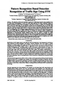
movements plus one “no movement” bilaterally, following the audio cues and the image displayed on the computer screen. These classes of the movements include 6 forearm movements and 15 hand gestures as listed in Table . The subjects could choose to give up if they were unable to finish a given motion task. The subjects were required to maintain each movement for 6 s with a moderate force and repeat 6 times with a rest time of 8 s between every two successive movements. In this study, a commercial wireless biological signal acquisition system (Delsys Inc, Boston, USA) and a pair of data gloves (Fifth Dimension Technologies, Iriven, USA ) were used to simultaneously record the EMG signals and kinematics information of both arms, respectively. For each subject, four parallel-bar EMG sensors were placed around the apex of the muscle belly in their arms, around 1-2 cm distal to the elbow crease, two were placed on the distal end over the flexor muscle and extensor muscle, and another two electrodes were placed on the thenar muscles and hypothenar muscles, respectively (Fig. 1). For comparison, another eight EMG sensors were also placed on the forearm and hand of the intact side at corresponding locations as on the affected side. In addition, subjects would wear a pair of gloves on both hands, as shown in Fig. 1. The data glove was used to measure the finger flexure as well as the abduction between the fingers with 14 flexure sensors. So a total of 8 channels of EMG signals and 14 channels of kinematics signals were recorded for each movement performed, for each arm. With the recording software, the EMG signals were filtered with a band-pass filter (20-450 Hz) and sampled at 2 kHz. The kinematics signals were scaled to 0-1 and sampled at 60Hz.
Figure 1. The placement of EMG electrodes and data glove wearing on both arm sides
B. Experiment Protocol TABLE I.
LIST OF THE MOTION CLASSES INVOLVED IN THE STUDY
Index 1 2 3 4 5 6 7 8 9 10 11
Movement Forearm flexion Forearm extension Forearm pronation Forearm supination Ulnar abduction Radial adduction Hand close Hand open Pinch grip Key grip Hook grip
Index 12 13 14 15 16 17 18 19 20 21 22
C. Data Pre-processing and Classification Considering that the major power (about 95%) of surface EMG signals is often below 400–500 Hz, the EMG signal recordings were down-sampled to 1 kHz to simplify data processing. To avoid mislabeling antagonist muscle activity as the actuated class, the active EMG data of each class were manually segmented.
Movement Tool grip Gun grip Thumb flexion Thumb extension Index flexion Thumb-index flexion Thumb-little flexion Ball grip Box grip Cylinder grip No movement
Before the experiment, we briefly explained the motion classes and experimental requirements to each of the subjects, and encouraged them to try 2-3 times to get familiar with the experimental procedure. During the experiment, they were instructed to perform 21 classes of forearm and hand
A sliding analysis window with a time length of 150ms and a time increment of 50ms was used in feature exaction. In this study, four commonly used time-domain features (mean absolute value (MAV), number of zeros crossings, number of slope sign changes, and waveform length) were extracted from each analysis window to represent the characteristics contained in the EMG recording. All these four features were also used for description of kinematics data. The feature matrix for each motion class was then fed into a classifier. For the simplicity and efficiency in computation, the linear
5919
discriminant analysis (LDA) pattern recogmt10n algorithm was used to build the classifier. Five-fold cross validation was used for model evaluation for each subject. The whole signals were randomly divided into five subsets and each time four of the five subsets were concatenated together to form a training dataset for building a classifier, and then the user-specific classifiers was tested with the remaining subset. In this study, the classification error was used as the measure index and a paired-t test was used for statistical analysis.
higher than that from the intact side when using kinematics signals only, but the difference was not significant (p-value=0.15). Classification Error Using EMG ~
~
0
itc 0
~
III. RESULTS
~
c
-2o_7-2tjf-·····-·····15.0
14. (
15.0 10.0 5.0
c3
0.0 ST01
Figure 3.
ST02
ST03
BI01
BI02
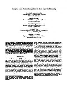
Comparison of classification performance for both arms of each subject using EMG.
Classification Error Using Kinemeics
~ ~
e
UJ c 0
~ ()
• Affected --1+.-7------------------
15.0
Gl Intact
5.0 0.0
Figure 4.
-····- ----r-1.-9----
10.0
cu
Cf) Cf)
- - Affected - - Intact
20.0
~
u
cu
20.0
Cl Intact
~ Cf)
x 10·3
::::i O"' Cf)
• Affected
()
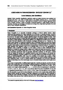
A. Comparison ofRaw Data from Bilateral Arms For each subject, the raw signals from both the intact and affected arms were compared. Fig. 2 shows the root mean square (RMS) of one-channel raw EMG signal acquired from the affected arm and the symmetrical location of the contralateral intact arm, respectively, when a subject (STOl) was performing "hand open" for six times. It can be seen from Fig. 2 that the EMG signals illustrated a consistent pattern in doing a movement by either his affected arm or intact arm. However, the RMS values of EMG from the affected side were obviously lower than those from the intact side due to the weakness of his affected arm. Similar EMG characteristics also were found in the other seven channels. 3
25.0
ST01
ST02
ST03
BI01
BI02
Comparison of classification performance for both arms of each subject using kinematics signals.
2
cu Q) ::2:
EMG Vesus Kinemics
0
~
0 0::
~
0
0
500
1000
1500
25.0
e 20.0
2000
UJ c
Figure 2. Root mean squares of one-channel raw EMG signal acquired from both the intact and affected arms. The blue and red waveforms denote the RMS from intact and affected arm, respectively.
• EMG DKinemics
15.0 1':9--
'14.f
15.0
0
10.0 ~ () -= 5.0 ·u; Cf) cu 0.0
B. Classification ofIntended Motion In the experiments of doing intended movements, the subjects STOl and ST03 could not move five fingers independently with their affected hand due to the observable weakness ofleft arm for STOl and spasticity ofright arm for ST03. STOl finished 15 of the 22 movements and ST03 finished 16 of the 22 movements. Subjects ST02, BIOl, BI02 finished 21, 22 and 21 classes, respectively. Fig. 3 shows the classification errors using EMG for each subject. It can be clearly seen that the classification errors from the affected side was significantly higher than those from the intact side for all subjects (p-value