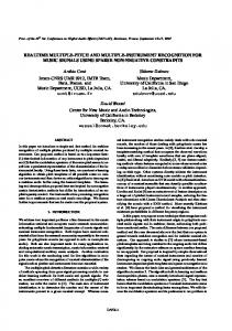
bins. Sliding windows consisting of 128 bins for each neuron were cross-correlated. .... created by holding the window for the reference neuron stationary while ...
Real-time Seizure Detection System using Multiple Single-Neuron Recordings Karen Moxon1,2, Valerie Kuzmick1, John Lafferty2, April Serfass1, Doug Szperka2, Benjamin Zale2, Jeremy Johnson2,3, Prawat Nagvajara2 1
School of Biomedical Engineering, Science and Health Systems, Drexel University, 3141 Chestnut Lane, Philadelphia, PA 2 Department of Electrical and Computer Engineering, Drexel University, 3141 Chestnut Lane, Philadelphia, PA 3 Department of Mathematics and Computer Science Drexel University, 3141 Chestnut Lane, Philadelphia, PA
Abstract- Approximately 20% of people diagnosed with epilepsy cannot be treated effectively. Consequently, there exists a significant need for alternative types of treatment. To aid in the effort of solving this problem, we developed a prototype system to detect changes in neural activity prior to the onset of a seizure. This system can be used as warning device or as part of a large system to terminate seizures in their initial stages via drug administration or nerve stimulation. The detection algorithm used data collected from intracranial electrodes. The waveforms were filtered and amplified to identify single neuron action potentials. The time of occurrence of each action potential for each neuron was then passed to a preprocessor algorithm that summed the data into 50ms time bins. Sliding windows consisting of 128 bins for each neuron were cross-correlated. The results were summed and the variance of the cross-correlation was used as a measure of global neuron correlation. The algorithm was implemented in a PC board and tested in rats treated with pentylenetetrazol (PTZ) a known seizure inducing drug. The system was 100% effective at detecting seizures approximately 4.6 seconds before seizure onset and had a false positive rate of 0.3%. Keywords – epilepsy; rat; neural system; neural control
I.
INTRODUCTION
Approximately 20% of patients with epilepsy do not respond to traditional treatment methods. Since the occurrence of seizures is spontaneous, often with no warning, a method for reliably anticipating the onset of a seizure would provide an opportunity for therapeutic intervention. Many investigations have attempted to use EEG data from surface or intracranial electrodes to capture dynamic changes in the neural signals that predict the onset of a seizure [1-2]. However, the EEG signals reflect global changes in neural activity and are often recorded far from the focal source of the seizure. Attempts to use these signals to anticipate the onset of a seizure have been mixed with generally low detection rate and high false positive rates [3]. Recent studies using multiple EEG recording sites suggest that there are unique characteristics of the epileptogenic network that can be detected prior to the onset of a seizure [45]. The EEG data just prior to the onset of a seizure show changes in the dynamic structure of the neural activity and these changes are sufficient to predict the onset of a seizure [6]. While these results are promising, additional study is
necessary to create a reliable and accurate system for therapeutic use. The underlying theory behind these detection algorithms is that the complexity of the neural signals decreases prior to the onset of a seizure [7]. By calculating complexity measures of the EEG waveform, dynamic changes can be detected. However, the reason for a decrease in complexity of the EEG signal is that neural activity becomes synchronous as the seizure develops. The EEG signals are measures of global neural activity and as such are only a reflection of this increased synchrony. Therefore, we propose to use signals recorded from single neurons to detect this synchrony. The advantages of using single neuron activity are threefold. First, the single neuron behavior at the seizure focal point represents the source of the EEG signal of interest for seizure detection. Second, measuring the synchrony of single neuron activity is computational more efficient than calculating complexity measures of the EEG waveform. Third, theoretically, the neural synchrony can be detected in the single neuron data long before the dynamic changes in the EEG are detectable. We have developed a method for detecting pre-seizure activity in rats by monitoring single neuron activity. It is well known that the EEG signal is a reflection of the underlying single-neuron activity. By recording neural activity from multiple, single-neurons, our results suggest that local changes in neural firing patterns can be detected and used to predict the onset of a seizure II.
METHODS
A. Chronic implantation of electrodes Two adult female Long-Evans rats were implanted with an eight channel electrode array to record single-neuron activity. All procedures and experiments were conducted in compliance with Drexel University animal use policies and were approved by the Drexel University Institutional Animal Care and Use Committee. The electrodes were implanted bilaterally into the temporal lobe of each rat. The rats were anesthetized with nebutal (50mg/kg). Small craniotomies were made in the skull over the implant site and the electrodes were slowly lowered into the neural tissue to a depth of 2.5 mm. Recordings were made throughout the
Report Documentation Page Report Date 25OCT2001
Report Type N/A
Title and Subtitle Real-time Seizure Detection System using Multiple Single-Neuron Recordings
Dates Covered (from... to) Contract Number Grant Number Program Element Number
Author(s)
Project Number Task Number Work Unit Number
Performing Organization Name(s) and Address(es) School of Biomedical Engineering, Science and Health Systems, Drexel University, 3141 Chestnut Lane, Philadelphia, PA
Performing Organization Report Number
Sponsoring/Monitoring Agency Name(s) and Address(es) US Army Research, Development & Standardization Group (UK) PSC 802 Box 15 FPO AE 09499-1500
Sponsor/Monitor’s Acronym(s) Sponsor/Monitor’s Report Number(s)
Distribution/Availability Statement Approved for public release, distribution unlimited Supplementary Notes Papers from the 23rd Annual International Conference of the IEEE Engineering in Medicine and Biology Society, October 25-28, 2001, held in Istanbul, Turkey. See also ADM001351 for entire conference on cd-rom., The original document contains color images. Abstract Subject Terms Report Classification unclassified
Classification of this page unclassified
Classification of Abstract unclassified
Limitation of Abstract UU
Number of Pages 4
implantation process to access electrode function. Small screws in the skull were used to anchor the electrodes, which were then cemented into place creating an electrode cap.
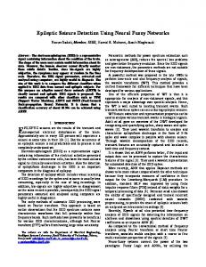
Figure 1 Binary data from the MNAP system that represented the time of occurence of action potentials for each single neuron were stored in registers. Then the number of action potentials during a 50 ms interval were summed to create a single bin. 128 bins representing 6.4 seconds of data were collected and represented a single window for cross correlation analysis.
B. Data Collection After two weeks, the rats were placed in a recording chamber and a headstage was connected to the electrode cap (Plexon, Inc., Dallas TX). The headstage transmitted neural signals from the rat to a Multi-Neuron data Acquisition Program (MNAP) that filtered and amplified the signal and discriminated single neuron action potentials from the analog signal. The times of occurrence of action potentials for each neuron were stored. During a recording session, five minutes of baseline data were collected and then the rats were given an injection of PTZ (40mg/kg). This dose of PTZ induced generalized seizure activity for up to 3 hours [8-9]. Continuous recording were made during the 3 hours post-injection. C. Behavioral Analysis During data acquisition, the animals were videotaped to monitor their behavior and to evaluate the onset of seizures. The videotapes were scored for each 30 msec frame as seizure or no-seizure as evaluated by the clonic jerking of the body and forelimbs. Half of this data was used to generate detection algorithm and the other half was used to test the algorithm. III. RESULTS A Preprocessing The raw data from the MNAP system consisted of M channels where M is the number of single neurons recorded per session. Data were represented at one millisecond (1 ms) time intervals and the occurrence of an action potential during that millisecond was represented as a 1 otherwise it
was a zero. The seizure detection unit summed the binary data over a 50 msec interval to create a single bin whose value represented the number of times the cell fired an action potential during that 50 ms interval (Fig. 1). A window was created that collected 128 bins, representing 6.4 seconds of data for each channel.
Figure 2 Schematic representation of the cross correlation method used to analyze the single neuron data and evaluate a seizure. This figure illustrates the pair-wise cross correlation for three neurons.
B. Signal Processing Solution The M windows, one for each neuron recorded, were pairwise cross correlated (Fig. 2) to create M choose 2 crosscorrelation vectors. The cross correlation vectors were created by holding the window for the reference neuron stationary while sliding each of the 128 bins of the window of the correlating neuron past the reference window one bin at a time. For each t, -127