J.W. Wheless, L.J. Willmore, J.I. Breier,. M. Kataki, J.R. Smith, D.W. King,. K.J. Meador, Y.D. Park, D.W. Loring,. G.L. Clifton, J. Baumgartner, A.B. Thomas,.
Interpretation of MEG spike source localization in frontal lobe epilepsy with multiple independent spike foci F. Tanaka1, H. Otsubo1, W.C. Gaetz1,2, and O.C. Snead1 1
2
1
Department of Neurology, The Hospital for Sick Children, Toronto; Department of Psychology, McMaster University, Hamilton, Ontario, Canada
Introduction
Magnetic source imaging using a whole-head MEG system provides a more accurate localization of epileptic focus than other routinely used noninvasive methods such as scalp video EEG and magnetic resonance imaging (MRI) [1-3]. However, MEG source localization, as estimated by the single dipole and spherical model, may not fully describe an epileptic region that includes extensive or multiple epileptic spike sources [4]. To localize the epileptogenic zone (EGZ), careful interpretation of MEG spike sources (MEG-SSs) is needed, particularly for patients with multiple independent spike foci (MISF). We report on three patients with MISF, revealed by their EEG and MEG, who underwent surgery for frontal lobe epilepsy and became seizure free for follow-up periods of 5-28 months. MEG findings were compared with the results of MRI, FDG, PET, intracranial invasive video EEG recording (IVEEG) using subdural grid electrodes, and surgery. We discuss here: (1) whether MEG-SSs can delineate the EGZ in a case with MISF, (2) whether MEGSSs can distinguish between the EGZ and the irritative zone (IZ).
2
Patient reports
Patient 1 was a 12 year-old boy (at the time of surgery) with an eight year history of medically refractory seizures consisting of a sensory aura in the right arm, eye deviation to the right side, right arm posturing, nocturnal hypermotor seizure, and secondary generalization. The scalp video EEG monitoring failed to lateralize his seizures, which showed generalized onset. MRI suggested an abnormal gyrus of thickened cortex in the right lateral frontal lobe. To localize the brain area responsible for seizure onset, an initial IVEEG was performed using subdural strip electrodes covering the bilateral fronto-parietal and temporal regions. Although synchronized interictal spikes were recorded from the bilateral hemispheres, all of the seizures originated from the left frontal region. Three
months later, the second IVEEG was performed using 108-channel subdural grid electrodes on the left hemisphere. Ictal discharges originated from the area around the subtle MRI lesion in the left lateral frontal lobe, and subsequently propagated to the left lateral parietal region where frequent interictal spikes were synchronized to the left frontal ictal onset region. To remove the EGZ, cortical excision and multiple subpial transection (MST) were performed on these regions. MST was performed on the areas where the language and motor cortices were observed by extra-operative functional mapping. The surgical specimen was reported to have tuberous sclerosis with the presence of balloon cells and form frost. Patient 2 was a 16 year-old boy (at the time of surgery) with developmental delay and nocturnal seizures since the age of two and half years. A scalp video EEG monitoring failed to lateralize his seizures because bilateral frontal and temporal epileptic discharges were recorded in both ictal and interictal states. MRI demonstrated a thickened cortex in the infero-medial part of the right frontal lobe. To lateralize the seizure onset, an initial IVEEG was performed using subdural strip electrodes placed over the bilateral fronto-temporal regions and depth electrodes placed in the bilateral mesial temporal regions. Ictal discharges were observed originating from the right frontal region, which then spread to the bilateral temporal regions. Nine months later, the second IVEEG was performed using 104-channel subdural grid electrodes over the right hemisphere, subdural strip electrodes over the right mesial and inferior frontal cortices, and depth electrodes in the bilateral temporal lobes. Ictal discharges were initiated from the right mesial and inferior frontal subdural strip electrodes, which then spread to the anterior part of the right frontal lobe. Twenty seconds after right frontal ictal onset, the bilateral temporal lobes became involved. Recordings of interictal spike discharges over the right anterior lateral frontal region were
synchronized with the spikes recorded from the right mesial and inferior frontal subdural strip electrodes. Unrelated intermittent spikes were also observed from the bilateral temporal lobes, where ictal onset was not identified. Once it was determined that the EGZ was localized to the right anterior frontal lobe, a right anterior frontal lobectomy was performed. Pathological results revealed focal cortical dysplasia distributed in the medial to inferior part of the right anterior frontal lobe where the MRI lesion was included. Patient 3 was a 15 year-old girl (at the time of surgery) with a history of complex partial seizures since the age of five. Seizures were characterized by fear feeling as aura, staring, screaming, and secondarily generalized as hypermotor seizures. The scalp video EEG monitoring suggested that her seizure originated from the left frontal region, with interictal spikes in the left temporal and the right parieto-occipital regions. MRI showed an abnormal gyrus and deep sulcus at the left frontal pole. To localize the EGZ, IVEEG was performed using 96-channel subdural grid electrodes over the left hemisphere, subdural strips over the left frontal pole, left mesial and inferior frontal cortices, depth electrodes in the left temporal lobe, and subdural strip electrodes over the right frontal, parietal, and occipital regions. Ictal discharges originated from the left frontal pole, and subsequently spread to the left anterior frontal lobe. Interictally, continuous polyspikes were seen at the left frontal pole. There were intermittent spikes over the left frontal, temporo-parieto-occipital, as well as the right parieto-occipital regions. A left anterior frontal lobectomy was performed to remove the EGZ. Pathological results revealed focal cortical dysplasia around the abnormal gyrus at the left frontal pole.
3
MEG recordings
Interictal MEG was performed at the Scripps Clinic Research Foundation in San Diego, CA, using whole-head Magnes 2500 WH (148-channel) biomagnetmeter system (4D neuroimaging, San Diego, CA). Concurrent with the magnetic recording, EEG was recorded using conventional 10-20 scalp electrodes and colloidian application (Neurofax 440A, Nihon Koden, Tokyo, Japan). The scalp EEG was simultaneously recorded to assist in spike detection. A physician or technician identified interictal spikes in the MEG and EEG recordings and triggered the collection of 6 seconds
epochs of data (5 seconds pre-trigger). Prior to analysis, the MEG and EEG waveforms were digitally filtered with a bandpass of 3 to 70 Hz to avoid collection and analysis of data contaminated by either artifact or sleep patterns. A single equivalent current dipole (ECD) model was used: ECDs were estimated by using an MRI-based spherical head model. The ECD source localization, orientation, and strength were computed with Biomagnetic Technologies software developed by 4D Neuroimaging. Dipoles with a correlation coefficient to the recorded field pattern larger than 98% and a physiologically realistic current magnitude (Q < 400 nAm) were selected. Somatosensory stimuli from pneumatically driven tactile stimulators were applied to the lower lip, first digit, and fifth digit. Data records between 45 and 75 ms after each stimulus were evaluated by means of a single equivalent current dipole (ECD) model. ECD sources of interictal spikes (MEG-spike sources) were superimposed on the subject’s MRI slices (axial, sagittal, and coronal) and also onto the cortical surface of the reconstructed threedimensional MR image. Three lipid markers were placed at fiducial points on the patient’s head during the magnetic resonance recording. The coordinates of the ECD sources were then transformed into the MRI frames.
4
MEG, MRI, and FDG PET findings
Patient 1 Table 1: MEG, MRI, and interictal FDG PET findings. EGZ: epileptogenic zone, IZ: irritative zone, Rt.: right, Lt.: left, contra: contralateral, lat: lateral, F: frontal lobe, P: parietal lobe, NA: not available. Epileptic Zones (location) number of MEG-SSs (location) MRI-lesion (location) FDG PEThypometabolism (location)
EGZ (Lt. latF-latP) 2* (Lt. latF-latP)
IZ (Rt. latF) 1 (Rt. latF)
abnormal gyrus (Lt. LatF) diffuse (Lt. latF-latP)
NA NA
*: There were two MEG-SSs in the left hemisphere; one MEG-SS overlapped a part area of the MRI lesion and another MEG-SS was located in the parietal area, which frequently generated interictal spikes synchronized to the left frontal area.
Patient 2 Table 2: MEG, MRI, and interictal FDG PET findings. ips: ipsilateral, ant: anterior, T: temporal lobe, H: hemispheric hypometabolism was observed on the same side. Epileptic Zones (location) number of MEG-SSs (location) MRI-lesion (location) FDG PEThypometabolism (location)
EGZ (Rt. antF) 4* (Rt. antF) Abnormal gyrus (Rt. inferomesial antF) focal (Rt. antF + H)
ips-IZ (Rt. T) 5 (Rt. T)
contra-IZ (Lt. T) >30 (Lt. T)
NA
NA
focal (Rt. T + H)
focal (Lt. T)
*: There were four MEG-SSs in the right frontal lobe, all of them were located in the white matter near the MRI lesion. Patient 3 Table 3: MEG, MRI, and interictal FDG PET findings. O: occipital lobe. Epileptic Zones (location) number of MEG-SSs (location) MRI-lesion (location) FDG PEThypometabolism (location)
EGZ (Lt. antF) 3* (Lt. antF) abnormal gyrus (Lt. antF) focal (Lt. antF + H)
ips-IZ (Lt. latFlatT-P-O) 3 (Lt. insula) NA diffuse (Lt. latFlatT-P-O + H)
contra-IZ P-O 5 (Rt. P-O) NA diffuse (Rt. P-O)
*: There were three MEG-SSs in the left hemisphere; all of them overlapped a part area of the MRI lesion. 5
Discussion
Our results showed that MEG-SSs identified the EGZ, but could not completely delineate the extent of the EGZ. In the case of epilepsy related to a single focal cerebral malformation, seizure generation has been shown to be associated with structural abnormalities [5-6] and that the EGZ may extend beyond the area of the cerebral malformation [5].
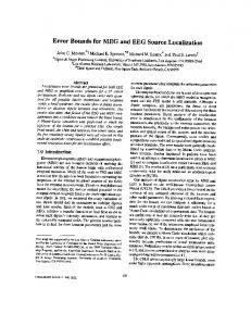
Rt.
Lt.
Figure 1: MEG-SSs were superimposed on a coronal T2-weighted MRI. MRI revealed an abnormal deep sulcus in the left anterior frontal lobe (red arrow). There were three MEG-SSs (white arrows) overlapping the MRI lesion. In Patients 1 and 3, MEG-SSs overlapped the MRI lesion, however, the extent of the EGZ could not be determined. Furthermore, in Patient 2, no MEGSSs overlapped the MRI lesion, although IVEEG showed that ictal discharges were arising from the area of MRI abnormality. In this case, MEG-SS localization might be dislocated to the white matter. Subdural electrode recording revealed multiple or extended epileptic sources in the right anterior frontal lobe in Patient 2. Because MEG data was calculated using a single ECD model, the frontal MEG-SSs might be dislocated to the center of these epileptic sources. Furthermore, the MRI lesion was located in the infero-mesial part of the anterior frontal lobe. MEG sensors can not cover inferior portions of the orbitofrontal regions, therefore a complete contour map for accurate ECD could not be made. We could not differentiate the EGZ from the IZ with the MEG findings, such as the number of MEG-SSs or the extent of MEG-SSs distribution. However, with the combined findings of MEG, MRI, and FDG PET, we were able to distinguish the EGZ from the IZ. In this study, findings from the MEG, MRI, and FDG PET co-existed in the EGZ (Table 1-3). The IZ of each patient was detected by MEG-SSs and/or FDG PEThypometabolism, and none of them showed abnormal MRI findings. A seizure free outcome was achieved with our patients by excision of only the EGZ, leaving the IZ intact. Even if the EEG and MEG show MISF, it is not necessary to remove the IZ for controlling seizures in some cases. A possible explanation for such a relationship between the EGZ and IZ is that epileptic discharges can propagate from EGZ to secondary area, which was recognized as the IZ. Such propagations can occur along the major fiber pathways (between the
cortices, between the hemispheres) and may even occur across regions without known major fiber pathways [7]. In conclusion, MEG is a sufficiently sensitive technique to detect the EGZ, as well as IZ. However, MEG can not distinguish between the EGZ and IZ. In this study, seizures were controlled by removal of the EGZ, while leaving the IZ intact. The combination of physiological (MEG), structural (MRI), and functional (FDG PET) information can provide a means of better diagnosis and treatment for some patients with MISF.
Acknowledgement We thank Mr. Andrew Wolf for editing this manuscript.
References 1. H. Stefan, S. Schneider, H. Feistel, G. Pawlik, P. Schüler, K. Abraham-Fuchs, T. Schlegel, U. Neubauer, and W. J. Huk, “Ictal and interictal activity in partial epilepsy recorded with multichannel magnetoelectroencephalography: correlation of electroencephalography/ electrocorticography, magnetic resonance imaging, single photon emission computed tomography, and positron emission tomography findings”, Epilepsia 33, 874-887, 1992. 2. R.C. Knowlton, K.D. Laxer, M.J. Aminoff, T.P.L. Roberts, S.T.C. Wong, and H.A. Rowley, “Magnetoencephalography in partial epilepsy: clinical yield and localization accuracy”, Annals of Neurology, 42, 622-631, 1997.
3. J.W. Wheless, L.J. Willmore, J.I. Breier, M. Kataki, J.R. Smith, D.W. King, K.J. Meador, Y.D. Park, D.W. Loring, G.L. Clifton, J. Baumgartner, A.B. Thomas, J.E.C. Constantinou, and A.C. Papanicolaou, “A comparison of magnetoencephalography, MRI, and V-EEG in patients evaluated for epilepsy surgery”, Epilepsia, 40, 931–941, 1999. 4. C.C. Gallen, E.C. Hirschkoff, and D.S. Buchanan, “Magnetoencephalography and magnetic source imaging – capabilities and limitations”, Neuroimaging Clinics of North America, 5, 227-249, 1995. 5. A. Palmini, F. Andermann, A. Olivier, D. Tampieri, Y. Robitaille, E. Andermann, and G. Wright, “Focal neuronal migration disorders and intractable partial epilepsy: a study of 30 patients”, Annals of Neurology, 30, 741-749, 1991. 6. R. Kuzniecky, J. M. Mounz, G. Wheatley, and R. Morawetz, “Ictal single-photon emission computed tomography demonstrates localized epileptogenesis in cortical dysplasia”, Annals of Neurology, 34, 627-631, 1993. 7. P. Jayakar, M. Duchowny, T.J. Resnick, and L.A. Alvarez, “Localization of seizure foci: pitfalls and caveats”, Journal of Clinical Neurophysiology, 8, 414-431, 1991.