1MIRALab - University of Geneva, Switzerland. 2University Hospital of Geneva, Radiology Department, Switzerland. 3University Hospital of Geneva, Orthopedic ...
EUROGRAPHICS 2009/ K. Bühler and D. Bartz
Medical Prize
Virtual Hip Joint: from Computer Graphics to Computer-Assisted Diagnosis C. Charbonnier1 , J. Schmid1 , F. Kolo-Christophe2 , N. Magnenat-Thalmann1 , C. Becker2 and P. Hoffmeyer3 1 MIRALab
- University of Geneva, Switzerland Hospital of Geneva, Radiology Department, Switzerland 3 University Hospital of Geneva, Orthopedic Department, Switzerland 2 University
Abstract Osteoarthritis (OA) is a major musculoskeletal disorder which causes are not always fully understood. Femoroacetabular impingements such as cam/ pincer cannot always explain observed OA in hips with normal morphology. This paper investigates the hypothesis of extreme repetitive movements as a source of cartilage degeneration. We present a clinical study conducted with professional ballet dancers and a methodology to perform functional simulations of the hip joint in extreme postures. Throughout the process, various computer graphics techniques are used, like motion capture, 3D body scanning and physically-based models. In addition to accelerate and strengthen some tasks, these techniques strongly participate in the clinical understanding of OA related to motion. Preliminary results have indeed shown a significant correlation between the location of impingements and radiologically observed damage zones in the labrum cartilage. Categories and Subject Descriptors (according to ACM CCS): I.3.3 [Computer Graphics]: Digitizing and scanning I.3.5 [Computer Graphics]: Physically based modeling I.6 [Simulation and Modeling]: Animation I.4 [Image Processing and Computer Vision]: Segmentation J.3 [Life and Medical Sciences]: Health
1. Introduction Osteoarthritis is a common cause of chronic hip pain. In some cases, hip OA can be explained by femoroacetabular impingements (FAI, i.e. bone collisions) caused by an abnormal morphology of the joint components: a non spherical head (cam FAI [PMD∗ 06, TGB∗ 08]) or an acetabular overcover (pincer FAI [RLK99, PMD∗ 06, TGB∗ 08]). Although the causes and effects of these FAI are well understood, some idiopathic OA is still routinely diagnosed. In this paper, the following hypothesis will be studied: can the idiopathic OA of some patients be explained by the practice of activities involving extreme and repetitive movements? To partially answer to this question, a clinical study with 30 duly informed and consenting professional ballet dancers is being conducted. The main goal is to find a relationship between their extreme recurrent motion and signs of emerging OA. We propose a methodology that is divided into 3 main parts: acquisition, modeling and analysis (Fig. 1). The main goal is to study the causes of hip OA based on c The Eurographics Association 2009.
a variety of computer graphics (CG) techniques, which play an important role in medical diagnosis and analysis. 2. From CG to medical modeling The first step aims at reconstructing 3D anatomical models from acquired medical datasets based on a segmentation procedure. While the usage of CG anatomical models is not recommended in the medical context, CG techniques can efficiently assist segmentation tasks (e.g., GPU-based approaches [HLSB04], deformable models methods applied both in CG [NMK∗ 06] and segmentation [MT96]). Another step consists in capturing motion by using standard Motion Capture technology. Before that, an anatomical calibration is executed to put in correspondence anatomical and motion frames by using the 3D body scanning technology. Finally, thanks to an optimized fitting algorithm which accounts for skin motion artifacts and anatomical constraints, the motion of the underlying anatomical models is accurately estimated from the optical markers. Each step will be now detailed.
C. Charbonnier et al. / Virtual Hip Joint Diagnosis MRI acquisition 3D body scanning
Anatomical modeling
Morphological Analysis Anatomical calibration Kinematical modeling
Motion capture
Acquisition
Modeling
Motion Analysis
Analysis
Figure 1: The pipeline of the methodology
2.1. Anatomical modeling The main idea of the proposed methodology is that 3D anatomical models can provide valuable insight into the understanding of hip pathology. In the domain of CG, various anatomical models were proposed (e.g., [SPCM97, TSB∗ 05]) with various degrees of realism. Some of the chosen simplifications or applied constraints (e.g., low number of elements to have an interactive visualization, simplified skeleton model for animation purpose) are not appropriate for medical purpose. As a result, the generation of 3D anatomical models requires a special care in case of medical analysis. Firstly, the 3D models must capture patient-specific details since those may be discriminant for a correct diagnosis. To achieve this, the models must be produced from appropriate medical acquisition modalities. We chose the magnetic resonance imaging (MRI) technique since it is not invasive, versatile and able to image simultaneously soft and bony tissues. We devised thus a protocol that offers a tradeoff between image quality and clinical constraints [Gil07]. Then, structures of interest must be accurately delineated in the medical datasets, such procedure being referred to as segmentation. Musculoskeletal segmentation is particularly challenging due to large anatomical differences existing among individuals and the presence of many artifacts in clinical MRI datasets. Our segmentation approach relies on 2-simplex discrete deformable models [Del99] that behave like a particles system. Each model represents a structure to segment (e.g., a bone). Each particle, which corresponds to a vertex of the deformable mesh, is a lumped mass subjected to forces. A first type of forces enforces a regularization, which relies on assumptions about surface regularity (smoothness and curvature) and shape priors. Then so-called external forces move particles toward anatomical boundaries based on image information such as image intensities and gradients. We exploit some CG advances on physically-based simulations of clothes [VMT05] to devise a method that uses a fast implicit numerical integration and efficient collision detection and response techniques. The proposed approach has been validated in various works [Gil07, SMT08] which reported a fast (≤ 15 mn) and accurate (1.5 mm error) segmentation approach for the muscles, cartilages, ligaments and bones of the thigh (Fig. 2a).
2.2. Anatomical calibration An anatomical calibration is required to put in correspondence anatomical and motion frames. To perform this step, most kinematic studies use a number of calibrated external anatomical landmarks (ALs) [CCCL95], which is not accurate due to ALs misplacement. Thus, our idea is to combine MRI and 3D body scan information to have a better approximation thanks to marker positions on the skin. Indeed, 3D body scanning is a modality that is traditionally used in CG to digitalize accurate skin models of the complete body (accuracy ≈ 1 mm). Therefore, the subjects underwent a 3D body scan (Vitus Pro, Vitronic, Germany) with the same markers setup used for the optical motion capture system. Thanks to this technology, the positions of the skin markers were easily identified on the resulting body scan mesh using a least-squares sphere fitting technique. Then, the body scan mesh was registered [CAVMT09] with the skin generated from MR images, performing the required calibration (Fig. 2a). Finally, the pelvic and femoral coordinates systems were computed from ALs [WSA∗ 02] defined directly on the 3D models of the bones and the hip joint center’s (HJC) position was evaluated using a functional method [Gil07]. 2.3. Motion estimation Optical motion capture systems are massively used by the computer animation community to animate virtual human bodies in a very realistic way. Since several years, the use of these systems in the medical field has also flowered. Unlike other motion acquisition devices (e.g., intra-cortical pins, external fixators), the optical system allows the recording of a large range of motion. Therefore, this technology is perfectly adapted to record the hip joint in extreme postures. For our study, the subjects were equipped with 6 skin markers on both thighs and 6 skin markers on the pelvis. Additional reflective markers were distributed over the body to confer a more complete visualization from general to detailed. Data from the subjects were acquired during dancing activities: lateral/ frontal split, developpé devant/ à la seconde, arabesque and grand plié. These movements have been chosen, because they require extreme hip flexion and/ or abduction. The markers trajectories were tracked with a Vicon system (Vicon MX 13i, Oxford Metrics, UK) using 8 infrared cameras, sampling at 120 Hz. Rigid motion of the bone segments are computed from the markers trajectories. Due to soft tissue deformation, skin markers are subject to large displacements during movement (e.g., 20 mm for a marker stuck on the thigh). The resulting estimations are thus embedded with soft tissue artifacts (STA). To overcome this issue, a nonlinear optimization algorithm [LT01] was used to find, for each segment and for each frame, the best rigid transformation that minimizes the error made globally on all the markers. Since it was observed that joint dislocation may occur due to STA, kinematic constraints allowing some shifts at the joint were also applied. c The Eurographics Association 2009.
C. Charbonnier et al. / Virtual Hip Joint Diagnosis
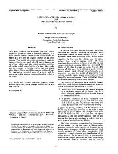
Figure 2: This image depicts a calibrated subject showing the markers setup with the reconstructed hip joints and the body scan mesh (a). The α angle (b) for this subject was normal (α = 37◦ ). Thus, the hip was not pathological but during the motion (developpé à la seconde), collisions were detected (c) at the location where lesions were diagnosed (d). The labral collisions (c) and the lesions (d) were both located in the posterosuperior area of the acetabular rim (yellow arrows).
This is actually done in two phases: 1) First, the HJC is kept fixed to avoid the dislocation 2) Then, our algorithm adjusts the HJC by detecting collisions among the articular bones, the goal being to reach the non-penetrating state. Indeed, although the HJC can be considered as fixed during low amplitude movements, this is not true for extreme motion. Thus, a potential subluxation may occur to avoid bones penetration. The validation of our method was obtained using marker position data collected during clinical motion patterns (abd/ add, flex/ ext, in/ ex rot) on 6 volunteers scanned with a dynamic MRI protocol [GPMTV05]. The kinematics derived from the marker position data was compared with that of the MRI bone tracking. The root mean square error was 0.59/0.24/0.4 mm in femur x/y/z translation and 3.86/1.71/0.55 deg in femur orientation. From these results, the STA errors for the femur were thus significantly reduced by the use of the proposed method.
3. Clinical analysis The anatomical models, the MRI data and the motion of the dancers are analyzed in a clinical way to better understand some mechanisms of OA.
3.1. Morphological analysis A morphological analysis was performed to evaluate the prevalence of the subjects’ hip joint. The morphology of the hip is well described by selected anatomical parameters. c The Eurographics Association 2009.
Two parameters are indicator of pincer FAI. The first parameter is the computation of the acetabular version [RLK99] that identifies the orientation of the opening of the acetabulum. Normal hips are inclined medially to the sagittal plane (anteversion), while abnormal hips are inclined laterally to the sagittal plane (retroversion). The second parameter refers to the depth of the acetabulum [PMD∗ 06]. If the acetabulum is too deep, pincer FAI are more favorable to occur. Finally, a standard parameter related to the femur geometry is the femoral alpha (α) neck angle that is used for detecting cam FAI [PMD∗ 06] (Fig. 2b). Deviation from the normal geometry is usually associated with larger α angles (> 60◦ ). Based on patient-specific data (MRI and 3D bones reconstruction), these standard measurement methods were implemented, improving the (subjective) reading of medical images. All the dancers’ hips were thus analyzed, according to those 3 anatomical parameters. No morphological abnormalities were detected and it was concluded that all the measured hips were anteverted, with a normal depth and an α angle in the normal range (30◦ < α < 55◦ ). The results were validated by radiological experts. 3.2. Radiological and motion analysis The subjects’ MR images were analyzed by the same radiological experts. For the majority of the dancers, labral lesions were diagnosed in the posterosuperior part of the acetabular rim. According to the morphological analysis, the dancer’s hips did not present any cam or pincer morphology. Therefore, to assess if repetitive extreme motion was at the origin
C. Charbonnier et al. / Virtual Hip Joint Diagnosis
of these lesions, the contact between the joint tissues (i.e., the cartilages and the bones) was simulated. While visualizing the subject’s hip joint in motion, collision detections were performed among the joint tissues [CLMT08]. Moreover, the surface-to-surface distance (i.e., penetration depth) was computed in order to estimate the overall impingement (Fig. 2c). The simulation results showed that strong collisions were observed when the subjects were performing extreme hip flexions or abductions. The labral collisions were located in the posterosuperior area of the acetabular rim, which corresponded with radiologically observed damage zones in the labrum (Fig. 2c,d). 4. Discussion and conclusion In this paper, a clinical study and a methodology to perform functional simulations of the hip joint in extreme postures has been described. For more than 50% of the dancers’ hips, lesions were diagnosed in the posterosuperior part of the acetabular rim. Thanks to the use of 3D anatomical models and classical CG technologies (i.e., 3D body scanning and motion capture), the conflict has been actively demonstrated in vivo and without surgery. Hence, there is little doubt that repetitive extreme hip motion could be a potential cause for the development of hip pain and OA. Apart from the assessment of idiopathic OA, our methodology has highlighted the maximum range of motion of the hip, providing us with an extreme kinematical model of the hip joint. Without the motion capture, the maximum hip range of motion would not have been clinically determinated with the same reliability. Therefore, this methodology paves the way to study other joints, in order to better understand the articular physiology and potential afflictions of mechanical etiologies. Acknowledgements This work is supported by the Co-Me project funded by Swiss National Research Foundation and by the 3D Anatomical Human project (MRTN-CT-2006-035763) funded by the European Union. We would like to thank Dr. J. Menetrey, L. Assassi, P. Volino, M. Arévalo, N. Cadi and J. Hausmann for their collaboration. We are also grateful to all volunteers from the ballet of the Great Theater of Geneva to have accepted to take part in the study.
References [CAVMT09] C HARBONNIER C., A SSASSI L., VOLINO P., M AGNENAT-T HALMANN N.: Motion study of the hip joint in extreme postures. The Visual Computer, In Press (2009). [CCCL95] C APPOZZO A., C ATANI F., C ROCE U. D., L EARDINI A.: Position and orientation of bones during movement: anatomical frame definition and determination. Clin Biomech 10 (1995), 171–178. [CLMT08] C HARBONNIER C., LYARD E., M AGNENATT HALMANN N.: Analysis of extreme hip motion in professional ballet dancers. In Proc of the 10th International Symposium on 3D Analysis of Human Movement, Amsterdam, Netherlands (October 2008).
[Del99] D ELINGETTE H.: General object reconstruction based on simplex meshes. Int J Comput Vis 32, 2 (1999), 111–146. [Gil07] G ILLES B.: Anatomical and Kinematical Modelling of the Musculoskeletal System from MRI. PhD thesis, Université de Genève, 2007. [GPMTV05] G ILLES B., P ERRIN R., M AGNENAT-T HALMANN N., VALLÉE J.-P.: Bones motion analysis from dynamic MRI: acquisition and tracking. Acad Radiol 12 (October 2005), 2385– 2392. [HLSB04] H ADWIGER M., L ANGER C., S CHARSACH H., B UEHLER K.: State of the Art Report 2004 on GPU-Based Segmentation. Tech. rep., VRVis, 2004. [LT01] L AWRENCE C., T ITS A.: A computationally efficient feasible sequential quadratic programming algorithm. SIAM J Optim 11, 4 (2001), 1092–1118. [MT96] M CINERNEY T., T ERZOPOULOS D.: Deformable models in medical images analysis: a survey. Med Image Anal 1, 2 (1996), 91–108. [NMK∗ 06] N EALEN A., M ULLER M., K EISER R., B OXERMAN E., C ARLSON M.: Physically based deformable models in computer graphics. Comput Graph Forum 25, 4 (2006), 809–836. [PMD∗ 06] P FIRRMANN C. W. A., M ENGIARDI B., D ORA C., K ALBERER F., Z ANETTI M., H ODLER J.: Cam and pincer femoroacetabular impingement: Characteristic mr arthrographic findings in 50 patients. J Radiol 240, 3 (2006), 778–785. [RLK99] R EYNOLDS D., L UCAS J., K LAUE K.: Retroversion of the acetabulum. a cause of hip pain. J of Bone Joint Surgery 81, 2 (1999), 281–288. [SMT08] S CHMID J., M AGNENAT-T HALMANN N.: Mri bone segmentation using deformable models and shape priors. In MICCAI 2008, Part I. LNCS (September 2008), Metaxas D., Axel L., Szekely G., Fichtinger G., (Eds.), vol. 5241, Springer-Verlag Berlin Heidelberg, pp. 119–126. [SPCM97] S CHEEPERS F., PARENT R., C ARLSON W., M AY S.: Anatomy-based modeling of the human musculature. In SIGGRAPH’97 (1997), pp. 163–172. [TGB∗ 08] TANNAST M., G ORICKI D., B ECK M., M URPHY S., S IEBENROCK K.: Hip damage occurs at the zone of femoroacetabular impingement. Clin Orthop Relat Res 466 (2008), 273– 280. [TSB∗ 05] T ERAN J., S IFAKIS E., B LEMKER S., N G -T HOWH ING V., L AU C., F EDKIW R.: Creating and simulating skeletal muscle from the visible human data set. IEEE Trans Visual Comput Graph 11 (2005), 317–328. [VMT05] VOLINO P., M AGNENAT-T HALMANN N.: Implicit midpoint integration and adaptive damping for efficient cloth simulation. Comput Animation Virt World 16, 3-4 (2005), 163– 175. [WSA∗ 02] W U G., S IEGLER S., A LLARD P., K IRTLEY C., L EARDINI A., ROSENBAUM D., W HITTLE M., D’L IMA D., C RISTOFOLINI L., W ITTE H., S CHMID O., S TROKES I.: ISB recommendation on definitions of joint coordinate system of various joints for the reporting of human joint motion - part I: Ankle, hip and spine. J Biomech 35, 4 (2002), 543–548.
c The Eurographics Association 2009.