D. Allemand, C. Ferrier-Pagès, P. Furla, F. Houlbrèque, S. Puverel, S. Reynaud, Ã. Tambutté, S. Tambutté and D. Zoccola,. Biomineralisation in reef-building ...
Mathematical Modeling of Calcification in Scleractinian Corals Jiangjun Cui1,
Jaap Kaandorp1
and
Denis Allemand2
1. Section Computational Science , University of Amsterdam(UvA), Kruislaan 403, 1098 SJ Amsterdam, The Netherlands 2. Centre scientifique de Monaco, avenue Saint Martin, MC 98000, Monaco, Principality of Monaco
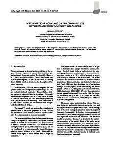
Figure 1. Left : Visualization of a computer tomography scan of a branching scleractinian coral (Madracis mirabilis). Middle: simulated nutrient-limited growth form of a coral[3]. Right: simulated nutrient and light limited growth form of a coral.
1. Introduction
4. Results
Many scleractinian corals, e.g. Stylophora pistillata, form an obligate symbiosis with zooxanthellae. Calcification in this coral is a highly biologically controlled mechanism regulated by the cellular supply of ions (Ca2+, HCO3-) and the organic matrix[1,2]. One of the most recent mechanism models for “light enhanced calcification” proposed by Denis et al. is based on the titration of H+ produced by calcification with the OH- produced by photosynthesis[1].
2. Objectives The aim of the project is to build a preliminary mathematical model of coral calcification in Stylophora pistillata which can be used to validate previously proposed mechanism of “light enhanced calcification”. Eventually we want to combine this model with models of growth and form of branching corals(Fig. 1. Left).
3. Methods Based on currently available experimental knowledge of the physiology of coral calcification [4], we can firstly build the geometry of our coral calcification problem as shown in Fig. 2. The mathematical modeling of calcium calcification is divided into four parts: photosynthesis modeling, respiration modeling, ion transportation modeling and precipitation modeling. Photosynthesis is simulated as a CO2 sink term in the oral tissue by zooxanthellae. Respiration is modeled as a HCO3- source term in the coral tissue. The CO2 assimilation rate versus irradiance is approximated by a function fitted to the experimental curve[2]. Different ion transporters are modeled by different mathematical terms according to their known characteristics[5]. Precipitation rate in virtual ECF (extracellular calcifying region) is modeled by a rate expression in an existent model for deep-sea coral calcification[7]. By using multi-compartmental modeling approach, eventually we derive a concise model consisting of 7 ODEs (ordinary differential equations, see below) to describe the whole calcification process.
Figure 3. Left : the simulated Ca2+ concentration has a reasonable steady state of around 10M (please note that free [Ca2+] in the normal cell is 50-200nM and 95-99% is bounded by Ca2+ buffer). Right: the simulated calcification rate reaches a steady state of 1 M/s which is comparable to the reported rate value for starved coral [2]. Simulations show that for various irradiance I (varying from 0 to 800 mol photon m-2 s-1), we always obtain the same curve.
Figure 4. Simulated calcification rates based on calcium signaling mechanism. If we assume that under lighting conditions, some signaling molecules diffuse through the coelenteron to increase the capacity of calcium channels on the aboral tissue, the simulated stable calcification rate does increase from 1 M/s (left figure, dark simulation) to 8 M/s (right figure, lighting simulation). Such calcium signaling mechanism assumption is based on some wellknown experimental observations, e.g., in mammalian cells responding to various extracellular stimuli, small IP3 molecules diffuse through the cytosol to open IP3R channel on ER membrane.
Conclusions The simulation results of our model seems to suggest that previously proposed mechanism model can not really explain “light enhanced calcification”. We propose a new mechanism of signaling molecules diffusing through the coelenteron to regulate the capacity of calcium channel in the aboral tissue. Future Works With close collaboration with pioneering coral experimentalists, we are preparing to further elaborate the model so that it can accurately approximate the real system. Obviously a lot of works need to be done (e.g., measuring ion transporters kinetics, detecting signal networks in the coral [6] and eventually establishing the full dynamics).
Figure 2. Geometry schematic of the model. It consists of five layers : sea water, oral tissue, coelenteron, aboral tissue and ECF (extracellular calcifying fluid) . Ca2+ in the sea water diffuses through oral tissue into coelenteron and is further transported into aboral tissue layer by L-type Ca2+ channel[1,4]. Ca2+ in the aboral tissue is eventually transported into ECF by Ca2+ ATPase which functions as an obligatory Ca2+/H+ exchanger with a probable stoichiometry of 1 to 1. The majority of HCO3- in the sea water (about 85%, the rest is through paracellular pathway which is not considered here) is transported into the oral tissue by a certain channel. Then part of HCO3- is taken by zooxanthellae through its CO2-concentrating mechanism to produce CO2 for the photosynthesis activity and secrete OH- into the coelenteron. The rest part of HCO3- further goes through the coelenteron into the oral tissue layer and is eventually transported into ECF by an unknown anion-exchanger. Ca2+ reacts with HCO3- to produce CaCO3 which is deposited into the skeleton. According to the mechanism model suggested by Denis et al.[1], H+ produced by this calcification process is removed from ECF by the Ca2+/H+ exchanger and is further transported into the coelenteron by a purported H+ATPase to neutralize with photosynthesis-induced OH-.
References 1. D. Allemand, C. Ferrier-Pagès, P. Furla, F. Houlbrèque, S. Puverel, S. Reynaud, É. Tambutté, S. Tambutté and D. Zoccola, Biomineralisation in reef-building corals: from molecular mechanisms to environmental control, C. R. Palevol 3 (2004) 453467. 2. F. Houlbrèque, É. Tambutté, D. Allemand and C. Ferrier-Pagès, Interactions between zooplankton feeding, photosynthesis and skeletal growth in the scleractinian coral Stylophora pistillata, J. Exp. Biol. 207 (2004) 1461-1469. 3. J.A. Kaandorp, P.M.A. Sloot, R.M.H. Merks, R.P.M. Bak and M.J.A. Vermeij, Morphogenesis of the branching reef coral Madracis mirabilis, Proc. Roy. Soc. B. 272:127-133, 2005 4. P. Furla, I. Glagani, I. Durand and D. Allemand, Sources and mechanisms of inorganic carbon transport for coral calcification and photosynthesis, J. Exp. Biol. 203 (2000) 3445-3457. 5. J. Cui and J.A. Kaandorp, Mathematical modeling of calcium homeostasis in yeast cells, Cell Calcium 39 (2006) 337-348. 6. D.J. Smith, D.J. Suggett and N.R. Baker, Is photoinhibition of zooxanthellae photosynthesis the primary cause of thermal bleaching in corals? Global Change Biol. 11 (2205) 1-11. 7. J.F. Adkins, E.A. Boyle, W.B. Curry and A. Lutringer, Stable isotopes in deep-sea corals and a new mechanism for “vital effects”, Geochim. Cosmochim. Acta 67 (2003) 1129-1143.
This work is carried out within the “mesoscale simulation paradigms for biological systems” funded by NWO.