acid (DNS) method (Rojas-Avelizapa et al., 1999). Under the assay conditions, ... DNS reagent was prepared by mixing distilled water 1416 ml with 10.6 g of 3,5-.
CHAPTER TWO Materials and method
2.1
Materials and Equipment
2.1.1
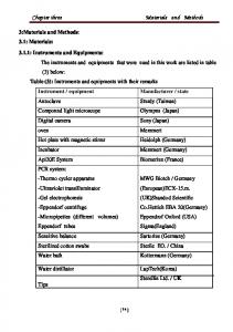
Chemical materials For the purpose of this research, every chemical used was of analytical grade
and purity, obtained mostly from Sigma-Aldrich (M) Sdn. Bhd. and Fisher (M) Sdn. Bhd. The chemicals and consumables used were: •
Chitin
•
(NH 4 ) 2 SO 4
•
MgSO 4 ·7H 2 O
•
KH 2 PO 4
•
K 2 HPO 4
•
Agar
•
Potato Dextrose Agar
•
Yeast extract
•
Distilled water
•
HCL
•
Sodium phosphate buffer
•
Bovine serum albumin
•
Coomassie Brilliant Blue G-250
•
95% ethanol
•
Phosphoric acid
•
Whatman #1 paper
•
NaOH 18
•
3,5 dinitrosalycilic acid
•
N-acetyl-d-glucosamine (NAG)
•
Phenol
•
Glass wool
•
NaKC 4 H 4 O 6
•
Na metabisulfite
•
Petri-dishes
2.1.2
Biological materials In this study, six strains of Geomyces spp. (Table 2.1) were screened for their
ability to produce chitinase enzyme using chitinase detection agar. These isolates were obtained from fungal cultural collection from National Antarctic Research Center. The soil samples were collected during summer expedition, 2007 from Fildes Peninsula, King George Island, Antarctica. King George Island is the largest island within the South Shetland Islands archipelago, North-west of the Antarctica Peninsula in the maritime Antarctica (Figure 2.1). The island which hosts 53 active research stations, have variety of rocks, soil, shallow water habitats and terrestrial ecosystems, influenced by human impacts. For example, any human activity may cause a risk of ecosystem disturbance or introduce a new non-indigenous species (Tin et al., 2009). The exact place of collection was recorded with a Global Positioning System (GPS).
19
Table 2.1 Source of isolation of Geomyces strains. Strain number
AK07KGI601R3-1 sp. 1
Isolation site
Lake GFZ
AK07KGI902 R1-1 sp. 4 Minas River
AK07KGI501R2-1 sp. 1
----
Description
GPS
Victoria Lake, Beside lake, Hilly/slopy Rocky, Algea, Birds side (feather, bones)
S62°10´09.1˝
Mountin , Lagion ,Rocky, Ornithogentic site (skua and terns) Bird skeletons, Rocky, Sea side, Slopy (hill). Vegetated area.
AK07KGI402 R1-1 sp. 1
----
Skua bird side, Rocky, Hilly, Vegetated area, Sea side.
AK07KGI102 R1-4 sp. 5
Lake estralles
Highly contaminated, Small lake near by snow patch.
Minas River
Mountin , Lagion ,Rocky, Ornithogentic site (skua and terns)
AK07KGI902 R1-1 sp. 2
W58°55´35.7˝ S62°13´38.7˝ W58°58´07.5˝ S62°09´30.0˝ W58°56´15.2˝ S62°12´14.9˝ W58°58´00.8˝ S62°12´14.9˝ W58°58´00.8˝ S62°13´38.7˝ W58°58´07.5˝
20
Figure 2.1 Geographical map of King George Island.
2.2
Research Methodology
2.2.1
Media preparation for fungal cultivation Potato Dextrose Agar (PDA) media was prepared according to manufacturers
instruction. PDA is a basic culture medium and it is also used to maintain cultures of control microorganisms. In this study, it was used to revive the test microorganisms before further analysis. For preparation of PDA, 39 g of PDA was suspended into 1L of distilled water. The mixture was then heated and continuously stirred until the entire PDA was completely dissolved. Then it was autoclaved for 20 min. at 121˚C before being poured into sterile petri dishes. 2.2.2
Inoculum on solid growth media Inoculum in PDA media were prepared by transferring 3-4 colonies from fungal
culture collection using sterile forceps and incubated at 4˚C. Five replicates were prepared and the plates were examined of any visible growth every day for 2-4 weeks. 21
2.2.3
Colloidal chitin preparation Five grams of chitin powder from crab shells (C7170, Sigma–Aldrich Co., USA)
was gradually introduced into 60 ml concentrated HCl (Merck S.A.) and kept at room temperature, with vigorous stirring, for 1 h. The mixture was filtered through glass wool and the filtrate was added to a 200 ml of 50% ethanol which was thoroughly stirred during the process. The ensuing precipitate was transferred to a glass funnel with filter paper (80 g/m) and washed with sterile distilled water until the colloidal chitin became neutral (pH 7.0). The colloidal chitin (C1) from filter paper was removed with a spatula, weighted and stored in the dark at 4°C (Skujins et al., 1965).
2.2.4
Preliminary screening for chitinase Preliminary screening of the fungi species (six organisms) was carried out in
order to ascertain and select the organisms with best chitinolytic activity potential. The following steps were taken; 1) Fungi were grown in selective medium (pH6.0) which contains of 1% colloid chitin, 0.5% (NH 4 ) 2 SO 4 , 0.05% MgSO 4 .7H 2 O, 0.24% KH 2 PO 4 , 0.06% K 2 HPO 4 .3H 2 O and 1.5% agar. The medium was adjusted to pH6 and sterilized by autoclaving at 121˚C for 15 min (Xia et al., 2009). 2) For each isolate, three replicates were prepared. Plates were incubated at 4°C. 3) Chitinase activity was visualized based on the formation of clearing or fluorescing zone around the fungal colony. 4) Measurement of Extracellular enzyme activity on the plate was described by Bradner et al. (1999). For each of the replicates, the diameter of the growth colony and activity zone was measured in two dimensions at 90° to each other and the values averaged. The index of relative enzyme activity (RA) was calculated by: 22
Relative enzyme activity 5) For the screening it was determined that a RA value of 1 or greater was classified as having significant enzyme activity (Duncan et al., 2008). 6) As it is known Geomyces spp. is psychrotolerant so, these procedures were repeated for five replicate at 250C.
2.2.5
Synthetic medium for chitinsae production in shake flask culture Fungi that showed good chitinolytic activity during the preliminary screening for
chitinase were selected for extraction of enzyme. Therefore, the following steps were taken; 1) Fungi isolates that were grown on colloid chitin agar were selected. 2) 100 ml of chitin broth was prepared (per litre: 15 g colloidal chitin; 0.5 g Yeast extract; 1 g (NH 4 ) 2 SO 4 ; 0.3 g MgSO 4 .7H 2 O; 1.36 g KH 2 PO 4 and pH adjusted to 5.5) (Rodriguez-Kabana et al., 1983). 3) Plugs of individual fungal colonies of each target species were inoculated into the 100 ml chitin broth for ten days. The mixture was incubated at 25˚C and agitated at 120 rpm aerobically on rotary shaker. After ten days, samples were filtered and centrifuged at 4000 rpm for 20 minutes. The supernatants were tested.
2.2.6
Quantitative assay-Bradford Technique Assay for total protein concentration was determined according to Bradford
(1976) using Bio-Rad protein dye reagent (Bio- Rad Laboratories GmbH, Germany) with bovine serum albumin (Sigma) as standard. A standard protein solution was prepared by dissolving 1mg of Crystal bovine serum album in 1 ml of distilled water to 23
obtain a final protein concentration of 1 mg/ mL. This was then kept in aliquot using microcentrifuge tube (1.5 ml), and stored at 4ºC. Bradford reagent was then prepared by dissolving 100 mg Coomassie Brilliant Blue G-250 in 50 ml 95% ethanol followed with addition of 100 ml 85% (w/v) phosphoric acid and diluted to 1 liter. The dissolved dye solution was filtered through Whatman #1 paper just before use. Protein standard curve was plotted to determine the concentration of the protein content present in the sample. A standard curve was prepared using 0, 10, 20, 25, 30, 40, 50 µl of standard protein solution per 10 ml of distilled water, with the addition of 5 ml of Bradford reagent. The standard and samples were replicated to three tubes. However, the protein concentration was measured with a spectrophotometer to obtain 595 nm reading. 2.2.7
Sample Analysis for protein concentration based on Bradford Technique The protein concentration of the sample was determined via addition of 5ml
Bradford reagent, 75µl of distilled water and 25µl of sample extraction. This solution was then dispensed into vortex tubes then the absorbance at 595 nm was measured after 2 min and before 1 hr in 4.5 ml cuvettes against a reagent blank (Bradford, 1976).
2.2.8
Manufacture of chitinase assay-Sugar reduction Color reagent for measuring chitinase activity was prepared according to
manufacture instructions. The chitinolytic activity was determined by measuring the amount of the reducing sugar liberated from enzymatic hydrolysis of chitin. N-acetyle glucosamine was used to construct the standard curve by adopting 3,5 dinitrosalicylic acid (DNS) method (Rojas-Avelizapa et al., 1999). Under the assay conditions, one unit (U) of enzymatic activity using chitin as a substrate is defined as the liberation of 1 µmole of the product (estimated as NAG) per hour. The measures of these assays are release of NAG, a reducing sugar from the action of chitinase enzyme on chitin substrate. 24
DNS reagent was prepared by mixing distilled water 1416 ml with 10.6 g of 3,5Dinitrosalicylic acid and 19.8g of NaOH. To the dissolved mixture, 306g Rochelle salts (Na-K tartarate), 7.6 ml Phenol (melted at 50°C), and 8.3 g of Na metabisulfite were added (Miller, 1959). The standard curve was prepared by using series of concentrations of NAG; 0, 0.090, 0.107, 0.115, 0.122, 0.130, 0.145, 0.159 and 0.166 mg/ml with added to 1 ml of 0.2M sodium phosphate buffer of pH 6.5 and then heated for one hour at 50˚ C. Later 3 ml of DNS was added and boiled for 5 minutes. Then 0.2 ml of from the previous was taken and diluted with distilled water to 2.5 ml. Absorbance at 535 nm was taken and a curve of absorbance at 535nm versus NAG concentration was plotted to give standard curve of enzyme assay. Chitinase activity throughout the fermentation process was assayed with the above colorimetric method by measuring the amount of the reducing end group of NAG degraded from chitin (Thamthiankul et al., 2004). The reacting mixture contained 1 ml of culture supernatant from sample solution and 1 ml of a 10% (w/v) suspension of colloid chitin, in 0.2 M sodium phosphate buffer pH 6.5 was incubated at 50˚C for 1 hour. The reaction was terminated by adding 1 ml of 1% NaOH followed by boiling for 5 min. Tubes were centrifuged at 4000 rpm and the reducing sugars produced were determined in the supernatant. Then 1 ml of supernatant and 1 ml of 1% of DNS were mixed and incubated for 5 min in a boiling water bath. However, the NAG production was analyzed by obtaining spectrophotometer absorbance at 535 nm and determining the enzyme activity according to the standard curve of prepared enzyme absorbance at 535 nm (Rojas-Avelizapa et al., 1999).
25
2.2.9
Optimization of chitinase activity
The optima pH and temperature were determined by assaying the chitinase activity from Geomyces sp. 5 at different pH values and temperatures using colloid chitin as the substrate. The effect of pH on chitinolytic activity was studied with citrate-phosphate buffer (pH 4.5), a sodium phosphate buffer (pH 6.5), and Tris- HCL buffer (pH 8.5). The optimum temperature for chitinase activity was determined by performing the standard assay in the range of 28-50 ˚C. The ranges of temperature experimented were 28, 37 and 50 ˚C.
26