IEEE TRANSACTIONS ON BIOMEDICAL ENGINEERING, VOL. 61, NO. 11, NOVEMBER 2014
2679
A System and Method for Online High-Resolution Mapping of Gastric Slow-Wave Activity Simon H. Bull, Gregory O’Grady, Peng Du, and Leo K. Cheng∗
Abstract—High-resolution (HR) mapping employs multielectrode arrays to achieve spatially detailed analyses of propagating bioelectrical events. A major current limitation is that spatial analyses must currently be performed “off-line” (after experiments), compromising timely recording feedback and restricting experimental interventions. These problems motivated development of a system and method for “online” HR mapping. HR gastric recordings were acquired and streamed to a novel software client. Algorithms were devised to filter data, identify slow-wave events, eliminate corrupt channels, and cluster activation events. A graphical user interface animated data and plotted electrograms and maps. Results were compared against off-line methods. The online system analyzed 256-channel serosal recordings with no unexpected system terminations with a mean delay 18 s. Activation time marking sensitivity was 0.92; positive predictive value was 0.93. Abnormal slow-wave patterns including conduction blocks, ectopic pacemaking, and colliding wave fronts were reliably identified. Compared to traditional analysis methods, online mapping had comparable results with equivalent coverage of 90% of electrodes, average RMS errors of less than 1 s, and CC of activation maps of 0.99. Accurate slow-wave mapping was achieved in near real-time, enabling monitoring of recording quality and experimental interventions targeted to dysrhythmic onset. This work also advances the translation of HR mapping toward real-time clinical application. Index Terms—Dysrhythmia, gastric electrical activity, interstitial cells of Cajal, software, visualization.
I. INTRODUCTION ASTRIC motility is regulated by slow-wave activity, generated and propagated by interstitial cells of Cajal, and dysrhythmic slow-wave activity is associated with gastric
G
Manuscript received February 9, 2014; revised April 29, 2014; accepted March 31, 2014. Date of publication May 20, 2014; date of current version October 16, 2014. This work was supported in part by the NIH (R01 DK64775), in part by the Health Research Council, New Zealand, in part by a Marsden Fast-Start Grant, and in part by a New Zealand Postdoctoral Fellowship. Asterisk indicates corresponding author. S. H. Bull was with the Auckland Bioengineering Institute, University of Auckland, Auckland 1010, New Zealand. He is now with the Technical University of Denmark, Copenhagen 2800, Denmark (e-mail: simonhbull@ gmail.com). G. O’Grady was with the Auckland Bioengineering Institute, University of Auckland, Auckland 1010, New Zealand. He is now with Westmeade Hospital, Sydney, N.S.W 2145, Australia (e-mail:
[email protected]). P. Du is with the Auckland Bioengineering Institute, University of Auckland, Auckland 1010, New Zealand (e-mail:
[email protected]). ∗ L. K. Cheng is with the Auckland Bioengineering Institute, University of Auckland, Auckland 1010, New Zealand, and also with Department of Surgery,Vanderbilt University, Nashville, TN 37235 USA (e-mail:
[email protected]). This paper has supplementary material downloadable at http://ieeexplore.ieee.org (File size: 9.4 mb). Color versions of one or more of the figures in this paper are available online at http://ieeexplore.ieee.org. Digital Object Identifier 10.1109/TBME.2014.2325829
dysmotility [1], [2]. High-resolution (HR) electrical mapping enables the analysis of slow-wave activation sequences in spatiotemporal detail. Arrays of multiple electrodes are used to simultaneously or sequentially record extracellular potentials at multiple sites, so that propagation patterns can be reconstructed from the activation times detected at each electrode [3], [4]. HR mapping has been extensively applied to define normal and aberrant slow-wave patterns in animal models [5], [6], and recently in humans [7], [8]. A major current barrier to progress in experimental and clinical HR slow-wave mapping is the inability to perform analyses “online,” i.e., during the recordings. At present, only selected electrograms can be viewed during experiments, while all spatial and graphical representations of the data must be performed after recordings have been completed [9]. This restriction is a particularly significant limitation to trialing therapeutic investigations, because manipulations such as gastric pacing for dysrhythmia ideally require immediate feedback of the transient and dynamic responses in slow-wave activity [10]. In addition, suboptimal electrode positioning currently goes undetected during experiments, potentially compromising outcomes. Online electrophysiological analysis tools that allow near real-time HR mapping are in routine clinical usage in cardiology, enabling clinicians to diagnose and treat cardiac dysrhythmias in their intervention suites [11], [12]. This clinical paradigm is also a feasible vision for gastric dysrhythmia, and the development of an online analysis platform would be a significant translational step. However, existing cardiac approaches cannot simply be adapted for gastric slow-wave analyses, due to the differences in slow-wave propagation and signal characteristics [13], [14]. Recently, a series of studies have presented robust event identification, cycle partitioning, and mapping algorithms specific for enabling automated slow-wave analyses, but the complexity means their computational demand is unsuitable for online usage [9], [13], [14]. This study addresses the above problems by presenting a new system and method for online HR gastric slow-wave mapping. Specifically, this study presents the following advances: 1) a simplified, robust and computationally efficient adaptation of existing slow-wave identification and partitioning algorithms suitable for online use; 2) a statistical method for calculating the likelihood of accurate event detection occurring in a particular channel, allowing the online identification and elimination of channels containing false-positive (FP) data to improve mapping accuracy; and 3) a user-friendly software package for online mapping, which can be readily integrated with existing methods of HR data acquisition. The developed system and method was tested in vivo in a porcine model, and validated against existing
0018-9294 © 2014 IEEE. Personal use is permitted, but republication/redistribution requires IEEE permission. See http://www.ieee.org/publications standards/publications/rights/index.html for more information.
2680
IEEE TRANSACTIONS ON BIOMEDICAL ENGINEERING, VOL. 61, NO. 11, NOVEMBER 2014
Fig. 2. Detection of slow-wave events. (a) Filtered signal trace and (b) the corresponding “detection signal” gives a short positive response to the downward slope of extracellular gastric slow-wave events (indicating a wave front passing under the electrode), and a much smaller response to the alternative downward deflections of competing noise.
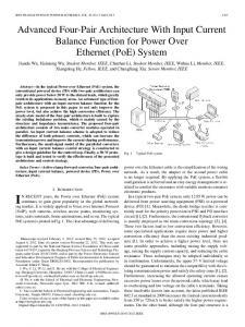
B. Event Detection Fig. 1. Flowchart illustrating the system developed to process online HR slowwave data. The signal input is shown in a light oval, the processing modules in rectangles, and the outputs in dark ovals.
off-line and manual analysis approaches. Aspects of this software and preliminary results have previously been reported in abstract form at an IEEE EMBS conference [15]. II. METHODS A flowchart outlining the system of online mapping developed here is illustrated in Fig. 1. Steps in the workflow are detailed in the subsequent sections. A. Filtering Sources of noise in extracellular recordings, such as cardiac activity (1 Hz), baseline drift (